 https://orcid.org/0000-0003-3624-1511
Abstract
Kolponomos newportensis is an enigmatic Miocene mammal allied to stem Pinnipedimorpha. It has been suggested that Kolponomos fed on hard-shelled benthic marine invertebrates by using its mandible as a wedge to dislodge its prey from the sea bottom by means of strong pull and torque forces. This unique feeding style was thought to originate from a singular case of mosaic convergence in mandible biomechanics between Kolponomos and the sabretoothed cat Smilodon, which complied with similarly strong torque forces when grappling with prey. As such forces must have reflected on the cranium as well, we hypothesize that the convergence between Kolponomos and the sabretoothed cats could have affected its shape. To test this hypothesis, we looked for patterns of morphological convergence in cranial shape between Kolponomos and sabretoothed cats. We found that Kolponomos is not distinctly closer to Smilodon than a number of other pinnipeds. Yet, local areas of shape convergence with Smilodon are observed in the canine area and the posterior part of the cranium, that is where the bite applies and the temporalis muscle is located, respectively. These results indicate that the mosaic convergence present between the mandibles of Kolponomos and Smilodon is partially reflected in the cranium as well.
INTRODUCTION
Convergence is among the best studied macroevolutionary patterns. It describes the independent appearance of similar phenotypes in distantly related lineages (Harmon et al., 2005; Losos, 2011). Among mammals, patterns of morphological convergence were reported for a variety of organisms and traits, such as elongated upper canines in different predatory species (Wroe et al., 2008; Melchionna et al., 2021), locomotory mode in Euarchontoglires (Geng et al., 2020) and sloths (Serio et al., 2020), high-crowned molars in grazers (Palmqvist et al., 2008; Gomes Rodrigues et al., 2017) and fused metatarsals in artiodactyls (Yohe & Solounias, 2020).
Although convergence is usually explained in terms of shared adaptations towards a common functional end, different phenotypes can produce highly similar functional outputs, known as many-to-one mapping of form to function (Alfaro et al., 2005; Wainwright, 2007; Losos, 2011; Renaud et al., 2018). This means that functional convergence might occur even in the absence of morphological convergence (Sansalone et al., 2020). In extinct species, the correct recognition of functional convergence is further complicated by the limited availability and incomplete preservation state of most fossil remains, and by the impossibility to perform in vivo observations. Therefore, palaeontologists usually infer functional performance by means of different approaches, such as indirect measurements of bite mechanics (Organ et al., 2011; Piras et al., 2013) and simulating bone loadings (Strait et al., 2013; Dickson & Pierce, 2019).
Recently, Tseng et al. (2016) described the feeding style of the extinct pinniped Kolponomos, reporting a rather unusual case of mosaic convergence (convergence limited to parts of a single anatomical structure) between Kolponomos and the sabretoothed cat Smilodon. Kolponomos was first described by Stirton (1960) from partial remains found in Lower Miocene marine deposits along the north-eastern margin of the Pacific Ocean in Oregon and Washington (Kolponomos clallamensis, Clallam Formation, Clallam Bay, Washington). Originally linked to Procyonidae, the attribution of Kolponomos to any specific group of Carnivora remained elusive. After the discovery of a nearly complete cranium with partial dentition, the mandible and a few post-cranial bones in the Miocene of the Nye Mudstone Formation, Oregon (Late Arikareean Land Mammal Age) a new species, K. newportensis, was erected and linked to Amphicyonidae (Tedford et al., 1994). The ancestry of Kolponomos was later revisited again and the genus was allied to stem Pinnipedimorpha (Paterson et al., 2020). Kolponomos probably had an amphibious lifestyle and fed on hard-shelled marine invertebrates living on rocky substrates. Tseng et al. (2016) suggested that during prey-capture Kolponomos used the anterior portion of its jaw as an anchoring fulcrum to dislodge shells from the hard bottom, whereas the broad occlusal surfaces on the cheek teeth served to crush the prey exoskeletons. By applying Finite Element Analysis (FEA) and geometric morphometrics (GM) to the mandibles of seven different mammal species, Tseng and co-authors found functional convergence between Kolponomos and Smilodon relative to the prey-capture phase, whereas the simulated prey-crushing phase indicated differences in structural stiffness-bite combinations between Kolponomos and any other taxon. The surprising convergence between Kolponomos and Smilodon was explained in terms of the way they both used their mandibles, wedging on prey items with their front teeth to withstand strong torque forces during biting. Both machairodontine (sabretooth) cats and Kolponomos possessed prominent mastoid processes on the cranium, suggesting the presence of massive atlanto-mastoid muscles to facilitate the head-flexing movements originating the torque (Antón et al., 2004; Tseng et al., 2016). The direct involvement of the cranium suggests that convergence can be identified in this structure as well, since the generation of strong muscle forces requires proper attachment areas for the muscles and a skull shape able to withstand reaction forces. Here, we address the role of cranial shape in morphological convergence between Kolponomos and sabretooth species (Barbourofelis fricki, Homotherium serum, Hoplophoneus primaevus, Smilodon fatalis, Smilodon californicus) while accounting for phylogeny. Our hypothesis is that morphological resemblance between Kolponomos and the sabretooths should be limited at best, given the presence of long and flattened upper canines exclusive to sabretooths and the complex, somewhat unique, feeding habits of Kolponomos. Yet, by mapping convergence on the 3D model of the cranium, which is now possible thanks to a recently developed method [conv.map (Melchionna et al., 2021)], we expect to find convergence in the posterior portion of the skull, which provides the area of attachment for the powerful neck and masticatory muscles that both Kolponomos and the sabretooths are inferred to have possessed, and at its front, where contact with the prey item applied.
MATERIAL AND METHODS
Data collection and preparation
We collected 159 3D cranial models belonging to different taxonomic groups: Barbourofelidae (N = 1), Canidae (N = 37), Felidae (N = 32), Herpestidae (N = 1), Hyaenidae (N = 4), Mephitidae (N = 2), Mustelidae (N = 23), Nimravidae (N = 2), Odobenidae (N = 2), Otariidae (N = 14), Phocidae (N = 20), Prionodontidae (N = 1), Ursidae (N = 11), Viverridae (N = 9), and Kolponomos newportensis. Overall, our data set included 98 species (both extant and extinct; see Supporting Information, Appendix S1; Table S1). Three-dimensional surfaces and/or CT scans used in this study belong to different online databases: Morphosource (https://www.morphosource.org/), Digimorph (http://digimorph.org/index.phtml), Phenome10K (https://www.phenome10k.org/), KUPRI (http://dmm.pri.kyoto-u.ac.jp/dmm/WebGallery/index.html), Sketchfab (https://sketchfab.com/), or were acquired by one of us (D.T.) from museum collections. The skull digital models of Yoshi garevskii and Dinofelis barlowi were kindly provided by Nikolai Spassov and Denis Geraads (Spassov & Geraads, 2015) and Justin Adams and colleagues (Adams et al., 2015), respectively. Surfaces were built from CT scans by using Amira (v.5.4.5, Visualization Sciences Group, 2013) and Geomagic Studio (v.2014.3.0.1781, Geomagic, 2014).
On each specimen, we placed a set of 37 landmarks by using the software Amira (Fig. 1, Table 1) to record cranial shape variation. A number of fossil specimens presented some missing parts and distortion due to taphonomic processes, which we accounted for before landmark positioning. The 3D skull model of Kolponomos newportensis (USNM215070) was taken from Tseng et al. (2016) and is the holotype of the species. To account for taphonomic effects on its cranial shape, we retrodeformed the digital model of K. newportensis by using the algorithm embedded in the R package ‘Morpho’, the retroDeformMesh function (Schlager et al., 2018). This function symmetrizes the 3D surface by using bilateral landmark information. To perform retrodeformation, we placed bilateral landmarks on the most recognizable and best-preserved parts of the skull (i.e. the frontal process of the zygomatic bone, the dental alveoli, the postorbital constriction). The retrodeformed digital model was further processed in Geomagic Studio to repair limited damage due to the taphonomic process. The 3D model was eventually smoothed and symmetrized by taking the best-preserved (right) side of the specimen as a reference (Fig. 2).
Landmark descriptions
| Number | Landmark definition |
|---|---|
| 1 and 2 | Alveolar margin at the posterior aspect of the last incisor (right and left side) |
| 3 and 4 | Alveolar margin at the anterior aspect of the canine (right and left side) |
| 5 and 6 | Frontal zygomatic process (right and left side) |
| 7 and 8 | Postorbital constriction (right and left side) |
| 9 and 10 | End of supraorbital margin (right and left side) |
| 11 and 12 | Margin of the nasal aperture (right and left side) |
| 13 and 14 | Infraorbital foramen (right and left side) |
| 15 and 16 | Alveolar margin at the posterior aspect of the last molar (right and left side) |
| 17 | Anterior edge of the premaxilla |
| 18 | Anterior point of midline between nasals |
| 19 | Topmost point of the nuchal crest |
| 20 | Interpremaxillary suture at the inferior margin of the nasal aperture |
| 21 and 22 | Anterior edge of the premaxilla-jugal suture |
| 23 and 24 | Root of the zygomatic arch (right and left side) |
| 25 and 26 | Lambdoid crest (right and left side) |
| 27 and 28 | Dorsalmost point of the mastoid process (right and left side) |
| 29 and 30 | Dorsalmost point of the acoustic meatus (right and left side) |
| 31 and 32 | Dorsalmost point of the frontal process of the zygomatic bone (right and left side) |
| 33 and 34 | Ventral intersection between the zygomatic arch and the maxilla |
| 35 and 36 | Alveolar margin at the posterior aspect of the canine (right and left side) |
| 37 | Juncture between incisor (internal side) |
| Number | Landmark definition |
|---|---|
| 1 and 2 | Alveolar margin at the posterior aspect of the last incisor (right and left side) |
| 3 and 4 | Alveolar margin at the anterior aspect of the canine (right and left side) |
| 5 and 6 | Frontal zygomatic process (right and left side) |
| 7 and 8 | Postorbital constriction (right and left side) |
| 9 and 10 | End of supraorbital margin (right and left side) |
| 11 and 12 | Margin of the nasal aperture (right and left side) |
| 13 and 14 | Infraorbital foramen (right and left side) |
| 15 and 16 | Alveolar margin at the posterior aspect of the last molar (right and left side) |
| 17 | Anterior edge of the premaxilla |
| 18 | Anterior point of midline between nasals |
| 19 | Topmost point of the nuchal crest |
| 20 | Interpremaxillary suture at the inferior margin of the nasal aperture |
| 21 and 22 | Anterior edge of the premaxilla-jugal suture |
| 23 and 24 | Root of the zygomatic arch (right and left side) |
| 25 and 26 | Lambdoid crest (right and left side) |
| 27 and 28 | Dorsalmost point of the mastoid process (right and left side) |
| 29 and 30 | Dorsalmost point of the acoustic meatus (right and left side) |
| 31 and 32 | Dorsalmost point of the frontal process of the zygomatic bone (right and left side) |
| 33 and 34 | Ventral intersection between the zygomatic arch and the maxilla |
| 35 and 36 | Alveolar margin at the posterior aspect of the canine (right and left side) |
| 37 | Juncture between incisor (internal side) |
Landmark descriptions
| Number | Landmark definition |
|---|---|
| 1 and 2 | Alveolar margin at the posterior aspect of the last incisor (right and left side) |
| 3 and 4 | Alveolar margin at the anterior aspect of the canine (right and left side) |
| 5 and 6 | Frontal zygomatic process (right and left side) |
| 7 and 8 | Postorbital constriction (right and left side) |
| 9 and 10 | End of supraorbital margin (right and left side) |
| 11 and 12 | Margin of the nasal aperture (right and left side) |
| 13 and 14 | Infraorbital foramen (right and left side) |
| 15 and 16 | Alveolar margin at the posterior aspect of the last molar (right and left side) |
| 17 | Anterior edge of the premaxilla |
| 18 | Anterior point of midline between nasals |
| 19 | Topmost point of the nuchal crest |
| 20 | Interpremaxillary suture at the inferior margin of the nasal aperture |
| 21 and 22 | Anterior edge of the premaxilla-jugal suture |
| 23 and 24 | Root of the zygomatic arch (right and left side) |
| 25 and 26 | Lambdoid crest (right and left side) |
| 27 and 28 | Dorsalmost point of the mastoid process (right and left side) |
| 29 and 30 | Dorsalmost point of the acoustic meatus (right and left side) |
| 31 and 32 | Dorsalmost point of the frontal process of the zygomatic bone (right and left side) |
| 33 and 34 | Ventral intersection between the zygomatic arch and the maxilla |
| 35 and 36 | Alveolar margin at the posterior aspect of the canine (right and left side) |
| 37 | Juncture between incisor (internal side) |
| Number | Landmark definition |
|---|---|
| 1 and 2 | Alveolar margin at the posterior aspect of the last incisor (right and left side) |
| 3 and 4 | Alveolar margin at the anterior aspect of the canine (right and left side) |
| 5 and 6 | Frontal zygomatic process (right and left side) |
| 7 and 8 | Postorbital constriction (right and left side) |
| 9 and 10 | End of supraorbital margin (right and left side) |
| 11 and 12 | Margin of the nasal aperture (right and left side) |
| 13 and 14 | Infraorbital foramen (right and left side) |
| 15 and 16 | Alveolar margin at the posterior aspect of the last molar (right and left side) |
| 17 | Anterior edge of the premaxilla |
| 18 | Anterior point of midline between nasals |
| 19 | Topmost point of the nuchal crest |
| 20 | Interpremaxillary suture at the inferior margin of the nasal aperture |
| 21 and 22 | Anterior edge of the premaxilla-jugal suture |
| 23 and 24 | Root of the zygomatic arch (right and left side) |
| 25 and 26 | Lambdoid crest (right and left side) |
| 27 and 28 | Dorsalmost point of the mastoid process (right and left side) |
| 29 and 30 | Dorsalmost point of the acoustic meatus (right and left side) |
| 31 and 32 | Dorsalmost point of the frontal process of the zygomatic bone (right and left side) |
| 33 and 34 | Ventral intersection between the zygomatic arch and the maxilla |
| 35 and 36 | Alveolar margin at the posterior aspect of the canine (right and left side) |
| 37 | Juncture between incisor (internal side) |
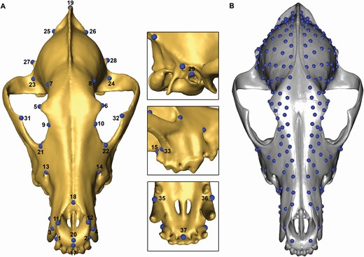
The landmark and semilandmark configurations used in the analyses. A, manually sampled landmarks (see Table 1 for the full landmark descriptions). B, semilandmark patches placed through the sliding procedure.

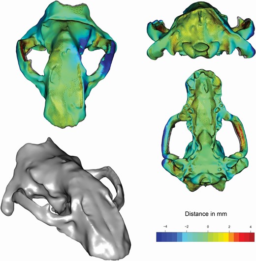
Restoration of Kolponomos newportensis (USNM215070). The coloured surfaces represent the retrodeformation result (through the application of the retroDeformMesh algorithm) while the reticulated surfaces represent the superimposed original shape of the 3D model. The most deformed areas were highlighted following the colour scale shown. The bottom left surface is the 3D mesh after the restoration with Geomagic Studio.

Some further fossil specimens, Dinofelis barlowi (DNMNH-BF55-22), Hoplophoneus primaevus (USNM-99), Smilodon californicus (BCGD-27000) and Yoshi garevskii (MMNH-Sk-69) bear traces of taphonomic distortion and are partially incomplete. For these particular specimens, we estimated the position of missing landmarks by using the fixLMtps and fixLMmirror functions from the R package ‘Morpho’ (Schlager, 2017). fixLMtps estimates missing landmarks through weighted nearest-neighbour interpolation combined with a thin-plate spline (TPS) deformation, while fixLMmirror estimates missing landmarks from their bilateral counterparts (Schlager, 2017). The former technique was applied in cases of damaged surfaces, the latter to missing portions of the skull for which the counterside portion was both present and undeformed.
In order to map patterns of convergence on the digital models (Melchionna et al., 2021), we placed 100 evenly-spaced semilandmarks on the left side of a reference sample and mirrored the semilandmarks on the right side. Eventually, we performed the sliding procedure on each specimen following the protocol included in ‘Morpho’ (Schlager, 2017). Some specimens (i.e. Aonyx capensis CMNH-VZ-17620, Zalophus californianus LEPBLBZ-BM-361-080300 and LEPBLBZ-CSL-484-230404) were excluded as they present bilaterally missing parts which prevent placing semilandmarks.
We used the complete set inclusive of landmarks and semilandmarks to perform Generalized Procrustes Analysis (GPA) to align, rotate and scale the landmark configurations. We performed a Principal Component Analysis (PCA) on the aligned configurations by using the R package ‘Morpho’. In order to perform the convergence analysis, we assembled a phylogenetic tree (Fig. 3) following the hypotheses proposed by Paterson et al. (2020) about the phylogenetic position of K. newportensis as stem pinnipeds, Tamagnini et al. (2021) for the living carnivorans, Melchionna et al. (2021) for the sabretooths, Raia et al. (2013) for Borophagus secundus, Wang et al. (2020) for Dinictis squalidens, Piras et al. (2018) for Dinofelis barlowi, Barrett (2016) for Hoplophoneus primaevus (Supporting Information, Appendix S1). The phylogenetic tree was assembled by using a new tool included in the R package ‘RRphylo’ (Castiglione et al., 2018) named tree.merger (Castiglione et al., 2022).
Morphological convergence test
To test for morphological convergence among the sabretooths and K. newportensis, we used the search.conv function included in the ‘RRphylo’ package (Castiglione et al., 2019). The function computes the angle θ between the vectors of PC scores for each pair of species as a measure of similarity. This angle is further divided by the patristic distance between the pair to provide a second measure of similarity accounting for the evolutionary time separating the species. Both metrics are compared to a family of 100 random values for the same metrics generated under the Brownian motion model of evolution to assess significance (Castiglione et al., 2019). Two different kinds of convergence can be tested in search.conv: (i) convergence among distinct monophyletic clades on the tree, and (ii) convergence among species sharing a particular ‘state’ (e.g. feeding habit, locomotory type). Here, we focused on the latter, erecting a ‘convergent’ state and collating S. fatalis, S. californicus, Hoplophoneus primaevus, Homotherium serum and Kolponomos newportensis under such a heading.
As some sabretooths (i.e. Smilodon, Homotherium) are phylogenetically close, trait similarity among them might be influenced by proximity rather than true convergence. To deal with this, search.conv tests for phylogenetic clustering (i.e. lower than expected by chance patristic distance among species falling under a given state) and removes tip species until clustering disappears. As the declustering procedure is random, the results may slightly change depending on which species is omitted. For this reason, we repeated search.conv ten times to test the reliability of results. Still, Tseng et al.’s study (2016) indicates Kolponomos to be functionally rather than morphologically convergent on Smilodon, and the PCA plots indicate Kolponomos stands well within the morphological variability of pinnipeds. Therefore, we also applied search.conv to a restricted number of pinniped species, selecting all pinnipeds which were found to be morphologically closer to the sabretooth, on average, than Kolponomos, as estimated by the angle θ between the vectors of PC scores between each pinniped and the vector of mean PC scores calculated over all sabretooth species. This allows us to test whether morphological similarity between Kolponomos and Smilodon is exceptional compared to other pinnipeds. GPA alignment, PCA and morphological convergence tests were repeated on the configurations inclusive of landmarks only and are presented in the Supporting Information.
Relative Warp Analysis and conv.map
To visually explore the patterns of morphological convergence we used conv.map, a novel method which charts the PC axes responsible for the greatest pairwise similarity among species directly on the 3D models (Melchionna et al., 2021). To apply conv.map, we used Relative Warp Analysis (RWA) to single-out the non-affine component of shape variation by means of partial warp scores (Rohlf & Bookstein, 2003). We performed RWA on full configurations (landmarks plus semilandmarks) by using the relWarps function of the R package ‘Morpho’ (Schlager, 2017).
RESULTS
By using the full, landmarks and semilandmarks configuration, the first two PCA vectors account for 32.09% and 18.87% of the total shape variation, respectively (Fig. 3A). Along PC1, there is a separation between Canidae, Viverridae, Felidae (at negative values) and Musteloidea (at positive values). Hyaenidae overlap with Canidae on the PC1/PC2 plot. Ursidae place close to Felidae and Musteloidea, with Helarctos malayanus being separated from other bears by virtue of its unusual cranial shape. Along PC2, Odobenidae, Otariidae and Phocidae are well distinguished from any other species. Felids with sabretooth morphologies, B. fricki and Hoplophoneus primaevus, are close to each other and separated from other feliforms and pinnipeds along PC2. The PCA plot of the landmark-only configurations (available in the Supporting Information) replicated the same general positioning of major groups in the PC1/PC2 biplot. Kolponomos places close to true sabretooths, but the same positioning is shared with other pinniped species. In keeping with this, we found the θ angle between the vector of sabretooths average PC scores and the vector of PC scores of eleven different pinnipeds to be lower than the corresponding angle between Kolponomos and the sabretooths (Tables 2, S2).
Pairwise θ angles among the pinnipeds closest to sabretooths
| S. fatalis | S. californicus | B. fricki | Homotherium serum | Hoplophoneus primaevus | Mean | |
|---|---|---|---|---|---|---|
| Halichoerus grypus | 73.36 | 71.13 | 33.24 | 76.18 | 75.88 | 65.96 |
| Mirounga angustirostris | 77.23 | 78.90 | 98.35 | 62.03 | 59.55 | 75.21 |
| Phoca vitulina | 93.91 | 96.77 | 88.43 | 88.44 | 34.64 | 80.44 |
| Neophoca cinerea | 79.48 | 81.08 | 106.02 | 27.43 | 121.74 | 83.15 |
| Pusa hispida | 101.15 | 104.02 | 102.06 | 77.54 | 39.53 | 84.86 |
| Monachus tropicalis | 96.80 | 97.35 | 50.85 | 70.98 | 111.81 | 85.56 |
| Phocarctos hookeri | 90.67 | 89.76 | 40.17 | 103.12 | 106.26 | 86.00 |
| Hydrurga leptonyx | 89.03 | 91.95 | 83.88 | 63.70 | 106.12 | 86.94 |
| Cystophora cristata | 79.66 | 81.76 | 113.33 | 84.61 | 75.42 | 86.95 |
| Eumetopias jubatus | 102.50 | 104.42 | 41.20 | 106.02 | 89.50 | 88.73 |
| Monachus monachus | 95.50 | 96.59 | 84.61 | 60.02 | 109.94 | 89.33 |
| Kolponomos newportensis | 79.45 | 76.49 | 66.80 | 120.75 | 111.64 | 91.03 |
| S. fatalis | S. californicus | B. fricki | Homotherium serum | Hoplophoneus primaevus | Mean | |
|---|---|---|---|---|---|---|
| Halichoerus grypus | 73.36 | 71.13 | 33.24 | 76.18 | 75.88 | 65.96 |
| Mirounga angustirostris | 77.23 | 78.90 | 98.35 | 62.03 | 59.55 | 75.21 |
| Phoca vitulina | 93.91 | 96.77 | 88.43 | 88.44 | 34.64 | 80.44 |
| Neophoca cinerea | 79.48 | 81.08 | 106.02 | 27.43 | 121.74 | 83.15 |
| Pusa hispida | 101.15 | 104.02 | 102.06 | 77.54 | 39.53 | 84.86 |
| Monachus tropicalis | 96.80 | 97.35 | 50.85 | 70.98 | 111.81 | 85.56 |
| Phocarctos hookeri | 90.67 | 89.76 | 40.17 | 103.12 | 106.26 | 86.00 |
| Hydrurga leptonyx | 89.03 | 91.95 | 83.88 | 63.70 | 106.12 | 86.94 |
| Cystophora cristata | 79.66 | 81.76 | 113.33 | 84.61 | 75.42 | 86.95 |
| Eumetopias jubatus | 102.50 | 104.42 | 41.20 | 106.02 | 89.50 | 88.73 |
| Monachus monachus | 95.50 | 96.59 | 84.61 | 60.02 | 109.94 | 89.33 |
| Kolponomos newportensis | 79.45 | 76.49 | 66.80 | 120.75 | 111.64 | 91.03 |
Pairwise θ angles among the pinnipeds closest to sabretooths
| S. fatalis | S. californicus | B. fricki | Homotherium serum | Hoplophoneus primaevus | Mean | |
|---|---|---|---|---|---|---|
| Halichoerus grypus | 73.36 | 71.13 | 33.24 | 76.18 | 75.88 | 65.96 |
| Mirounga angustirostris | 77.23 | 78.90 | 98.35 | 62.03 | 59.55 | 75.21 |
| Phoca vitulina | 93.91 | 96.77 | 88.43 | 88.44 | 34.64 | 80.44 |
| Neophoca cinerea | 79.48 | 81.08 | 106.02 | 27.43 | 121.74 | 83.15 |
| Pusa hispida | 101.15 | 104.02 | 102.06 | 77.54 | 39.53 | 84.86 |
| Monachus tropicalis | 96.80 | 97.35 | 50.85 | 70.98 | 111.81 | 85.56 |
| Phocarctos hookeri | 90.67 | 89.76 | 40.17 | 103.12 | 106.26 | 86.00 |
| Hydrurga leptonyx | 89.03 | 91.95 | 83.88 | 63.70 | 106.12 | 86.94 |
| Cystophora cristata | 79.66 | 81.76 | 113.33 | 84.61 | 75.42 | 86.95 |
| Eumetopias jubatus | 102.50 | 104.42 | 41.20 | 106.02 | 89.50 | 88.73 |
| Monachus monachus | 95.50 | 96.59 | 84.61 | 60.02 | 109.94 | 89.33 |
| Kolponomos newportensis | 79.45 | 76.49 | 66.80 | 120.75 | 111.64 | 91.03 |
| S. fatalis | S. californicus | B. fricki | Homotherium serum | Hoplophoneus primaevus | Mean | |
|---|---|---|---|---|---|---|
| Halichoerus grypus | 73.36 | 71.13 | 33.24 | 76.18 | 75.88 | 65.96 |
| Mirounga angustirostris | 77.23 | 78.90 | 98.35 | 62.03 | 59.55 | 75.21 |
| Phoca vitulina | 93.91 | 96.77 | 88.43 | 88.44 | 34.64 | 80.44 |
| Neophoca cinerea | 79.48 | 81.08 | 106.02 | 27.43 | 121.74 | 83.15 |
| Pusa hispida | 101.15 | 104.02 | 102.06 | 77.54 | 39.53 | 84.86 |
| Monachus tropicalis | 96.80 | 97.35 | 50.85 | 70.98 | 111.81 | 85.56 |
| Phocarctos hookeri | 90.67 | 89.76 | 40.17 | 103.12 | 106.26 | 86.00 |
| Hydrurga leptonyx | 89.03 | 91.95 | 83.88 | 63.70 | 106.12 | 86.94 |
| Cystophora cristata | 79.66 | 81.76 | 113.33 | 84.61 | 75.42 | 86.95 |
| Eumetopias jubatus | 102.50 | 104.42 | 41.20 | 106.02 | 89.50 | 88.73 |
| Monachus monachus | 95.50 | 96.59 | 84.61 | 60.02 | 109.94 | 89.33 |
| Kolponomos newportensis | 79.45 | 76.49 | 66.80 | 120.75 | 111.64 | 91.03 |
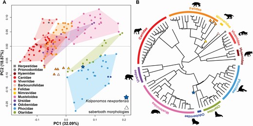
Morphospace defined by the two first PC axes and the phylogenetic tree. A, PC1 and PC2 axes. B, phylogenetic tree assembled for the analyses. The star represents the phylogenetic position of Kolponomos newportensis. The triangles represent the species with a sabretooth morphology, which are: Hoplophoneus primaevus (Nimravidae), Barbourofelis fricki (Barbourofelidae, pink triangle), Smilodon californicus and Smilodon fatalis and Homotherium serum (Felidae). The grey square represents Suricata suricatta and the grey circle represents Prionodon linsang.

We tested convergence between true sabretooths and all of the 12 selected pinnipeds, applying search.conv ten times separately to each of them. We found the highest frequency of positive evidence for morphological convergence relative to Kolponomos (five times out of ten, seven by using landmarks only, see Supporting Information) and the hooded seal Cystophora cristata which were found to converge on sabretooths half of the time. The Australian sea lion (Neophoca cinerea), Steller sea lion (Eumetopias jubatus) and ringed seal (Pusa hispida) were found to converge on sabretooths four out of ten times (Table 3). The corresponding figures by using landmarks only are: Neophoca, four times; Eumetopias six times (see Supporting Information, Table S3). Pusa and Cystophora were not selected for the landmark-only convergence test as they are not morphologically closer to sabretooths than Kolponomos by using the landmark configuration.
Output from search.conv analysis. Angle state is the mean θ angle between species assigned to the state ‘convergent’. Convergence is reported as the percentage of significant instances of convergence found
| Angle state | Percentage of issues of convergence found | |
|---|---|---|
| Halichoerus grypus | 56.45 | 20 |
| Neophoca cinerea | 57.24 | 40 |
| Mirounga angustirostris | 58.42 | 30 |
| Kolponomos newportensis | 58.70 | 50 |
| Cystophora cristata | 60.71 | 50 |
| Phocarctos hookeri | 63.09 | 30 |
| Hydrurga leptonyx | 64.35 | 30 |
| Monachus monachus | 65.76 | 30 |
| Phoca vitulina | 68.32 | 0 |
| Eumetopias jubatus | 69.49 | 40 |
| Monachus tropicalis | 69.75 | 10 |
| Pusa hispida | 70.77 | 40 |
| Angle state | Percentage of issues of convergence found | |
|---|---|---|
| Halichoerus grypus | 56.45 | 20 |
| Neophoca cinerea | 57.24 | 40 |
| Mirounga angustirostris | 58.42 | 30 |
| Kolponomos newportensis | 58.70 | 50 |
| Cystophora cristata | 60.71 | 50 |
| Phocarctos hookeri | 63.09 | 30 |
| Hydrurga leptonyx | 64.35 | 30 |
| Monachus monachus | 65.76 | 30 |
| Phoca vitulina | 68.32 | 0 |
| Eumetopias jubatus | 69.49 | 40 |
| Monachus tropicalis | 69.75 | 10 |
| Pusa hispida | 70.77 | 40 |
Output from search.conv analysis. Angle state is the mean θ angle between species assigned to the state ‘convergent’. Convergence is reported as the percentage of significant instances of convergence found
| Angle state | Percentage of issues of convergence found | |
|---|---|---|
| Halichoerus grypus | 56.45 | 20 |
| Neophoca cinerea | 57.24 | 40 |
| Mirounga angustirostris | 58.42 | 30 |
| Kolponomos newportensis | 58.70 | 50 |
| Cystophora cristata | 60.71 | 50 |
| Phocarctos hookeri | 63.09 | 30 |
| Hydrurga leptonyx | 64.35 | 30 |
| Monachus monachus | 65.76 | 30 |
| Phoca vitulina | 68.32 | 0 |
| Eumetopias jubatus | 69.49 | 40 |
| Monachus tropicalis | 69.75 | 10 |
| Pusa hispida | 70.77 | 40 |
| Angle state | Percentage of issues of convergence found | |
|---|---|---|
| Halichoerus grypus | 56.45 | 20 |
| Neophoca cinerea | 57.24 | 40 |
| Mirounga angustirostris | 58.42 | 30 |
| Kolponomos newportensis | 58.70 | 50 |
| Cystophora cristata | 60.71 | 50 |
| Phocarctos hookeri | 63.09 | 30 |
| Hydrurga leptonyx | 64.35 | 30 |
| Monachus monachus | 65.76 | 30 |
| Phoca vitulina | 68.32 | 0 |
| Eumetopias jubatus | 69.49 | 40 |
| Monachus tropicalis | 69.75 | 10 |
| Pusa hispida | 70.77 | 40 |
To visually inspect which parts of the skull show the highest convergence with Smilodon (which is limited to five species for graphical purposes) we selected hooded seal, sea lion, ringed seal and Kolponomos (Fig. 4, the corresponding figures with Neophoca and then Pusa replacing Eumetopias are reported in the Supporting Information, Figs S2–S3). As expected, convergence between Smilodon and Kolponomos involves both the facial and neurocranial portions of the skull, and in particular the dorsal surface of the posterior region of the cranium, and the alveolar area around the canines. Still, they share a narrow postorbital constriction. In addition, by selecting PC axes responsible for convergence, conv.map indicates Kolponomos shows a higher degree of convergence with Smilodon than to other pinnipeds, and none of the latter shows any particularly intense pattern of convergence on the sabretooth, especially considering that some convergence for these pinnipeds (Neophoca, Cystophora) is concentrated in the nasal area which is absent in Kolponomos (Fig. 4; Supporting Information, Figs S2–S3).
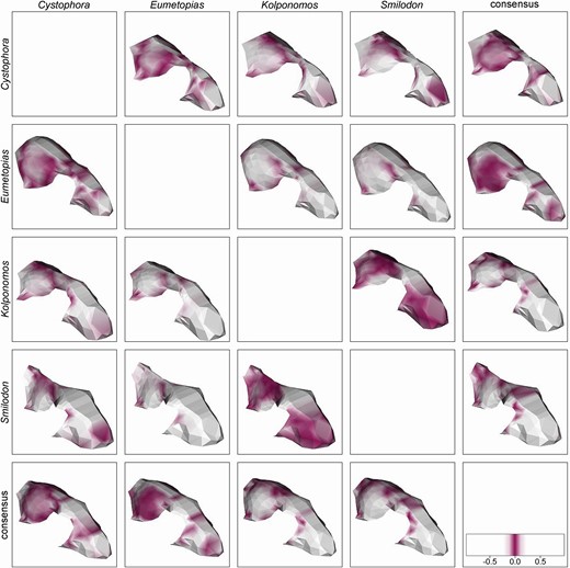
Visualization of the pairwise comparison between Cystophora, Eumetopias, Kolponomos, Smilodon and the consensus. The colour gradient indicates local differences between the two surfaces (the scale bar was rescaled to the range 0–1). In each case, differences between the two taxa are displayed on a reconstruction of the taxon named on the left.

DISCUSSION
Tseng et al. (2016) recently reported on a rather unusual case of mosaic convergence between the stem pinniped Kolponomos and the sabretooth cat Smilodon. Their analysis indicates that such convergence is essentially functional and relates to the way Smilodon and Kolponomos had to withstand strong torquing loads when biting prey with the infamous canines or whilst dislodging marine shelled organisms fastened to the sea bottom, respectively. Although Tseng et al. (2016) focused on the mandible, the attachment areas of the masticatory and neck muscles and the loadings they originate pertain to the cranium (Salesa et al., 2005). Hence, we scanned this structure seeking for any pattern of convergence between Kolponomos and the sabretooth species. We found that Kolponomos skull shape falls well within the morphological variability of otariids, in line with its phylogenetic position (Paterson et al., 2020). Although Kolponomos is not closer in skull shape to Smilodon and the other sabretooths than other pinnipeds (Steller’s sea lion, Eumetopias jubatus, in particular was found to converge on the sabretoothed cats nearly as frequently as Kolponomos by either using the full set of landmarks and semilandmarks or landmarks only, Table 3, Supporting Information, Table S3) it remains the most ‘sabretooth-like’ among pinnipeds, and the closest to Smilodon and the other sabretooths in absolute terms in PC plots (Fig. 3). It is possible to speculate the morphological proximity between otariids and sabretooths (Fig. 3; Supporting Information, Fig. S1) may have facilitated convergence between Smilodon and Kolponomos, providing the latter with the basic morphological equipment to deal with the especially demanding loadings originated at biting. We took advantage of a recently implemented software tool which allows to chart phenotypic resemblance directly on the digital models (Melchionna et al., 2021) to inspect which part of the cranium of Kolponomos resembles sabretooths the most. Kolponomos and Smilodon shapes are similar in the alveolar area around the canines, around the postorbital constriction, and in the vast area of attachment of the temporalis profundus muscle in the temporalis fossa (Wroe et al., 2013). In Smilodon as well as in other sabretooths, the low coronoid reduced the mechanical advantage of the temporalis (McHenry et al., 2007) which is compensated for by the increased strength in the neck musculature allowing strong lateral head movements during biting (Antón et al., 2004). Tseng et al. (2016) confirmed similar bite mechanics in Kolponomos, which further shares with sabretooths the derived shape of the mastoid process to provide attachment for an (inferred) massive atlanto-mastoid muscle, whereas it maintains a prominent coronoid process in agreement with its phylogenetic position. This in turn provides convergence between Kolponomos and Smilodon in a localized area of the mandible, as driven by functional adaptation to withstand strong torque loadings at biting. The cranium seems to conform to this pattern of mosaic convergence. The areas of this structure which converge the most between Kolponomos and Smilodon clearly relate to the main jaw-closing muscle and the canine area, which are massive in both species and represent the portion of the cranium most solicited at biting. These findings indicate mosaic convergence between Kolponomos and the sabretooth extend to both components of the skull. They also highlight that convergence is a complex evolutionary pattern, whereby morphological resemblance does not grant adaptation to shared functional ends (Sansalone et al., 2020). Taking into consideration that even divergent morphologies may provide functional convergence (Alfaro et al., 2005; Wainwright, 2007; Losos, 2011; Renaud et al., 2018), we emphasize the importance of visualizing patterns of morphological resemblance directly on the phenotype, as a mean to seek after functional explanations that may pertain to biomechanical considerations. The particular case of the enigmatic Kolponomos further reminds us that convergence in response to similar adaptive solicitations may be limited to restricted regions of an anatomical structure, which highlights the importance of accurately mapping the potential patterns of convergence on the phenotype of interest.
SUPPORTING INFORMATION
Additional Supporting Information may be found in the online version of this article at the publisher’s web-site:
Figure S1. Morphospace relative to the PC1 and PC2 axes of the landmarks data. Kolponomos is indicated by a star, sabertooths are indicated by triangles.
Figure S2. Visualization of the pairwise comparisons between Cystophora, Neophoca, Kolponomos, Smilodon and the consensus. The colour gradient indicates local differences between the two surfaces (the scale bar was rescaled to the range 0–1). In each case, differences between the two taxa are displayed on a reconstruction of the taxon named on the left.
Figure S3. Visualization of the pairwise comparison between Cystophora, Pusa, Kolponomos, Smilodon and the consensus. The colour gradient indicates local differences between the two surfaces (the scale bar was rescaled to the range 0–1). In each case, differences between the two taxa are displayed on a reconstruction of the taxon named on the left.
Table S1. Specimens included in the analyses.
Table S2. Pairwise θ angle among pinnipeds closest to sabertooths when landmark configuration set is taking into account.
Table S3. Output from search.conv analysis for the landmark sets. Angle state is the mean θ angle between species belonging to the given state ‘convergent’. Convergence is reported as percentage of significant convergence found.
ACKNOWLEDGEMENTS
A number of pinniped and felid specimens included in the present study are from the Ph.D. database of D.T., whose Ph.D. project received support from ‘Avvio alla Ricerca 2019 and 2020’ funding, which is financed by the University of Rome ‘La Sapienza’. D.T. also received support from the SYNTHESYS Access Programme that is financed by the European Community Research Infrastructure Action under the FP7 (ES-TAF-2750 awarded to D.T.). We owe a huge debt of thanks to all curators, collection managers and staff, whose help and support was fundamental for the sampling operations. In particular, with anticipated apologies for certainly forgetting to explicitly mention several of the many people to whom thanks are owed, we wish to thank: Susana Fraile, Jorge Morales, Géraldine Veron, Aurélie Verguin, Riccardo Castiglia, Cristiano Dal Sasso, Pierfilippo Cerretti, Adriano De Faveri, Saverio Bartolini Lucenti, Paolo Agnelli, Tony Parker, Itatí Olivares and Agustín Ruella. We also thank Filippo Galimberti and Simona Sanvito for sharing some 3D models of pinnipeds collected by the Elephant Seals Research Group. We are deeply indebted to Stephen Wroe for sharing a number of digital models, part of the collection of the FEAR Laboratory at the University of New England, Arimdale, Australia. We are especially grateful to Stephan Lautenschlager and the three anonymous reviewers for their kind comments on the manuscript and for providing helpful insight through the revision process. The authors declare no conflicts of interest.
DATA AVAILABILITY
The data underlying this article are available on Zenodo at https://doi.org/10.5281/zenodo.6457753.