2025 ESC/EACTS Guidelines for the management of valvular heart disease: Developed by the task force for the management of valvular heart disease of the European Society of Cardiology (ESC) and the European Association for Cardio-Thoracic Surgery (EACTS)

European Heart Journal, Volume 46, Issue 44, 21 November 2025, Pages 4635–4736, https://doi.org/10.1093/eurheartj/ehaf194
Published:
29 August 2025

Table of contents

  • 1. Preamble 4640

  • 2. Introduction 4642

  •  2.1. What is new 4643

  • 3. The Heart Team and Heart Valve Centre 4649

  •  3.1. The Heart Valve Network 4649

  •   3.1.1. Composition of the Heart Team 4650

  •   3.1.2. Procedural volume and clinical outcomes 4651

  • 4. Imaging of patients with valvular heart disease 4652

  •  4.1. Initial valve assessment 4652

  •  4.2. Associated diseases and conditions 4653

  •  4.3. Evaluation of valvular heart disease dynamics and variability 4653

  •  4.4. Assessment of extravalvular cardiac consequences from valvular heart disease 4653

  •  4.5. Evaluation of eligibility, planning, and guiding of interventions 4653

  • 5. Clinical evaluation of patients with valvular heart disease 4653

  •  5.1. Clinical examination 4653

  •  5.2. Assessment of comorbidities and risk stratification 4653

  •  5.3. Biomarkers 4654

  •  5.4. Exercise testing 4655

  •  5.5. Invasive investigations 4655

  •   5.5.1. Coronary angiography 4655

  •   5.5.2. Cardiac catheterization 4655

  •  5.6. Patient-centred care and shared decision-making 4655

  • 6. Management of conditions associated with valvular heart disease 4655

  •  6.1. Diagnosis and management of coronary artery disease 4655

  •  6.2. Atrial fibrillation 4657

  •  6.3. Cancer and radiation therapy 4657

  •  6.4. Prophylaxis of rheumatic fever 4658

  •  6.5. Cardiogenic shock and acute heart failure 4658

  •  6.6. Palliative care 4658

  • 7. Aortic regurgitation 4658

  •  7.1. Prevalence and aetiology 4658

  •  7.2. Evaluation 4658

  •  7.3. Medical therapy 4660

  •  7.4. Indications for intervention 4660

  •  7.5. Follow-up 4660

  •  7.6. Special patient populations 4662

  • 8. Aortic stenosis 4662

  •  8.1. Prevalence and aetiology 4662

  •  8.2. Evaluation 4662

  •   8.2.1. Echocardiography and cardiac computed tomography 4662

  •   8.2.2. Additional diagnostic and prognostic parameters 4663

  •   8.2.3. Procedural planning 4663

  •  8.3. Medical therapy 4663

  •  8.4. Indication for intervention 4665

  •   8.4.1. Symptomatic severe aortic stenosis 4665

  •   8.4.2. Asymptomatic severe aortic stenosis 4665

  •   8.4.3. Moderate aortic stenosis 4665

  •  8.5. Treatment options 4667

  •   8.5.1. The mode of intervention in candidates for a bioprosthesis 4667

  •    8.5.1.1. Age and life expectancy 4668

  •    8.5.1.2. Anatomical features 4668

  •    8.5.1.3. Lifetime management 4668

  •  8.6. Follow-up 4671

  • 9. Mitral regurgitation 4671

  •  9.1. Primary mitral regurgitation 4671

  •   9.1.1. Prevalence and aetiology 4671

  •   9.1.2. Evaluation 4671

  •    9.1.2.1. Echocardiography and right heart catheterization 4671

  •    9.1.2.2. Biomarkers 4671

  •    9.1.2.3. Cardiac magnetic resonance and computed tomography 4672

  •    9.1.2.4. Genetic evaluation 4674

  •   9.1.3. Medical therapy 4674

  •   9.1.4. Indications for intervention 4674

  •   9.1.5. Follow-up 4675

  •  9.2. Secondary mitral regurgitation 4675

  •   9.2.1. Prevalence and aetiology 4675

  •   9.2.2. Evaluation 4675

  •   9.2.3. Definition of atrial secondary mitral regurgitation 4675

  •   9.2.4. Management of ventricular secondary mitral regurgitation 4676

  •    9.2.4.1. Medical and device therapy 4676

  •    9.2.4.2. Indications for intervention 4677

  •    9.2.4.3. Follow-up 4678

  •   9.2.5. Management of atrial secondary mitral regurgitation 4679

  •    9.2.5.1. Medical therapy and rhythm management 4679

  •    9.2.5.2. Indications for intervention 4679

  •    9.2.5.3. Follow-up 4679

  • 10. Mitral stenosis 4679

  •  10.1. Prevalence and aetiology 4679

  •  10.2. Rheumatic mitral stenosis 4679

  •   10.2.1. Evaluation 4679

  •   10.2.2. Medical therapy 4680

  •   10.2.3. Indications for intervention 4680

  •   10.2.4. Follow-up 4680

  •  10.3. Degenerative mitral stenosis with mitral annular calcification 4680

  •   10.3.1. Evaluation 4681

  •   10.3.2. Indications for intervention 4681

  • 11. Tricuspid regurgitation 4682

  •  11.1. Prevalence and aetiology 4682

  •  11.2. Evaluation 4683

  •  11.3. Medical therapy 4683

  •  11.4. Indications for intervention 4683

  •   11.4.1. Surgery 4683

  •    11.4.1.1. Patients without indication for left-sided valve surgery 4685

  •    11.4.1.2. Patients with indication for left-sided valve surgery 4685

  •   11.4.2. Transcatheter techniques 4686

  • 12. Tricuspid stenosis 4687

  •  12.1. Prevalence and aetiology 4687

  •  12.2. Evaluation 4687

  •  12.3. Medical therapy 4688

  •  12.4. Indications for intervention 4688

  • 13. Multiple and mixed valvular heart disease 4688

  •  13.1. Prevalence and undertreatment 4688

  •  13.2. Evaluation and diagnostic pitfalls 4688

  •  13.3. Indications for intervention 4688

  •   13.3.1. Multiple valvular heart disease 4688

  •   13.3.2. Mixed aortic valve disease 4690

  •   13.3.3. Mixed mitral valve disease 4690

  •  13.4. Follow-up 4690

  • 14. Management of patients with prosthetic valves or valve repair 4690

  •  14.1. Choice of prosthetic valve 4690

  •  14.2. Follow-up of patients with prosthetic valves 4691

  •  14.3. Antithrombotic therapy in patients with treated valvular heart disease 4691

  •   14.3.1. Mechanical heart valves 4691

  •    14.3.1.1. Post-operative anticoagulation and therapeutic targets 4691

  •    14.3.1.2. Prevention and management of bleeding 4693

  •    14.3.1.3. Management of anticoagulation therapy before and after non-cardiac invasive procedures 4693

  •   14.3.2. Biological heart valves 4695

  •    14.3.2.1. Patients with a surgical biological heart valve and no indication for oral anticoagulation 4695

  •    14.3.2.2. Patients with a transcatheter heart valve and no indication for oral anticoagulation 4695

  •    14.3.2.3. Patients with a surgical biological heart valve and an indication for oral anticoagulation 4696

  •    14.3.2.4. Patients with a transcatheter biological heart valve and an indication for oral anticoagulation 4696

  •  14.4. Management of prosthetic valve dysfunction and complications 4697

  •   14.4.1. Structural valve deterioration 4697

  •   14.4.2. Non-structural valve dysfunction 4698

  •    14.4.2.1. Prosthesis–patient mismatch 4698

  •    14.4.2.2. Paravalvular leak and haemolysis 4698

  •   14.4.3. Endocarditis 4698

  •   14.4.4. Valve thrombosis 4698

  •    14.4.4.1. Hypo-attenuated leaflet thickening 4698

  •    14.4.4.2. Clinically significant valve thrombosis 4698

  • 15. Management during non-cardiac surgery 4700

  •  15.1. Pre-operative evaluation 4700

  •  15.2. Specific valve lesions 4700

  •   15.2.1. Aortic stenosis 4700

  •   15.2.2. Mitral stenosis 4701

  •   15.2.3. Aortic and mitral regurgitation 4701

  •  15.3. Peri-operative monitoring 4702

  • 16. Management of valvular heart disease during pregnancy 4702

  •  16.1. Management before pregnancy 4702

  •  16.2. Management during pregnancy 4703

  •   16.2.1. Patients with native valve disease 4703

  •   16.2.2. Patients with prosthetic valves 4703

  • 17. Sex-specific considerations in patients with valvular heart disease 4703

  •  17.1. Aortic valve disease 4703

  •  17.2. Mitral valve disease 4704

  •  17.3. Tricuspid valve disease 4704

  • 18. Key messages 4704

  • 19. Gaps in evidence 4705

  • 20. ‘What to do’ and ‘What not to do’ messages from the Guidelines 4707

  • 21. Evidence tables 4710

  • 22. Data availability statement 4710

  • 23. Author information 4710

  • 24. Appendix 4710

  • 25. References 4711

Tables of Recommendations

  • Recommendation Table 1 — Recommendations for the management of chronic coronary syndrome in patients with valvular heart disease (see also Supplementary data online, Evidence Table 1) 4656

  • Recommendation Table 2 — Recommendations for the management of atrial fibrillation in patients with native valvular heart disease (see also Supplementary data online, Evidence Tables 2 and 3) 4657

  • Recommendation Table 3 — Recommendations on indications for intervention in severe aortic regurgitation (see also Supplementary data online, Evidence Tables 4–8) 4662

  • Recommendation Table 4 — Recommendations on indications for intervention in symptomatic and asymptomatic severe aortic stenosis, and recommended mode of intervention (see also Supplementary data online, Evidence Tables 9–13) 4670

  • Recommendation Table 5 — Recommendations on indications for concomitant aortic valve replacement at the time of coronary artery bypass grafting or ascending aorta surgery 4671

  • Recommendation Table 6 — Recommendations on indications for intervention in severe primary mitral regurgitation (see also Supplementary data online, Evidence Tables 14–16) 4674

  • Recommendation Table 7 — Recommendations on indications for intervention in secondary mitral regurgitation (see also Supplementary data online, Evidence Tables 17–20) 4679

  • Recommendation Table 8 — Recommendations on indications for percutaneous mitral commissurotomy, mitral valve surgery, and transcatheter intervention in clinically severe rheumatic and degenerative mitral stenosis (see also Supplementary data online, Evidence Table 21) 4682

  • Recommendation Table 9 — Recommendations on indications for intervention in tricuspid regurgitation (see also Supplementary data online, Evidence Tables 22 and 23) 4687

  • Recommendation Table 10 — Recommendations on indications for intervention in tricuspid stenosis 4688

  • Recommendation Table 11 — Recommendations on indications for surgery of concomitant left-sided valvular heart disease 4690

  • Recommendation Table 12 — Recommendations on indications for intervention in patients with mixed moderate aortic stenosis and moderate aortic regurgitation (see also Supplementary data online, Evidence Table 24) 4690

  • Recommendation Table 13 — Recommendations for prosthetic valve selection 4691

  • Recommendation Table 14 — Recommendations for the management of antithrombotic therapy in patients with a mechanical heart valve 4693

  • Recommendation Table 15 — Recommendations for the management of antithrombotic therapy in patients with a mechanical heart valve undergoing elective non-cardiac surgery or invasive procedures 4694

  • Recommendation Table 16 — Recommendations for the management of antithrombotic therapy in patients with a biological heart valve or valve repair 4696

  • Recommendation Table 17 — Recommendations for the management of prosthetic valve dysfunction (see also Supplementary data online, Evidence Table 25) 4700

List of tables

  • Table 1 Classes of recommendations 4641

  • Table 2 Levels of evidence 4641

  • Table 3 New recommendations 4643

  • Table 4 Revised recommendations 4645

  • Table 5 Requirements for a Heart Valve Centre 4651

  • Table 6 Complex procedures ideally performed in the most experienced Heart Valve Centres 4651

  • Table 7 Clinical and echocardiographic criteria predicting outcome improvement in patients with severe ventricular secondary mitral regurgitation undergoing mitral transcatheter edge-to-edge repair 4678

  • Table 8 Contraindications for percutaneous mitral commissurotomy in rheumatic mitral stenosis 4682

  • Table 9 Echocardiographic pitfalls, robust measures, and complementary multimodality imaging parameters in multiple or mixed valvular heart disease 4689

  • Table 10 International normalized ratio targets and therapeutic ranges for patients with a mechanical heart valve 4693

  • Table 11 Peri-operative management of antithrombotic treatment in patients with a mechanical heart valve undergoing non-cardiac surgery based on type of procedure and underlying risk 4694

  • Table 12 Criteria for the diagnosis of moderate or severe aortic and mitral haemodynamic valve deterioration 4697

  • Table 13 ‘What to do’ and ‘what not to do’ 4707

List of figures

  • Figure 1 The Heart Valve Network 4650

  • Figure 2 Integrative imaging assessment of patients with valvular heart disease 4652

  • Figure 3 Central illustration. Patient-centred evaluation for treatment of valvular heart disease 4654

  • Figure 4 Imaging assessment of patients with aortic regurgitation 4659

  • Figure 5 Management of patients with aortic regurgitation 4661

  • Figure 6 Integrative imaging assessment of patients with aortic stenosis 4664

  • Figure 7 Management of patients with severe aortic stenosis 4666

  • Figure 8 Aortic valve treatment options 4667

  • Figure 9 Factors to be considered when selecting the mode of intervention for aortic stenosis 4669

  • Figure 10 Echocardiographic assessment of patients with mitral regurgitation 4672

  • Figure 11 Management of patients with severe primary mitral regurgitation 4673

  • Figure 12 Most frequently used criteria for the diagnosis of atrial secondary mitral regurgitation 4676

  • Figure 13 Treatment of severe secondary mitral regurgitation without concomitant coronary artery disease 4677

  • Figure 14 Management of clinically severe rheumatic mitral stenosis (mitral valve area ≤1.5 cm2) 4681

  • Figure 15 Echocardiographic and invasive assessment of tricuspid regurgitation 4684

  • Figure 16 Stepwise evaluation of patients with tricuspid regurgitation 4685

  • Figure 17 Management of patients with tricuspid regurgitation 4686

  • Figure 18 Antithrombotic therapy following mechanical heart valve implantation 4692

  • Figure 19 Antithrombotic therapy following biological heart valve implantation or surgical valve repair 4695

  • Figure 20 Management of left-sided obstructive and non-obstructive mechanical heart valve thrombosis 4699

  • Figure 21 Management of non-cardiac surgery in patients with severe aortic stenosis 4701

  • Figure 22 The Pregnancy Heart Team model of care 4702

Abbreviations and acronyms

     
  • 2D

    Two-dimensional

  •  
  • 3D

    Three-dimensional

  •  
  • 4D

    Four-dimensional

  •  
  • ACE-I

    Angiotensin-converting enzyme-inhibitor

  •  
  • ACTIVATION

    PercutAneous Coronary inTervention prIor to transcatheter aortic VAlve implantaTION trial

  •  
  • AF

    Atrial fibrillation

  •  
  • AO

    Aorta

  •  
  • AP

    Anteroposterior

  •  
  • AR

    Aortic regurgitation

  •  
  • ARB

    Angiotensin receptor blocker

  •  
  • AS

    Aortic stenosis

  •  
  • ASA

    Acetylsalicylic acid

  •  
  • ATLANTIS

    Anti-Thrombotic Strategy to Lower All Cardiovascular and Neurologic Ischemic and Hemorrhagic Events after Trans-Aortic Valve Implantation for Aortic Stenosis

  •  
  • AU

    Agatston units

  •  
  • AV

    Aortic valve

  •  
  • AVA

    Aortic valve area

  •  
  • AVAi

    Aortic valve area indexed for body surface area

  •  
  • AVATAR

    Aortic Valve Replacement Versus Conservative Treatment in Asymptomatic Severe Aortic Stenosis

  •  
  • AVCS

    Aortic valve calcium score

  •  
  • AVr

    Aortic valve repair

  •  
  • BASILICA

    Bioprosthetic or native Aortic Scallop Intentional Laceration to prevent Iatrogenic Coronary Artery obstruction

  •  
  • BAV

    Bicuspid aortic valve

  •  
  • BHV

    Biological heart valve

  •  
  • BNP

    Brain natriuretic peptide

  •  
  • BP

    Blood pressure

  •  
  • BSA

    Body surface area

  •  
  • CABG

    Coronary artery bypass grafting

  •  
  • CAD

    Coronary artery disease

  •  
  • CCT

    Cardiac computed tomography

  •  
  • CCTA

    Coronary computed tomography angiography

  •  
  • CHA2DS2-VASc

    Congestive heart failure or left ventricular dysfunction, hypertension, age ≥75 (doubled), diabetes, stroke (doubled), vascular disease, age 65–74, sex category (female)

  •  
  • CI

    Confidence interval

  •  
  • CIED

    Cardiac implantable electronic device

  •  
  • CKD

    Chronic kidney disease

  •  
  • CMR

    Cardiac magnetic resonance

  •  
  • COAPT

    Cardiovascular Outcomes Assessment of the MitraClip Percutaneous Therapy for Heart Failure Patients with Functional Mitral Regurgitation

  •  
  • CRT

    Cardiac resynchronization therapy

  •  
  • CT

    Computed tomography

  •  
  • CW

    Continuous wave

  •  
  • CYP

    Cytochrome P

  •  
  • DAPT

    Dual antiplatelet therapy

  •  
  • DEDICATE

    Randomized, Multicenter, Event-Driven Trial of TAVI versus SAVR in Patients with Symptomatic Severe Aortic-Valve Stenosis

  •  
  • DOAC

    Direct oral anticoagulant

  •  
  • DSE

    Dobutamine stress echocardiography

  •  
  • DVI

    Doppler velocity index

  •  
  • EACTS

    European Association for Cardio-Thoracic Surgery

  •  
  • EARLY TAVR

    Evaluation of TAVR Compared to Surveillance for Patients with Asymptomatic Severe Aortic Stenosis trial

  •  
  • ECG

    Electrocardiogram

  •  
  • EDV

    End-diastolic velocity

  •  
  • ENVISAGE-TAVI AF

    Edoxaban vs. Standard of Care and Their Effects on Clinical Outcomes in Patients Having Undergone Transcatheter Aortic Valve Implantation–Atrial Fibrillation

  •  
  • EOA

    Effective orifice area

  •  
  • EROA

    Effective regurgitant orifice area

  •  
  • ESC

    European Society of Cardiology

  •  
  • EuroSCORE

    European System for Cardiac Operative Risk Evaluation

  •  
  • EVoLVeD

    Early Valve Replacement guided by Biomarkers of LV Decompensation in Asymptomatic Patients with Severe Aortic Stenosis

  •  
  • FAC

    Fractional area change

  •  
  • FFR

    Fractional flow reserve

  •  
  • FWS

    Free wall strain

  •  
  • GALILEO

    Global multicenter, open-label, randomized, event-driven, active-controlled study comparing a rivAroxaban-based antithrombotic strategy to an antipLatelet-based strategy after transcatheter aortIc vaLve rEplacement (TAVR) to Optimize clinical outcomes trial

  •  
  • GDMT

    Guideline-directed medical therapy

  •  
  • GLS

    Global longitudinal strain

  •  
  • h

    Hour

  •  
  • HALT

    Hypo-attenuated leaflet thickening

  •  
  • HF

    Heart failure

  •  
  • HFpEF

    Heart failure with preserved ejection fraction

  •  
  • HFrEF

    Heart failure with reduced ejection fraction

  •  
  • HR

    Hazard ratio

  •  
  • HTx

    Heart transplantation

  •  
  • INR

    International normalized ratio

  •  
  • IU

    International unit

  •  
  • KCCQ

    Kansas City Cardiomyopathy Questionnaire

  •  
  • LA

    Left atrium/left atrial

  •  
  • LAAO

    Left atrial appendage occlusion

  •  
  • LAAOS

    Left Atrial Appendage Occlusion Study

  •  
  • LAMPOON

    Laceration of the Anterior Mitral leaflet to Prevent Outflow ObstructioN

  •  
  • LAVI

    Left atrial volume index

  •  
  • LMWH

    Low-molecular-weight heparin

  •  
  • LV

    Left ventricle/left ventricular

  •  
  • LVAD

    Left ventricular assist device

  •  
  • LVEF

    Left ventricular ejection fraction

  •  
  • LVESD

    Left ventricular end-systolic diameter

  •  
  • LVESDi

    Left ventricular end-systolic diameter indexed to BSA

  •  
  • LVESVi

    Left ventricular end-systolic volume indexed to BSA

  •  
  • LVOT

    Left ventricular outflow tract

  •  
  • MA

    Mitral annulus

  •  
  • MAC

    Mitral annular calcification

  •  
  • MATTERHORN

    Multicenter, Randomized, Controlled Study to Assess Mitral Valve Reconstruction for Advanced Insufficiency of Functional or Ischemic Origin trial

  •  
  • MHV

    Mechanical heart valve

  •  
  • MITRA-FR

    Percutaneous Repair with the MitraClip Device for Severe Functional/Secondary Mitral Regurgitation trial

  •  
  • mPAP

    Mean pulmonary artery pressure

  •  
  • MR

    Mitral regurgitation

  •  
  • MRI

    Magnetic resonance imaging

  •  
  • MS

    Mitral stenosis

  •  
  • M-TEER

    Mitral transcatheter edge-to-edge repair

  •  
  • MV

    Mitral valve

  •  
  • MVA

    Mitral valve area

  •  
  • MVHD

    Multiple valvular heart disease

  •  
  • N

    No

  •  
  • NCS

    Non-cardiac surgery

  •  
  • NOTION

    Nordic Aortic Valve Intervention trial

  •  
  • NT-proBNP

    N-terminal pro-B-type natriuretic protein

  •  
  • NYHA

    New York Heart Association

  •  
  • OAC

    Oral anticoagulation

  •  
  • PAWP

    Pulmonary artery wedge pressure

  •  
  • PCI

    Percutaneous coronary intervention

  •  
  • PET

    Positron emission tomography

  •  
  • PH

    Pulmonary hypertension

  •  
  • PHT

    Pressure half-time

  •  
  • PISA

    Proximal isovelocity surface area

  •  
  • P mean

    Mean pressure gradient

  •  
  • PMC

    Percutaneous mitral commissurotomy

  •  
  • PMR

    Primary mitral regurgitation

  •  
  • POPular TAVI

    Antiplatelet Therapy for Patients Undergoing Transcatheter Aortic Valve Implantation

  •  
  • PPM

    Prosthesis–patient mismatch

  •  
  • PROM

    Patient-reported outcome measure

  •  
  • PVL

    Paravalvular leak

  •  
  • PVR

    Pulmonary vascular resistance

  •  
  • RA

    Right atrium/right atrial

  •  
  • RCT

    Randomized controlled trial

  •  
  • RECOVERY

    Randomized Comparison of Early Surgery versus Conventional Treatment in Very Severe Aortic Stenosis

  •  
  • RESHAPE-HF2

    Randomized Investigation of the MitraClip Device in Heart Failure: 2nd Trial in Patients with Clinically Significant Functional Mitral Regurgitation

  •  
  • RF

    Regurgitant fraction

  •  
  • RHC

    Right heart catheterization

  •  
  • RHD

    Rheumatic heart disease

  •  
  • RV

    Right ventricle/right ventricular

  •  
  • RVEF

    Right ventricular ejection fraction

  •  
  • RVol

    Regurgitant volume

  •  
  • SAM

    Systolic anterior movement

  •  
  • SAPT

    Single antiplatelet therapy

  •  
  • SAV

    Surgical aortic valve

  •  
  • SAVR

    Surgical aortic valve replacement

  •  
  • SGLT2i

    Sodium–glucose co-transporter 2 inhibitor

  •  
  • SMR

    Secondary mitral regurgitation

  •  
  • SPAP

    Systolic pulmonary artery pressure

  •  
  • STS

    Society of Thoracic Surgeons

  •  
  • STS-PROM

    Society of Thoracic Surgeons predicted risk of mortality

  •  
  • SVD

    Structural valve deterioration

  •  
  • SVi

    Stroke volume index

  •  
  • TAPSE

    Tricuspid annular plane systolic excursion

  •  
  • TAV

    Transcatheter aortic valve

  •  
  • TAVI

    Transcatheter aortic valve implantation

  •  
  • TDI

    Tissue Doppler imaging

  •  
  • TEER

    Transcatheter edge-to-edge repair

  •  
  • THV

    Transcatheter heart valve

  •  
  • TMVI

    Transcatheter mitral valve implantation

  •  
  • TOE

    Transoesophageal echocardiography

  •  
  • TR

    Tricuspid regurgitation

  •  
  • TRILUMINATE

    Clinical Trial to Evaluate Cardiovascular Outcomes in Patients Treated With the Tricuspid Valve Repair System trial

  •  
  • TRISCEND

    Edwards EVOQUE Transcatheter Tricuspid Valve Replacement: Pivotal Clinical Investigation of Safety and Clinical Efficacy using a Novel Device

  •  
  • TS

    Tricuspid stenosis

  •  
  • TTE

    Transthoracic echocardiography

  •  
  • TTR

    Time in therapeutic range

  •  
  • TV

    Tricuspid valve

  •  
  • UFH

    Unfractionated heparin

  •  
  • VCA

    Vena contracta area

  •  
  • VHD

    Valvular heart disease

  •  
  • VKA

    Vitamin K antagonist

  •  
  • Vmax

    Peak transvalvular velocity

  •  
  • VSARR

    Valve-sparing aortic root replacement

  •  
  • VTE

    Venous thromboembolism

  •  
  • VTI

    Velocity time integral

  •  
  • WHO

    World Health Organisation

  •  
  • WU

    Wood unit

  •  
  • Y

    Yes

1. Preamble

Guidelines evaluate and summarize available evidence with the aim of assisting health professionals in proposing the best diagnostic or therapeutic approach for an individual patient with a given condition. ESC/EACTS Guidelines are intended for use by health professionals but do not override their individual responsibility to make appropriate and accurate decisions in consideration of each patient's health condition and in consultation with the patient or the patient's caregiver where appropriate and/or necessary. It is also the health professional's responsibility to verify the rules and regulations applicable in each country to drugs and devices at the time of prescription and to respect the ethical rules of their profession.

ESC Guidelines represent the official position of the ESC on a given topic. Guideline topics are selected for updating after annual expert review of new evidence conducted by the ESC Clinical Practice Guidelines (CPG) Committee. ESC Policies and Procedures for formulating and issuing ESC Guidelines can be found on the ESC website (https://www.escardio.org/Guidelines/Clinical-Practice-Guidelines/Guidelines-development/Writing-ESC-Guidelines).

This guideline updates and replaces the previous version from 2021. This Task Force was selected by the ESC and the EACTS to include professionals involved with the medical care of patients with this pathology and to include patient representatives and methodologists. The selection procedure included an open call for authors and aimed to include members from across the whole of the ESC region and from relevant ESC Subspecialty Communities. Consideration was given to diversity and inclusion.

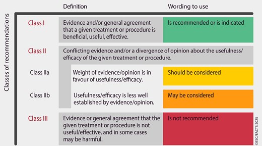
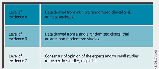
Guidelines Task Forces perform a critical review and evaluation of the published literature on diagnostic and therapeutic approaches including assessment of the risk–benefit ratio. Recommendations are based on major randomized trials and relevant systematic reviews and meta-analyses, when available. Systematic literature searches are conducted in cases of controversy or uncertainty to ensure that all key studies were considered. For recommendations related to diagnosis and prognosis, additional types of evidence are included, such as diagnostic accuracy studies and studies focused on the development and validation of prognostic models. The strength of each recommendation and the level of evidence supporting it are weighed and scored according to pre-defined criteria as outlined in Tables 1 and 2. Patient-Reported Outcome Measures (PROMs) and Patient-Reported Experience Measures (PREMs) are also evaluated when available as the basis for recommendations and/or discussion in these guidelines.

Table 1

Classes of recommendations

Table 2

Levels of evidence

Evidence tables summarizing key information from relevant studies are generated to facilitate the formulation of recommendations, to enhance comprehension of recommendations after publication, and to reinforce transparency in the guidelines development process. The tables are published in their own section of the guidelines and reference specific recommendation tables.

After an iterative process of deliberations, a first Task Force vote on all recommendations is conducted prior to the initiation of rounds of review. A second Task Force vote on all recommendations is conducted after the final round of review and revision. For each vote, the Task Force follows ESC voting procedures and all recommendations require at least 75% agreement among voting members to be approved. Voting restrictions may be applied based on declarations of interests.

The writing and reviewing panels provide declaration-of-interest forms for all relationships that might be perceived as real or potential sources of conflicts of interest. Their declarations of interest are reviewed according to the ESC declaration-of-interest rules, which can be found on the ESC website (http://www.escardio.org/doi) and are compiled in a report published in a supplementary document with the guidelines. Funding for the development of these ESC/EACTS Guidelines was derived entirely from the ESC and the EACTS with no involvement of the healthcare industry.

The ESC CPG Committee supervises and co-ordinates the preparation of new guidelines and approves their publication. In addition to review by the ESC CPG Committee, these ESC/EACTS Guidelines underwent multiple rounds of double-blind peer review on a dedicated online review platform. The review was conducted by topic experts, including members from ESC National Cardiac Societies, EACTS Network of National Cardiac Surgery Societies and from relevant ESC Subspecialty Communities. The Guideline Task Force considered all review comments and was required to respond to all those classified as major. After appropriate revisions, the Task Force, the ESC CPG Committee members and the EACTS Council members approved the final document for publication in the European Heart Journal and in the European Journal of Cardio-Thoracic Surgery.

Unless otherwise stated, the guideline content refers to sex, understood as the biological condition of being male or female, defined by genes, hormones, and sexual organs. Off-label use of medication may be presented in this guideline if a sufficient level of evidence shows that it can be considered medically appropriate for a given condition. However, decisions on off-label use must be made by the responsible health professional giving special consideration to ethical rules concerning healthcare, the specific situation of the patient, patient consent, and country-specific health regulations.

2. Introduction

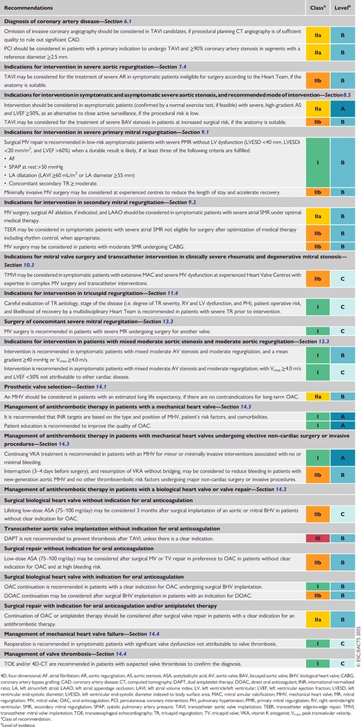
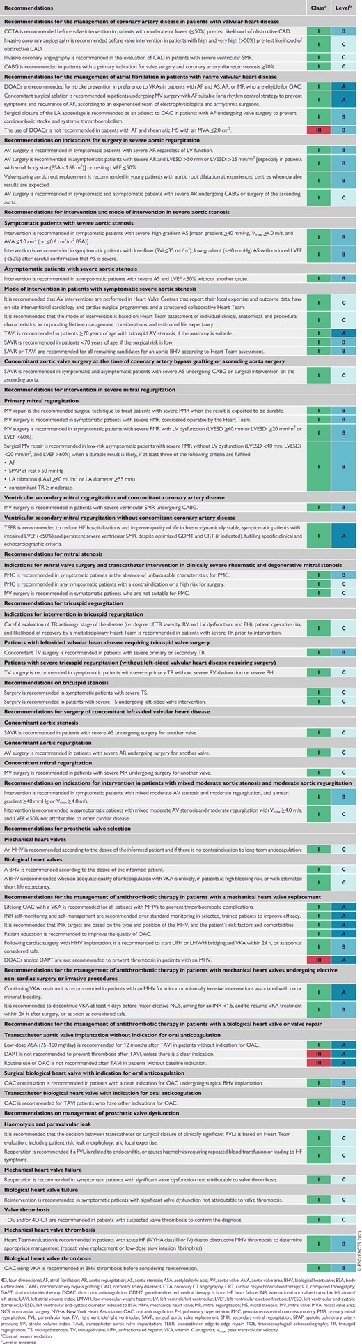
New evidence has accumulated since the publication of the 2021 European Society of Cardiology (ESC)/European Association for Cardio-Thoracic Surgery (EACTS) Guidelines for the management of valvular heart disease, leading to the need for new recommendations (Table 3 New recommendations) and revision of existing recommendations (Table 4 Revised recommendations) concerning the following topics:

  • The importance of shared and patient-centred decision-making by multidisciplinary expert Heart Teams working within a regional network has been reinforced. Patients with complex conditions or requiring complex procedures should be referred to high-volume centres, where corresponding expertise is concentrated to ensure high-quality treatment.

  • Advanced imaging modalities—such as three-dimensional (3D) echocardiography, cardiac computed tomography (CCT), and cardiac magnetic resonance (CMR) imaging—have gained importance and become a central aspect in the screening and evaluation of patients with valvular heart disease (VHD).

  • Emphasis is put on the importance of correctly assessing the cause(s) and mechanism(s) of all valve diseases. In particular, the distinction between atrial and ventricular secondary mitral regurgitation (SMR) has clear implications in terms of prognosis and management.

  • New evidence has been published regarding the benefits of intervention for the treatment of severe aortic stenosis (AS) irrespective of symptoms, left ventricular ejection fraction (LVEF), and flow reserve.

  • The criteria used for decision-making concerning the optimal modality of AS treatment [transcatheter aortic valve (AV) implantation (TAVI) or surgical AV replacement (SAVR)] based on a Heart Team approach have been refined, including the combination of key aspects such as age, procedural risk, and anatomical suitability, incorporating estimated life expectancy and lifetime management considerations.

  • Further randomized evidence confirming the mid-term safety and efficacy of TAVI in low-risk patients has been published.

  • The indications for TAVI in patients with bicuspid AV (BAV) stenosis or severe aortic regurgitation (AR) at high surgical risk, based on anatomical suitability and a comprehensive Heart Team evaluation, are discussed.

  • Several advancements have been made regarding the treatment of patients with primary mitral regurgitation (PMR): refinement of the criteria for intervention in asymptomatic patients; demonstration of the value of minimally invasive mitral valve (MV) surgery to reduce the length of hospital stay and accelerate recovery; and large-scale data confirming the role of transcatheter edge-to-edge repair (TEER) in high-risk patients.

  • Longer-term follow-up data and two new randomized controlled trials (RCTs) concerning the management of patients with ventricular SMR have been published.

  • The evidence for the treatment of tricuspid valve (TV) disease is growing—including new randomized data supporting concomitant TV repair during left-sided valve surgery, and transcatheter options (repair and replacement) that reduce tricuspid regurgitation (TR), promote reverse right ventricular (RV) remodelling, and improve quality of life compared with medical treatment.

  • Efforts have been made to provide improved guidance regarding the diagnostic steps and management of patients with multiple and mixed VHD.

  • Definitions of structural valve deterioration (SVD) have been updated and unified.

  • The recommendations concerning the use of direct oral anticoagulants (DOACs) in patients with VHD have been updated, and the importance of education and (self-)monitoring is emphasized.

  • Sex-specific considerations in patients with VHD have been extended and regrouped into a new dedicated section (see Section 17).

Because of demographic changes, patients with VHD frequently present with concomitant cardiovascular diseases, increasing the complexity of treatment strategies. These Guidelines focus on acquired VHD and do not deal in detail with overlapping cardiovascular diseases such as infective endocarditis,5 chronic coronary syndrome,6 and atrial fibrillation (AF),7 as well as all scenarios of aortic or congenital disease,8,9 because these topics are covered in separate Guidelines.

The 2025 ESC/EACTS Guidelines for the management of valvular heart disease aim to be concise, focused on relevant issues for clinicians and patients, and to provide clear and simple practical recommendations, assisting healthcare providers in their daily clinical decision-making. A compilation of the evidence considered for new recommendations, or those with an updated class of recommendation or level of evidence, can be consulted online (see Supplementary data online, Evidence Tables). The Task Force for these Guidelines acknowledges that multiple factors influence and ultimately determine the most appropriate treatment of individual patients within a given community. These factors include the availability of equipment and technology, and the expertise and volumes, in complex procedures, such as valve repair or transcatheter interventions. Moreover, given the lack of evidence on some topics related to VHD, several recommendations are the result of expert consensus opinion. Therefore, deviations from these Guidelines may be appropriate in certain clinical circumstances, and decision-making should always be based on a collaborative, multidisciplinary Heart Team approach centred on individual characteristics, needs, and prognosis, as well as the preferences of the informed patient.

2.1. What is new

Table 3

New recommendations

Table 4

Revised recommendations

3. The Heart Team and Heart Valve Centre

3.1. The Heart Valve Network

Despite increasing attention within the medical community, VHD continues to be underdiagnosed and undertreated in the general population, and public awareness remains low.10–13 Beside screening using auscultation and imaging when appropriate, the co-ordinated implementation of Heart Teams, Heart Valve Centres, and Heart Valve Networks at a local level represents an essential step to timely diagnose and treat patients with VHD.

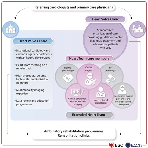
An integrated regional Heart Valve Network, incorporating outpatient Heart Valve Clinics (for initial diagnosis and ongoing surveillance) and specialist Heart Valve Centres (for advanced imaging and surgical or transcatheter intervention), allows optimal patient care through timely access to specialist assessment, accurate diagnosis, improved decision-making, and matching of patients to healthcare providers with appropriate expertise, experience, and resources (Figure 1).12 In addition, dedicated Heart Valve Clinics ensure consistent application of clinical guidelines, efficient use of resources, and overall high-quality patient care, which in turn may improve outcomes.14,15 Medical goals include careful clinical and echocardiographic evaluation, monitoring at appropriate time intervals (so-called ‘watchful waiting’), application of guideline-directed medical therapy (GDMT), timely referral, and post-procedural follow-up.15,16 Broader aims include patient education, the training of physicians and nurse specialists, swift and efficient access to specialist care, and recruitment into clinical trials.
The Heart Valve Network.
Figure 1

The Heart Valve Network.

HF, heart failure; VHD, valvular heart disease.

Heart Valve Centres should ensure that their facilities match institutional and local statutory requirements (Table 5), report procedural volumes and outcomes, and monitor treatment quality. They hold responsibility for the training and education of surgeons, interventional and imaging cardiologists, dedicated nurses, and allied professionals.17,18 Expertise in the surgical management of coronary artery disease (CAD), vascular diseases, and complications must be available. New techniques should be taught by trained mentors using simulator models, when feasible, to minimize learning-curve effects. More broadly, Heart Valve Centres co-ordinate the management of patients with VHD across the entire Heart Valve Network, supporting services at community level, encouraging early referral, and promoting education and communication with other medical departments, referring cardiologists, primary care physicians, and rehabilitation clinics.

Table 5

Requirements for a Heart Valve Centre

Requirements
Centre performing heart valve procedures with on-site interventional cardiology and cardiac surgery departments providing 24-h/7-day services.
Heart Team core members: Cardiologist with imaging expertise, interventional cardiologist, cardiac surgeon.
Additional specialists, if required (Extended Heart Team): Specialized nursing personnel, HF specialist, electrophysiologist, cardiovascular anaesthetist, geriatrician, and other specialists (e.g. intensive care, vascular surgery, infectious diseases, neurology, radiology).
The Heart Team must meet on a regular basis and work according to locally defined standard operating procedures and clinical governance arrangements.
A hybrid cardiac catheterization laboratory is desirable.
High volume for hospital and individual operators.
Multimodality imaging (including advanced echocardiography, CCT, CMR, and nuclear techniques) and expertise in peri-procedural imaging guidance of surgical and transcatheter procedures.
Heart Valve Clinic for outpatient assessment and follow-up.
Data review: continuous monitoring, evaluation, and reporting of procedural volumes and quality indicators, including clinical outcomes, as well as PROMs complemented by local/external audits.
Education programmes targeting primary care and referring physicians, operators, and diagnostic and interventional imaging specialists.

CCT, cardiac computed tomography; CMR, cardiac magnetic resonance; HF, heart failure; PROM, patient-reported outcome measure.

© ESC/EACTS 2025

3.1.1. Composition of the Heart Team

The Heart Team is now an established feature of VHD programmes that has been formally endorsed by previous ESC/EACTS Guidelines19,20 and corresponding organizations worldwide.17

The value of the Heart Team approach has become increasingly apparent as options for the treatment of VHD have extended to include high-risk and inoperable patients (most of whom now undergo transcatheter interventions), and low-risk and asymptomatic patients (who derive prognostic benefit from increasingly safe procedures). Despite significant accumulation of data concerning the management of VHD over the last two decades, many patients in daily practice have clinical characteristics that do not match those of participants included in clinical trials. The Heart Team approach is therefore particularly helpful when there is uncertainty or a lack of strong evidence.

The Heart Team meeting facilitates balanced presentation of all appropriate options for medical, interventional, and surgical treatment, using tools and techniques for shared decision-making. The patient's preference plays a central role in this process, although the Heart Team recommendation should be based upon key objective medical considerations (particularly the relative risks and benefits of any procedure).

Meetings should take place on a regular basis with standardized minimum datasets (to ensure that all relevant information is available) and appropriate administrative support (often provided by specialist nurses with expertise in the care of patients with VHD). In-person meetings of the full Heart Team may not be feasible for every patient, and local standardized protocols may be implemented to facilitate swift decision-making for specific cohorts (e.g. elderly TAVI candidates or young patients with BAV disease). Equally, the need for Heart Team evaluation should not paralyse clinical decision-making, and ad hoc discussions remain appropriate in urgent situations.

Core members of the Heart Team include the cardiologist treating the patient (who is best placed to present their case and act as their advocate), specialists in advanced cardiovascular imaging and peri-procedural guiding,21,22 surgeons, and interventional cardiologists with training and expertise in surgical and transcatheter valve procedures. Specialized nursing personnel play an essential role to improve patient information and education, as well as co-ordinate work-up and management steps in high-volume centres (Figure 1; Table 5). Cardiologists with expertise in heart failure (HF) and electrophysiology, as well as geriatricians, cardiovascular anaesthetists, and intensivists involved in peri-procedural care, should also be available to facilitate the discussion of particularly complex clinical scenarios when needed (extended Heart Team) (Figure 1; Table 5).

3.1.2. Procedural volume and clinical outcomes

The correlation between high institutional (and individual operator) volume and best procedural outcomes is intuitive, yet complex. Nevertheless, there is evidence of such a relationship for many cardiovascular procedures including SAVR,23,24 surgical MV repair,25,26 mitral and tricuspid TEER,27–30 and TAVI (particularly in centres with an associated high-volume SAVR programme).31–33 Studies have shown that an annualized operator volume of approximately 25 surgical mitral valve procedures,26 50 TAVIs (∼100 per centre),33 a cumulative experience of ∼50 M-TEER procedures per operator/centre,27 and a site volume of more than 20 T-TEERs/year30 are associated with improved technical and clinical outcomes. Higher institutional surgical volume is associated with lower complication rates,34–36 improved management,37 and better infrastructural support.38,39

National procedural activity varies widely between high-, middle-, and low-income countries,40 and it is therefore difficult to provide recommendations concerning the precise number of institutional or operator procedures that is required for high-quality care, excellent facilities, and processes. Instead, a network approach that highlights the importance of centres performing a high volume of procedures (e.g. based upon quartiles in individual countries or regions) seems more suitable, with complex procedures concentrated in the centres with the highest volumes (Table 6). Internal quality assessment (see dedicated document concerning TAVI from the ESC41), systematic recording, and public availability of the volume and outcome data of the performed procedures are essential. Participation in national or international registries should be encouraged. These considerations are of particular importance regarding asymptomatic low-risk patients (where low mortality and procedural safety are paramount), those with multiple comorbidities (where the need for multidisciplinary collaboration is essential), and new techniques with a steep learning curve (where better results may be obtained at experienced centres).

Table 6

Complex procedures ideally performed in the most experienced Heart Valve Centres

Transcatheter interventionsSurgical interventions
  • Transfemoral TAVI in patients with high-risk features:

    • –

      Low coronary ostia

    • –

      Difficult femoral anatomy

    • –

      Bicuspid valve

    • –

      Severe calcification protruding into the LVOT

    • –

      Severe LV and/or RV impairment

    • –

      Pure AV regurgitation

    • –

      Multiple valve disease

    • –

      Complex coronary artery disease

    • –

      Severe extracardiac disease (e.g. renal failure, PH)

  • Non-transfemoral TAVI

  • Valve-in-valve (including TAV-in-TAV)

  • All leaflet modification procedures (BASILICA, LAMPOON etc.)

  • PVL closure procedures

  • Complex M-TEERa

  • Redo M-TEER procedures

  • Tricuspid or mitral valve-in-ring or valve-in-valve, valve-in-MAC

  • TMVI

  • All tricuspid procedures

  • High-risk procedures (especially in patients with LV and/or RV impairment)

  • Redo procedures

  • Minimally invasive and robotic valve surgery

  • Complex MV repair

    • –

      Barlow disease

    • –

      Anterior or bileaflet prolapse

    • –

      High risk of SAM

    • –

      Severe MAC

  • AV repair

  • Ross procedure

  • Valve surgery combined with complex surgery of the aorta

  • Endocarditis surgery

AV, aortic valve; BASILICA, Bioprosthetic or native Aortic Scallop Intentional Laceration to prevent Iatrogenic Coronary Artery obstruction; LAMPOON, Laceration of the Anterior Mitral leaflet to Prevent Outflow ObstructioN; LV, left ventricular/left ventricle; LVOT, left ventricular outflow tract; MAC, mitral annular calcification; M-TEER, mitral transcatheter edge-to-edge repair; MV, mitral valve; PH, pulmonary hypertension; PVL: paravalvular leak; RV, right ventricular/right ventricle; SAM, systolic anterior movement; TAV, transcatheter aortic valve; TAVI, transcatheter aortic valve implantation; TMVI, transcatheter mitral valve implantation.

© ESC/EACTS 2025

There is a pressing need to ensure higher dispersion and adoption of interventions for VHD, especially (but not exclusively) in middle- and lower-income countries where rheumatic heart disease (RHD) remains the principal cause of VHD.42,43 Key strategies include awareness programmes, increased public and medical education, simplified and improved diagnostic tools, and measures to reduce costs and facilitate access to evidence-based treatment options.44

4. Imaging of patients with valvular heart disease

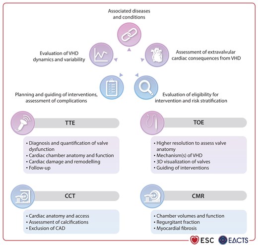
Multimodality imaging is now the standard approach in VHD management to determine the pathophysiology, assess severity, plan interventions, and identify complications (Figure 2). The use of imaging for the assessment of each specific valve lesion is described in the corresponding sections. The role of imaging is transversal from diagnosis to follow-up and should encompass an integrative assessment.
Integrative imaging assessment of patients with valvular heart disease.
Figure 2

Integrative imaging assessment of patients with valvular heart disease.

3D, three-dimensional; CAD, coronary artery disease; CCT, cardiac computed tomography; CMR: cardiac magnetic resonance; TOE; transoesophageal echocardiography; TTE, transthoracic echocardiography; VHD, valvular heart disease.

4.1. Initial valve assessment

Comprehensive transthoracic echocardiography (TTE) is the first-line examination to confirm valve dysfunction, and determine the aetiology, mechanism, and severity of VHD, as well as cardiac chamber anatomy and damage.45,46 It should be performed by properly trained imagers.47–49 Quantitative imaging analysis (as opposed to visual) should be the goal in all patients with relevant VHD, complemented by qualitative and semi-quantitative evaluation. The severity of VHD should be assessed using an integrative approach of all criteria checked for consistency. When TTE is of poor quality or inconclusive, transoesophageal echocardiography (TOE) and/or additional diagnostic imaging modalities should be applied (e.g. calcium scoring and anatomy of the valve using CCT). In specific clinical scenarios [e.g. thrombosis, prosthetic valve dysfunction, endocarditis, mitral stenosis (MS), assessment of MV or TV anatomy], TOE has a central diagnostic role.50,51 Moreover, in patients with regurgitant lesions, particularly AR, CMR has gained key value in clinical practice.

4.2. Associated diseases and conditions

Imaging plays a crucial role in identifying associated diseases and conditions. The presence of concomitant left ventricular (LV) systolic or diastolic dysfunction, RV systolic dysfunction, red flags indicating cardiomyopathies (e.g. amyloidosis, hypertrophic cardiomyopathy), and aortopathy should prompt further examinations to ensure optimal risk stratification and VHD management. In addition to TTE, advanced imaging modalities, like CCT, may be required to assess the aorta, single-photon emission computed tomography (CT) for detection of ischaemia/necrosis, positron emission tomography (PET) for inflammation, nuclear scintigraphy for cardiac amyloidosis, or CMR for tissue characterization.

4.3. Evaluation of valvular heart disease dynamics and variability

Serial imaging studies to detect changes over time, or variability due to haemodynamic conditions or initiation/up-titration of medical therapy, are of utmost importance to guide decisions. The dynamic pattern of VHD may convey additional prognostic information. Exercise echocardiography helps to identify the cause of dyspnoea, unveil symptoms in apparently asymptomatic patients, identifies dynamic changes of VHD severity, and can contribute to refinement of the indication for an intervention, especially for AS and mitral regurgitation (MR).52

4.4. Assessment of extravalvular cardiac consequences from valvular heart disease

Several studies investigating different valve lesions have established the relevance of extravalvular cardiac damage in terms of prognosis,53–55 recovery after intervention,56 and quality of life.57 The presence of LV hypertrophy, left atrium (LA) dilatation, LV or RV dysfunction and/or remodelling, myocardial fibrosis, and pulmonary hypertension (PH) provide important prognostic information, and may influence the timing and type of treatment. Although not necessarily chronological in their order of appearance, the understanding of cardiac damage, particularly damage involving the LV, is essential to guide appropriate medical therapy before and after any intervention. Transthoracic echocardiography (TTE) including global longitudinal strain (GLS) and CMR can be particularly useful in that regard.58–60

4.5. Evaluation of eligibility, planning, and guiding of interventions

Transoesophageal echocardiography (TOE) (preferentially 3D) is the preferred tool for the assessment of suitability for aortic, mitral, and TV repair.45,49,61–63 Risk stratification for an intervention should integrate all the points mentioned above.

Transoesophageal echocardiography (TOE), including standard 3D views, is also the modality of choice to guide atrioventricular transcatheter interventions and should be performed by specially trained interventional echocardiographers.61,64 Cardiac computed tomography (CCT) is frequently used to evaluate the relationship of the valve with adjacent structures [e.g. coronary arteries and left ventricular outflow tract (LVOT)], the extension of calcification [e.g. in mitral annular calcification (MAC)], and for sizing of the prosthesis. CT angiography is frequently employed to evaluate the anatomy of surgical, arterial, or venous access routes and detect cardiac or extracardiac complications (e.g. bleeding or embolic events). Coronary CT angiography (CCTA) is increasingly utilized to assess the presence of CAD.

5. Clinical evaluation of patients with valvular heart disease

5.1. Clinical examination

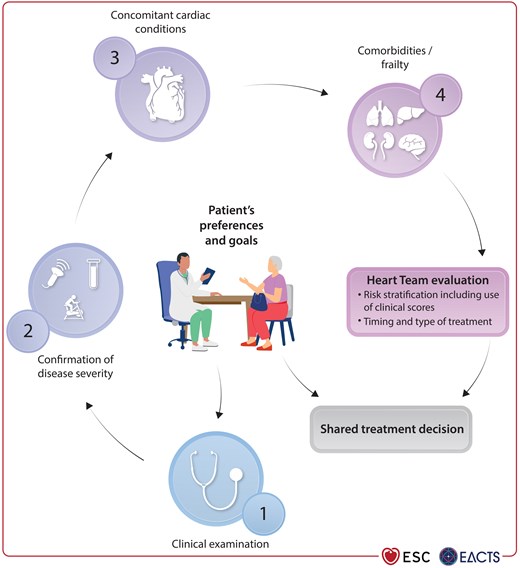
Patients with VHD can be either asymptomatic or present with a wide spectrum of symptoms, including acute or chronic HF. An initial meticulous history and a comprehensive physical examination of the patient with auscultation, documentation of clinical signs of HF such as dyspnoea, impaired physical capacity and fatigue, peripheral oedema, and pleural effusion, as well as a systematic frailty assessment, are crucial.65 In addition, comorbidities and coexisting cardiac conditions should be documented (Figure 3). Particular attention should be given to recent changes in symptoms or physical findings indicating a potential worsening of the valve lesion or ventricular function.
Central illustration. Patient-centred evaluation for treatment of valvular heart disease.
Figure 3

Central illustration. Patient-centred evaluation for treatment of valvular heart disease.

5.2. Assessment of comorbidities and risk stratification

Risk stratification of patients with VHD has been mainly developed based on surgical populations. The European System for Cardiac Operative Risk Evaluation II (EuroSCORE II)66,67 (https://www.euroscore.org) and the Society of Thoracic Surgeons predicted risk of mortality (STS-PROM) score68 (http://riskcalc.sts.org/stswebriskcalc/calculate) are the most commonly used scoring systems to estimate surgical risk. Both risk scores have been calibrated to predict post-operative outcomes.69–71 The STS-PROM score is dynamic to account for changes in patient risk profiles, the type of procedure (aortic, mitral, and tricuspid), and outcomes over time. The outdated (logistic) EuroSCORE I model systematically overestimated surgical mortality.70,72

In patients considered for TAVI, surgical risk scores have lower accuracy and tend to overestimate the risk of events.73–75 Discrepancies between observed and predicted peri-procedural mortality after TAVI using surgical risk scores point towards a need for TAVI-specific scores. Models predicting short- and medium-term survival specifically designed for TAVI are rarely used in daily clinical practice due to limitations of their predictive performance.76–79 Specific scores have also been developed for patients undergoing mitral TEER (M-TEER),80–82 but the high heterogeneity of the population with MR limits their external validity and therefore routine clinical use so far.83 Recently, a dedicated clinical score has been calibrated and validated to stratify the risk associated with first-time and repeat isolated TV surgery (TRI-SCORE; https://www.tri-score.com/), and its use should be encouraged for patients with TV disease,84,85 as an alternative to the more complex Society of Thoracic Surgeons (STS) score for isolated TV surgery (https://isolatedtvsurgcalc.research.sts.org/).

Other cardiac conditions and comorbidities—such as CAD, concomitant multiple valve and aortic disease, and RV dysfunction, as well as chronic kidney disease (CKD)—are not always appropriately captured in risk models, even if they are known to impact outcomes.86 Similarly, specific unfavourable characteristics like porcelain aorta, mobile aortic atheroma, and previous mediastinal radiation therapy increase the surgical risk, and therefore may favour transcatheter treatment options. Frailty, including nutritional state, represents another important determinant of outcomes after valve interventions,87–89a which can be evaluated using appropriate tools as summarized in a recent consensus statement.90 Several methods have been proposed, from the simple Fried Frailty Index to more complex scores91 such as the Hospital Frailty Risk Score, which has been validated in a large cohort of TAVI and M-TEER patients.92

The use of PROMs that engage patients in the co-evaluation of their health and wellbeing is encouraged. Several scoring systems have been proposed and validated for reproducibility and association with clinical outcomes, like the Quality of Recovery 15-item PROM or the Kansas City Cardiomyopathy Questionnaire (KCCQ).93–95

5.3. Biomarkers

Biomarker levels indicating either cardiac wall stress [e.g. brain natriuretic peptide (BNP)] or myocardial damage (e.g. troponin), in asymptomatic and symptomatic patients, may help to monitor VHD progress and determine the most appropriate timing of intervention. In patients with VHD, the natriuretic peptide ratio [the ratio of measured BNP or N-terminal pro-B-type natriuretic protein (NT-proBNP) to upper limit of normal for age, sex, and assay] has been shown to be a powerful, independent, and incremental predictor of mortality.96–98 In patients undergoing AV replacement, the accumulation of several elevated biomarkers of cardiovascular stress was associated with higher all-cause and cardiovascular mortality, and a higher rate of repeat hospitalization.99,100

5.4. Exercise testing

Because of the slow progression of valve lesions, patients may gradually limit their activity levels over several years and deny having actual symptoms that can be unmasked by exercise testing.12 This is particularly important in cases of AS, because once symptoms occur there is a sharp increase in the risk of sudden cardiac death, unless a valve intervention is performed.101–104 Exercise testing may provide additional information about the haemodynamic severity of VHD and help determine the risk and optimal timing of intervention by objectively evaluating functional capacity.101,103 Cardiopulmonary exercise testing has a prognostic role and can assist decision-making in patients with VHD of intermediate severity, particularly those with asymptomatic AR.101,105,106

Exercise echocardiography is used for the assessment of LV global and segmental function, pulmonary artery pressure, and aortic and mitral pressure gradients.101,107 It also documents exercise-induced increase of MR and TR severity, especially in patients with secondary disease.108,109 Prognostic impact has been shown mainly for AS and MR.110,111 Misconceptions regarding its risk and tolerability contribute to the overall underuse of exercise testing in patients with VHD,12 despite data confirming its safety in most asymptomatic patients.112,113

5.5. Invasive investigations

5.5.1. Coronary angiography

Coronary artery assessment is recommended to evaluate the need for revascularization when valve surgery or an intervention is planned. The information regarding the existence of concomitant CAD should be available at the time of Heart Team discussion. CCTA is recommended as an alternative to coronary angiography to rule out CAD in patients who are at low or moderate risk of obstructive CAD.

Coronary blood flow and fractional flow reserve (FFR) are altered in the setting of concomitant VHD, and functional haemodynamic assessment of CAD in these patients is not well established.114–118

5.5.2. Cardiac catheterization

Right heart catheterization (RHC) should be performed in patients with equivocal echocardiographic findings, particularly those with MV disease, as well as in all candidates for the treatment of severe TR. In exceptional cases with unclear AS severity, it can be combined with measurements of the transaortic gradients allowing estimation of the aortic valve area (AVA). Right heart catheterization contributes to assess the repercussions of any left-sided VHD or LV impairment on the pulmonary circulation and right side of the heart. It provides information regarding volume state, cardiac output, and vascular resistance, differentiating between pre- and post-capillary PH, and should be ideally performed in euvolaemia.

Measurements of the pulmonary capillary wedge pressure v-wave can inform about MR severity, but are neither sensitive nor specific, and may also be increased if the compliance of the LA is reduced or in case of diastolic LV dysfunction, as in patients with MS or chronic HF.119

Similarly, the height of the right atrium (RA) v-wave, and the pressure curve mimicking the RV pattern (‘ventricularization’ of the RA pressure), are signs of relevant TR, which are frequently accompanied by increased RV end-diastolic pressure in case of associated RV dysfunction. In patients with severe TR, pulmonary vascular resistance (PVR) should be calculated to unmask pulmonary vascular disease, which may not be captured by echocardiography due to RV systolic dysfunction or underestimation of pulmonary pressures because of TR.

5.6. Patient-centred care and shared decision-making

Given that treatment of VHD typically involves several modalities and specialities, and may result in a complex and sometimes time-consuming decision-making process, patient education and information, using online material and face-to-face conversations, are essential at each step. A clearly defined point of contact for all questions relating to the disease or type of treatments should be communicated to the patient and their relatives.120 The symptomatic and prognostic benefits, as well as the advantages and disadvantages of any treatment option, should be presented in an open and evidence-based manner. This includes mortality and risks of reintervention and complications (also over the long term), as well as recovery time and the need for cardiac, and if necessary psychological, rehabilitation until return to physical activity and work. Other issues to be discussed before the procedure include the need for oral anticoagulation (OAC) and its monitoring, as well as the noise generated by mechanical heart valves (MHVs). Information regarding centre experience and volume for a specific procedure should be provided. Misconceptions (e.g. subjective overestimation of the risk of surgery) should be addressed and potential interactions with individual lifestyle factors—including social activities, family and professional life, and hobbies—should be discussed in detail.

The Heart Team recommendation regarding the treatment and its modality must be based on evidence and anatomical considerations, balancing the risks and benefits of available treatment options.16 The patient and patient's relatives need to be well informed about the rationale leading to the Heart Team recommendation, and given ample time to share personal preferences.121 At the end of the process, a shared decision is made between the treating team and the informed patient and relatives (Figure 3).

6. Management of conditions associated with valvular heart disease

6.1. Diagnosis and management of coronary artery disease

The presence of CAD plays an important role in decision-making regarding the timing and modality of treatment, and should be assessed before Heart Team discussion. In patients with a low or moderate pre-test likelihood of obstructive CAD (≤50%), CCTA is recommended to rule out relevant CAD with high sensitivity.122–124

Several studies have investigated the value of CCT angiography for CAD screening in elderly TAVI candidates. While sensitivity for the detection of obstructive CAD is high (95%–97%), specificity (68%–73%) is modest, mainly explained by the high prevalence of coronary artery calcification and AF in patients with severe AS, which limit imaging resolution and interpretability.125,126 If CCT angiography obtained during standard pre-TAVI evaluation is of sufficient quality to exclude relevant CAD, omission of invasive coronary angiography should be considered.125–129

The value of invasive functional haemodynamic assessment of CAD in patients with severe AS may be limited, because AS impacts coronary haemodynamics. Therefore, caution is warranted in the interpretation of functional measurements in the presence of severe AS until more data are available.115,116

Recommendations for the management of chronic CAD associated with VHD are provided below (Recommendation Table 1), as well as in dedicated guideline documents.6 The indications for coronary artery bypass grafting (CABG) in patients undergoing surgery for the treatment of VHD are mainly based on observational data, which do not provide detailed information on the degree of stenosis and the complexity of CAD.130 It has been demonstrated that subendocardial blood flow in the myocardium improves early after SAVR, most likely due to improved cardiac output and reduction of LV wall stress.131 The presence of CAD is associated with peri-operative and late adverse events in patients with AS undergoing SAVR132 that likely outweigh the increased risk of peri-procedural adverse events of combined SAVR and CABG compared with isolated SAVR. Indeed, in a large observational study, patients with CAD demonstrated better long-term survival after combined SAVR and CABG compared with SAVR alone, despite longer cross-clamp times.133 In patients with a primary indication for valve surgery, CABG is recommended in patients with coronary artery stenosis of ≥70% and should be considered in those with stenosis of ≥50%–70%, given the opportunity of concomitant full revascularization.

Recommendation Table 1

Recommendations for the management of chronic coronary syndrome in patients with valvular heart disease (see also Supplementary data online, Evidence Table 1)
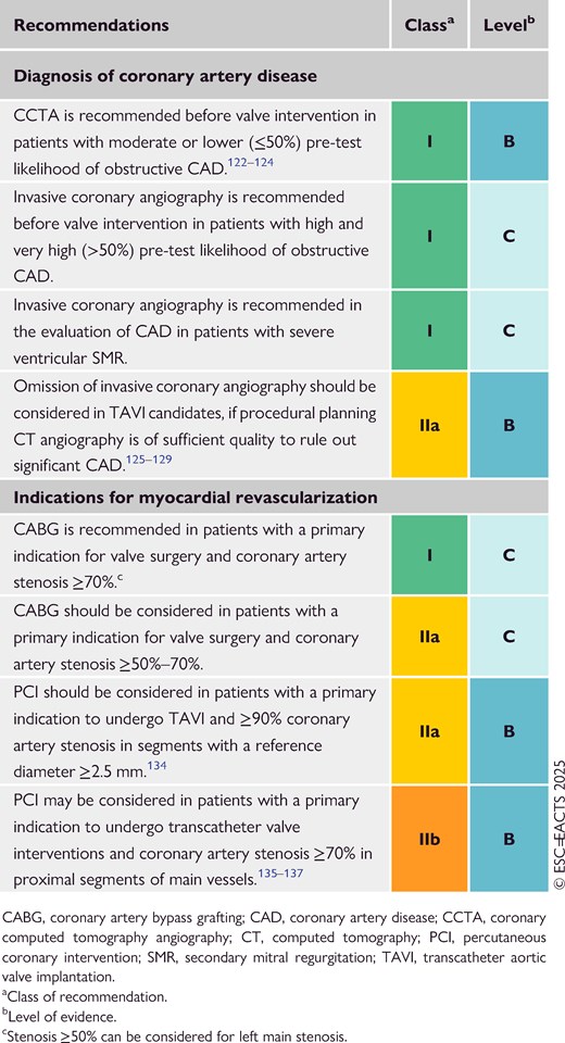
The impact of CAD in patients undergoing TAVI remains under investigation. The randomized Nordic Aortic Valve Intervention (NOTION)-3 trial compared a strategy of routine percutaneous coronary intervention (PCI) versus conservative management in 455 patients with severe symptomatic AS undergoing TAVI, who also had stable CAD and at least one stenosis of ≥90% based on visual angiographic assessment or FFR of ≤0.80 in a segment with ≥2.5 mm reference diameter.134 Percutaneous coronary intervention was associated with lower risk of a composite endpoint event including all-cause death, myocardial infarction or urgent revascularization at a median follow-up of 2 years. Exploratory analyses suggest that the increased risk in the conservative treatment arm was driven by differences in the risk of myocardial infarction and urgent revascularization in patients with a diameter stenosis of ≥90%, rather than those with positive FFR and stenosis of <90%. The risk of bleeding was higher in the PCI than in the conservative treatment group. Another multicentre RCT, the PercutAneous Coronary inTervention prIor to transcatheter aortic VAlve implantaTION (ACTIVATION) trial, was discontinued due to slow recruitment.135 In this underpowered and thus inconclusive trial, a routine PCI strategy of ≥70% stenoses in main epicardial vessels (or ≥50% if protected left main or vein graft) did not meet non-inferiority compared with conservative CAD treatment with respect to the composite of all-cause death and rehospitalization; moreover, PCI was associated with higher bleeding rates. Observational data show that TAVI can be performed safely in patients with untreated CAD with low short- and long-term rates of acute coronary syndrome and unplanned coronary revascularization.135–139 In two recent meta-analyses of mostly observational data, PCI was not associated with a mortality benefit in patients with chronic CAD undergoing TAVI.140,141

Optimal timing of PCI in patients undergoing TAVI remains yet to be determined. In NOTION-3, PCI was performed before TAVI in the majority of the patients (concomitantly or shortly after in only 26% of the patients). Decision-making concerning the timing of PCI should take into account the type of valve used for TAVI and the complexity of the coronary lesions.142,143 Valves with a high frame—particularly in the context of a narrow aortic root, commissural misalignment, or valve-in-valve procedures—can pose challenges for coronary access following TAVI.144,145 The presence of significant CAD should therefore be considered for optimal transcatheter valve selection and reinforces the importance of optimized implantation technique and commissural alignment.143,146 Several RCTs comparing the value and timing of PCI with medical therapy are ongoing (NCT04634240, NCT04310046, and NCT05078619).

Based on the available data, PCI should be considered in patients with a primary indication to undergo TAVI and high-grade (≥90%) coronary artery stenosis in large vessels of ≥2.5 mm. In patients with stenosis of ≥70%, PCI may be considered based on symptom status.142,143 In patients with ischaemic ventricular SMR, surgical revascularization of CAD has been associated with MR reduction147 and favourable clinical outcomes in observational studies.148,149 According to very limited data, improvement of SMR may occur in a minority of patients (about one-third) after PCI, which may therefore be considered prior to MV intervention.150

For patients with VHD presenting with acute coronary syndrome, treatment decisions should be made according to the most recent ESC Guidelines.151 In patients presenting with non-ST-segment elevation acute coronary syndrome, it may be particularly challenging to determine the leading cause of elevated troponin levels, which are also frequently observed in decompensated VHD. Therefore, the treatment strategy should be determined by the Heart Team, taking into account symptoms, as well as coronary, valve, and access anatomy.136,138,139

6.2. Atrial fibrillation

The interplay between AF and VHD is complex, and has an essential role in the prognosis and evolution of VHD during the patient's lifetime. Valvular heart disease is independently associated with AF and almost one-third of patients with AF have a history of VHD.152 Conversely, AF is the main trigger of the development of atrial secondary MR and TR. In a recent cohort study, 8% of individuals with AF developed moderate or severe TR within 3 years of follow-up compared with only 2% in those in sinus rhythm.153 While disturbed annular dynamics have been postulated,150,154 the exact pathophysiology leading to secondary atrioventricular VHD in some, but not all patients with AF remains largely unclear.

Detailed recommendations on the anticoagulation management of patients with VHD and AF are provided below (Recommendation Table 2) and in Section 14, as well as in specific Guidelines.7 Patients with a combination of VHD and AF have a high incidence of thromboembolic or bleeding complications.155 DOACs have replaced vitamin K antagonists (VKAs) in most clinical scenarios and are recommended for patients with VHD presenting with AF, except for patients with a MHV or mitral stenosis with a valve area ≤2.0 cm2. The use of apixaban,156 dabigatran,157 edoxaban,158 and rivaroxaban159 is supported by subgroup analyses of large RCTs. Recommendations regarding the antithrombotic treatment of patients with MHVs and biological heart valves (BHVs) are described in Section 14 of these Guidelines.

Recommendation Table 2

Recommendations for the management of atrial fibrillation in patients with native valvular heart disease (see also Supplementary data online, Evidence Tables 2 and 3)
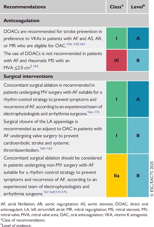
In the Left Atrial Appendage Occlusion Study (LAAOS) III trial, surgical left atrial appendage occlusion (LAAO) in patients with AF and a CHA2DS2-VASc [congestive heart failure or left ventricular dysfunction, hypertension, age ≥75 (doubled), diabetes, stroke (doubled), vascular disease, age 65–74, sex category (female)] score of ≥2 points undergoing cardiac surgery was associated with a 33% reduction of the risk of stroke or systemic embolism at a mean follow-up of 3.8 years.160 These findings were confirmed by a large meta-analysis including four RCTs.161 A subanalysis of LAAOS III showed that the benefit of LAAO remains consistent irrespective of the use of VKAs or DOACs, as well as in the absence of OAC (although representing only 10% of the included population).162 In an RCT including patients with severe AS and AF undergoing TAVI, concomitant transcatheter LAAO was non-inferior to medical therapy with respect to a composite primary endpoint including all-cause mortality, stroke, and major bleeding at 2 years. Of note, rates of major or life-threatening bleeding were similar in the two groups and arterial or venous thromboembolisms more frequent in the TAVI/LAAO arm, leaving uncertainty regarding the usefulness of combining both procedures.163

6.3. Cancer and radiation therapy

Valvular heart disease is commonly associated with cancer and represents a well-known long-term side effect of intensive radiation therapy176 for treatment of Hodgkin or non-Hodgkin lymphoma, breast cancer, and other thoracic malignancies.177 Clinically significant VHD most commonly appears decades after radiotherapy. The incidence of radiation-induced VHD is increasing owing to longer survival of patients with cancer. Risk factors are summarized in Supplementary data online, Table S1. Patients at risk should be screened for VHD using TTE 10 years after radiation exposure and followed up every 5 years thereafter.178

Radiotherapy may lead to aortic and/or valvular calcification, CAD of proximal segments, restrictive cardiomyopathy, pericardial adhesions and calcifications with constriction, restrictive lung disease, chest wall scarring, and impaired wound healing, especially in patients who underwent radiation by means of older techniques (>20 years ago). The above factors complicate any surgical approach and increase the operative risk, which is underestimated by traditional risk scores.179–181 TAVI is proposed as an alternative for patients presenting with radiation-induced AS in the 2022 ESC Guidelines on cardio-oncology182 based on favourable, but limited, observational data,183,184 because this category of patients has been excluded from RCTs.183,184 Furthermore, M-TEER in patients presenting with radiation-induced MV disease with MR is often limited by thickened leaflets with restricted movement and subsequent risk of iatrogenic stenosis.

In patients with active or stable cancer and severe AS, both TAVI and SAVR can be considered based on life expectancy, age, prognosis, and disability following cancer treatment, with a trend towards more TAVI utilization.185 TAVI procedural complication rates appear similar compared with those of control subjects without cancer.186 To avoid futility, treatment decisions discussed by the Heart Team should involve the treating oncologists.5,182

6.4. Prophylaxis of rheumatic fever

Rheumatic heart disease remains the most common cause of death from VHD worldwide.187 Prevention should preferentially target the first occurrence of acute rheumatic fever. Correct diagnosis and early antibiotic treatment of group A Streptococcus throat or skin infection is key for primary prevention. Large-scale screening combined with prophylaxis in children or adolescents with latent RHD appears to be an effective strategy to reduce the risk of disease progression and RHD prevalence.188–190 In patients with established RHD, secondary long-term prophylaxis with benzathine benzyl penicillin 1.2 million international units (IU) every 3–4 weeks over 10 years is recommended to prevent recurrent episodes, especially in children and adolescents. Long-term prophylaxis into adulthood should be considered in high-risk patients according to the severity of VHD and exposure to group A Streptococcus.191,192

6.5. Cardiogenic shock and acute heart failure

Acute presentation or decompensation of VHD can result in cardiogenic shock due to rapid haemodynamic deterioration and altered cardiac function. Alternatively, pre-existing VHD can be a bystander of an acute cardiovascular condition further exacerbating circulatory impairment until the occurrence of cardiogenic shock. In this setting, assessment of the severity of VHD, as well as its contribution to acute HF, may be difficult. Evidence concerning acute VHD management in this context is scarce and only exists for the MV and AV.193,194

Hospital admission for acute decompensated AS is a frequent problem seen in up to 25% of AS hospitalizations.195,196 However, only a minority of these patients (1.6%–3.2%) present with cardiogenic shock.197,198 While intensive care treatment remains the cornerstone of haemodynamic stabilization and support, an intervention should be considered early, because it represents the only way to reverse progressive organ dysfunction due to low cardiac output. Balloon aortic valvuloplasty has been used in this context in the past, but has been largely replaced by TAVI in recent years, due to the high risk of severe AR and mortality in the acute setting.199–201 Several large observational studies have established the feasibility of TAVI in patients with cardiogenic shock with similar device success, even if LVEF is low, although 30-day mortality remains higher (13%–19%) compared with routine TAVI.194,197,198 Surgery represents the preferred treatment in patients with acute AR, while TAVI has only been described in individual cases or patients with a failed surgical valve (valve-in-valve). Fast pacing over a temporary pacemaker lead shortens the diastole and may temporarily improve haemodynamics until the intervention.5,8

Except for papillary muscle rupture, acute PMR rarely leads to cardiogenic shock and may be best treated by surgical valve repair or replacement. In contrast, increasing evidence supports the use of M-TEER in patients with acute ventricular SMR, particularly following acute myocardial infarction, due to lower mortality compared with surgery or medical treatment in propensity-matched analyses.194,202,203 This strategy might also be helpful to facilitate weaning from mechanical circulatory support.

6.6. Palliative care

In some patients with advanced VHD not qualifying for surgical or transcatheter therapies, medical HF treatment remains the only, and sometimes best, available option. These patients usually present late, have extensive cardiac damage, develop terminal HF during the natural evolution of VHD, and are ineligible for mechanical circulatory support or heart transplantation (HTx). Early implementation of expert multidisciplinary palliative and end-of-life care, with the support of HF specialists,204–206 reduces the number of hospitalizations and improves quality of life and symptom burden, in particular dyspnoea, pain, and anxiety.207 Continuous co-ordination between all involved subspecialties, and transparent communication with the patient and their relatives, are key to ensure high quality of care.

7. Aortic regurgitation

7.1. Prevalence and aetiology

Chronic AR is mainly due to intrinsic abnormalities of the AV cusps and/or secondary to progressive dilatation of the aortic root and/or ascending aorta. In high-income countries, degenerative changes are the leading cause of AR, while RHD is more frequent in middle- and low-income countries.12 Acute presentations are usually related to infective endocarditis or extension of aortic dissection into the aortic root. Chronic pure severe AR is more frequent in men and is associated with BAV and concomitant dilatation of the aorta in more than one-half of cases.208

7.2. Evaluation

During stepwise AR evaluation, the following aspects should be addressed: the severity of AR, its mechanism, and aetiology; the haemodynamic impact on LV function and pulmonary pressure; and evaluation of the ascending aorta. While echocardiography is the first-line modality, CMR and CCT are more accurate for the measurement of specific parameters (Figure 4). The results of the evaluation need to take into consideration the haemodynamic condition of the patient, particularly the blood pressure (BP), since high pressures can lead to overestimation of the regurgitant volume (RVol).
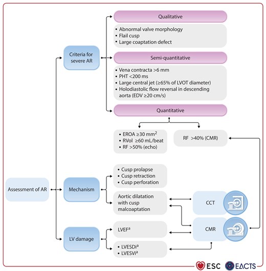
Imaging assessment of patients with aortic regurgitation.
Figure 4

Imaging assessment of patients with aortic regurgitation.

AR, aortic regurgitation; CCT, cardiac computed tomography; CMR, cardiac magnetic resonance; EDV, end-diastolic velocity; EROA, effective regurgitant orifice area; LV, left ventricle/left ventricular; LVEF, left ventricular ejection fraction; LVESDi, left ventricular end-systolic diameter indexed to body surface area; LVESVi, left ventricular end-systolic volume indexed to body surface area; LVOT, left ventricular outflow tract; PHT, pressure half-time; RF, regurgitant fraction; RVol; regurgitant volume. aSee Recommendation Table 3 for specific cut-offs.

Assessment of AR severity with TTE follows an integrative approach considering qualitative, semi-quantitative, and quantitative parameters, but remains challenging.45 Consequences of AR on LV size and function must be carefully assessed. Cut-offs for intervention are mostly based on two-dimensional (2D) echocardiographic measurements. However, 3D echocardiography and CMR allow more accurate evaluation of LV volumes and LVEF than 2D echocardiography, and are useful in borderline cases (Figure 4).45,209 Strain imaging can be helpful in identifying subclinical LV dysfunction209–211 and can therefore influence the optimal timing of intervention. Reduced longitudinal strain and contractile reserve at stress echocardiography,212 elevated biomarkers (BNP),213,214 and the presence of myocardial fibrosis detected by CMR need to be integrated in the decision-making process, even if not entirely validated yet.209

Given its close relationship with AV function, accurate measurements of the aortic diameter are required at all levels: the annulus, sinuses of Valsalva, sinotubular junction, and ascending aorta.8,215,216 The largest diameter is used to indicate the specific aortic phenotype: root phenotype, ascending phenotypes, and extended or mixed forms (see Supplementary data online, Figure S1).8 The mechanism of AR and aortic diameters determine suitability for AV sparing or repair.217,218

The morphology of the AV represents a critical aspect in the diagnosis and treatment of AR. Pure AR in the context of BAV uncommonly manifests with normal aortic diameters, being more often associated with a dilated ascending aorta and/or root. It is important to define the valve phenotype to determine the repair probability and long-term result of AV repair or AV-sparing procedures. The degree of symmetry is an important predictor of BAV reparability with better long-term results in more symmetric phenotypes.219–221

7.3. Medical therapy

Medical therapy, especially angiotensin-converting enzyme-inhibitors (ACE-Is) or dihydropyridine calcium channel blockers, may provide symptomatic improvement in individuals with chronic severe AR for whom surgery is not feasible or contraindicated. The value of ACE-Is or dihydropyridines in delaying surgery in the presence of moderate or severe AR in asymptomatic patients has not been established, and their use is not recommended for this indication. The use of beta-blockers increases the length of the diastole and therefore the RVol, and should be used with caution if indicated for another reason. However, beta-blockers can be used along with ACE-Is or angiotensin receptor blockers (ARBs) after surgery, if indicated (systolic HF or heart rate control).222,223

7.4. Indications for intervention

Acute severe AR usually requires immediate surgery depending on the aetiology, such as infective endocarditis or spontaneous, traumatic, or iatrogenic aortic dissection.5,8 Surgery for the treatment of chronic severe AR is indicated depending on symptoms and/or the effects of the RVol on LV size and function (see Recommendation Table 3 and Figure 5). The presence of associated aortic dilatation dictates surgery, irrespective of AR severity. When the patient is symptomatic and AR severe, surgery is recommended unless the anticipated surgical risk is prohibitive.224–228 Concomitant surgical treatment of severe AR is also recommended, irrespective of symptoms, in patients requiring CABG, ascending aorta surgery, or any other cardiac surgical procedures.229,230
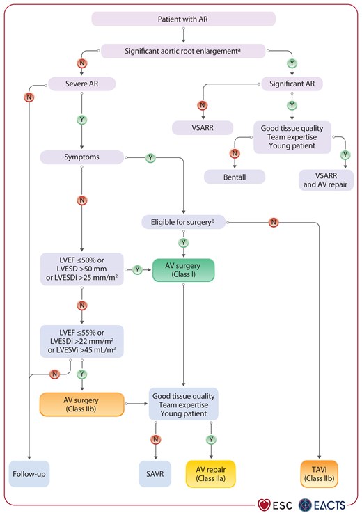
Management of patients with aortic regurgitation.
Figure 5

Management of patients with aortic regurgitation.

AR, aortic regurgitation; AV, aortic valve; LVEF, left ventricular ejection fraction; LVESD, left ventricular end-systolic diameter; LVESDi, left ventricular end-systolic diameter indexed to body surface area; LVESVi, left ventricular end-systolic volume indexed to body surface area; SAVR, surgical aortic valve replacement; TAVI, transcatheter aortic valve replacement; VSARR, valve-sparing aortic root replacement. aIndications for surgery on the root/ascending aorta are described in the 2024 ESC Guidelines for the management of peripheral arterial and aortic diseases.8  bConcomitant replacement of the aortic root or ascending aorta should be considered if the maximal diameter is ≥45 mm and the predicted surgical risk is low.

Recommendation Table 3

Recommendations on indications for intervention in severe aortic regurgitation (see also Supplementary data online, Evidence Tables 4–8)
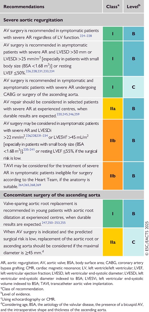
For the asymptomatic patient with severe AR, indications for surgery are based on the degree of functional impairment of the LV [LVEF ≤50%, left ventricular end-systolic diameter (LVESD) >50 mm, or left ventricular end-systolic diameter indexed to body surface area (BSA) (LVESDi) >25 mm/m2, especially in those with small BSA (BSA <1.68 m2) and elderly patients with low ventricular compliance].226,228,231–234 If surgery is deemed low risk, there is observational evidence from echocardiographic studies that early intervention might be beneficial for long-term prognosis when LVEF is ≤55%, LVESDi is >22 mm/m2, and/or left ventricular end-systolic volume indexed to BSA (LVESVi) is >45 mL/m2.235–239 A volumetric cut-off value of LVESVi ≥43 mL/m2 using CMR was recently proposed to guide the management of asymptomatic patients240,241 and appears to have better predictive value than LV diameter.242 Surgery may also be discussed in selected low-risk asymptomatic patients with significant LV dilatation (left ventricular end-diastolic diameter >65 mm) and progressive increase of LV diameters and/or decrease of LVEF during follow-up. Exercise testing should be performed, when feasible, in patients with severe AR who do not report symptoms and do not meet criteria for surgery.212

AV replacement is still the standard surgical approach in most AR cases (see Section 14.1 regarding prosthesis type considerations). However, owing to a better understanding of the pathophysiology of the aortic root (see Supplementary data online, Figure S1) paralleled with favourable long-term results, valve-sparing aortic root replacement (VSARR) and AV repair are increasingly performed in centres with appropriate expertise (Figure 5).243–255 In patients with root enlargement and good tissue quality (i.e. pliable AV cusps with normal motion), a valve-sparing procedure has been demonstrated to be superior to the use of a composite valve graft (Bentall procedure) in terms of long-term mortality and overall morbidity (thromboembolism and endocarditis, with similar need for reoperation),250–253,255–257 and should be therefore favoured by experienced centres, in particular in patients with an estimated long life expectancy.258 Valve preservation or valve repair should also be considered for patients with BAV based on age, anatomical presentation, and centre experience (Figure 5).220,245,246,259

When performed by experienced surgeons in well-selected young individuals, pulmonary autograft implantation (Ross operation) may also be a good alternative to prosthetic valve replacement.260–263

TAVI may be considered at experienced centres for selected patients with AR who are ineligible for surgery. The use of non-dedicated transcatheter valves for this indication is off-label and associated with an increased risk of valve malpositioning and residual AR, with consecutively higher rates of second valve implantation (about 10%) or surgical conversion, as compared with TAVI in AS.264–268 Dedicated devices appear to minimize the risk of valve migration and residual AR in selected patients, but are associated with a high new permanent pacemaker implantation rate (24%).267–269

Aortic dilatation is closely linked to AR. Dedicated ESC Guidelines give guidance regarding the evaluation and management of aortic root and ascending aortic dilatation.8 The aortic phenotype (see Supplementary data online, Figure S1), degree and rate of progression of aortic dilatation, and the underlying aetiology all affect timing of surgery, with the main indication being maximum aortic diameter.270–272 Dilation of the aortic root, which typically occurs in Marfan syndrome and other patients with connective tissue disease, has a worse prognosis compared with isolated dilatation of the ascending aorta and requires closer surveillance.270–272 Surgery is recommended in all patients with a maximal aortic root or ascending aneurysm diameter of ≥55 mm. In the presence of additional risk factors, a threshold of 50 mm may be considered for selected low-risk patients treated at experienced centres.8 If the patient has an established indication for AV surgery (due to AR or AS), concomitant surgery of the aortic root or the ascending aorta should be considered at a diameter of ≥45 mm. This threshold has been more clearly demonstrated in patients with BAV and should also be based on the patient’s height273 or specific intraoperative findings, such as the shape and thickness of the aortic wall.

7.5. Follow-up

A multimodality imaging approach45,274 and biomarkers like BNP213,214 might help identify patients at increased risk of LV damage early and guide the appropriate timing of intervention. Yearly follow-up is recommended in asymptomatic patients with severe AR. Closer follow-up (3–6 months) is recommended for those approaching thresholds for surgery, or showing a progressive dilatation of the LV or decrease of LVEF. Cardiac magnetic resonance can be especially useful in such a setting. Patients with moderate AR should be followed on a yearly basis, with echocardiography performed every 2 years.

When a dilated ascending aorta is initially diagnosed by TTE, a multislice electrocardiographic-triggered CCT/CMR scan is recommended to confirm maximal diameter, rule out isolated single sinus dilatation, and provide a baseline reference. When the baseline aortic diameter is >45 mm, a second TTE examination is recommended at 6 months to confirm the stability of the finding, followed by serial examinations on a yearly basis thereafter.275 Any increase of >3 mm should be validated by CT angiography/CMR and compared with baseline data.275 After repair of the ascending aorta, patients with Marfan syndrome and other connective tissue diseases remain at risk for dissection of untreated portions of the aorta, and require lifelong regular multidisciplinary follow-up at an expert centre.8

7.6. Special patient populations

Patients with concomitant VHD and those with AS combined with AR are discussed in Section 13.3.3. In patients with moderate AR and indication for CABG or MV surgery, the decision to treat the AV should be discussed by the Heart Team based on the aetiology of AR and other clinical factors, like the estimated life expectancy and the operative risk, as data show that progression of moderate AR may be very slow.276

The presence of aortic dilation and AR in asymptomatic patients poses the problem of limiting the level of physical activity, but consistent data are lacking. Current recommendations for participation in competitive sport are restrictive, especially regarding isometric exercise in patients with connective tissue disease,277 while a more liberal approach is likely to be appropriate in other patients.

Given the familial risk of thoracic aortic aneurysms, screening with appropriate imaging studies and testing for genetic abnormalities in first-degree relatives is indicated in patients with connective tissue disease.278 Since aortic dilation is present in ∼10% of first-degree relatives of patients with a BAV, it is also considered appropriate to encourage echocardiographic screening in this specific population.275,279

8. Aortic stenosis

8.1. Prevalence and aetiology

Even if AS is the most common primary valve lesion referred for intervention in Europe and North America,12 underdiagnosis and undertreatment remain relevant concerns.10 Degenerative pathogenesis with cusps calcification is most common in developed countries and prevalence is rising rapidly because of the ageing population.11,12,187 BAVs, or more rarely unicuspid AVs, are prone to earlier degeneration, constitute the dominant valve morphology in younger patients requiring AV replacement, and are frequently associated with dilatation of the aortic root or ascending aorta.280,281 In low- and middle-income countries, rheumatic aetiology remains frequent and AS usually presents combined with rheumatic MV disease.282

8.2. Evaluation

8.2.1. Echocardiography and cardiac computed tomography

Aortic stenosis is a disease slowly evolving from mild to severe valve obstruction as a consequence of increasing valve fibrosis and calcification, although progression accelerates as haemodynamic severity increases.283

Echocardiography is key to confirm the diagnosis and allows comprehensive assessment of the anatomy and severity of stenosis. Evaluation of the haemodynamic consequences on cardiac function and geometry, and the detection of aortic pathology or concomitant valve disease, provide important prognostic information that may influence management.284 Staging of extravalvular damage has been proposed,53 but it may be difficult to attribute other cardiac abnormalities to AS itself, because comorbidities are frequent in patients with AS and the observed damage may not occur in the expected chronological sequence. However, the detection of concomitant cardiac conditions [e.g. HF with preserved EF (HFpEF), amyloidosis, or hypertrophic cardiomyopathy] may help to optimize medical treatment before and after valve intervention.285,286

Current European recommendations for the echocardiographic grading of AS rely on measurement of the mean pressure gradient (most robust parameter), peak transvalvular velocity (Vmax), and effective AVA. Although AVA is theoretically the ideal parameter for assessing severity, there are numerous technical limitations associated with its calculation.284,287

Aortic stenosis may be further categorized according to flow state based on stroke volume index (SVi) when there is discordance between echocardiographic parameters (Figure 6). A threshold of 35 mL/m2 is conventionally accepted to discern low from normal flow, although sex-specific thresholds have been proposed.292
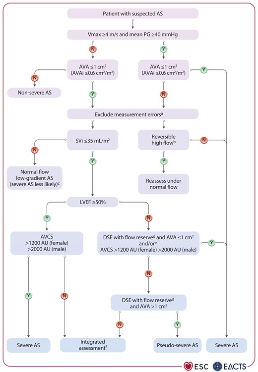
Integrative imaging assessment of patients with aortic stenosis.
Figure 6

Integrative imaging assessment of patients with aortic stenosis.

AS, aortic stenosis; AU, Agatston units; AV, aortic valve; AVA, aortic valve area; AVAi; aortic valve area indexed for body surface area; AVCS, aortic valve calcium score; CCT, cardiac computed tomography; CT, computed tomography; DSE, dobutamine stress echocardiography; LVEF, left ventricular ejection fraction; LVOT, left ventricular outflow tract; MRI, magnetic resonance imaging; mean PG, mean pressure gradient; SVi, stroke volume indexed for BSA; TOE, transoesophageal echocardiography; TTE, transthoracic echocardiography; Vmax, peak transvalvular velocity. aIn particular, verify LVOT diameter and multiwindow Doppler interrogation. bHigh flow may be reversible (anaemia, hyperthyroidism, or arteriovenous fistulae). Upper limit of normal flow using pulsed Doppler: cardiac index 4.3 L/min/m2, SVi 58 mL/m2.288  cAvailable evidence refers to patients with preserved LVEF. Check for bradycardia or uncontrolled hypertension, which may lead to prolonged ejection time and reduced flow rate. Depending on symptoms, integrated assessment complemented by CCT AV calcium scoring may be pursued. dFlow reserve: ≥20% increase in stroke volume in response to low-dose dobutamine or if change in stroke volume of 10%–20%, calculate projected AVA.289,290  eIf one test is not conclusive, complement diagnostics with the other test. fBased on clinical judgement (typical symptoms without other explanation), morphological valve changes, LV hypertrophy (in absence of coexistent hypertension), and consistent findings using different modes of assessment [TTE and TOE, invasive assessment, AV planimetry by CT or MRI (cut-off 1.2 cm2)]291.

Concordant criteria:

  • High-gradient AS [mean gradient ≥40 mmHg, Vmax ≥4.0 m/s, AVA ≤1 cm2 (or ≤0.6 cm2/m2)] is considered severe irrespective of LV function and flow conditions.

Discordant criteria:

  • Low-flow, low-gradient AS with reduced LVEF (mean gradient <40 mmHg, AVA ≤1 cm2, SVi ≤35 mL/m2, LVEF <50%).

  • Low-flow, low-gradient AS with preserved LVEF (mean gradient <40 mmHg, AVA ≤1 cm2, SVi ≤35 mL/m2, LVEF ≥50%).

  • Normal-flow, low-gradient AS with preserved EF (mean gradient <40 mmHg, AVA ≤1 cm2, SVi >35 mL/m2, LVEF ≥50%).

  • Discordant high-gradient AS (mean gradient ≥40 mmHg, AVA >1 cm2).

Patients with discordant normal-flow, low-gradient AS usually have moderate stenosis.293–295 Discordant high-gradient AS is considered severe if not caused by a reversible high-flow status.296–298

In patients with low-flow, low-gradient AS with reduced LVEF, dobutamine stress echocardiography (DSE) can help to discriminate between pseudo-severe and true severe AS in the presence of flow reserve (increase in stroke volume of ≥20%).289,299

Cardiac computed tomography calcium AV scoring is readily available and provides important adjunctive information in patients with low-flow, low-gradient AS because it correlates with haemodynamic severity, progression, and clinical outcomes.300,301 Values of >2000 Agatston units (AU) in men and >1200 AU in women indicate severe AS with high sensitivity and specificity (∼85%).302,303 While higher thresholds (men >3000 AU, women >1600 AU) are very specific, severe AS becomes unlikely in patients with calcium AV scoring of <1600 AU in men and <800 AU in women.284,302,303 Cautious interpretation is required in patients who can develop severe AS without pronounced AV calcification such as in BAV, concomitant amyloidosis, and predominantly fibrotic stenosis associated with post-rheumatic, radiation-induced and inflammatory disease.304–308

In low-flow, low gradient AS with reduced LVEF, CCT AV calcium scoring and DSE provide complementary information. If findings are equivocal, an integrated assessment considering all available clinical, morphological, and haemodynamic factors is required.

8.2.2. Additional diagnostic and prognostic parameters

The ratio of the LVOT to the AV Doppler jet velocity time integral (VTI, dimensionless index or velocity ratio) does not require calculation of LVOT area and may assist evaluation when other parameters are equivocal (<0.25 suggests that severe AS is highly likely).309

Assessment of GLS can be useful for risk stratification310 and evaluation of extravalvular cardiac damage.311,312 It provides additional information regarding LV function and a threshold of −15% may contribute to identifying patients with severe asymptomatic AS at increased risk of clinical deterioration or premature mortality.59

Estimated valvuloarterial impedance has been shown to be prognostic of adverse clinical outcomes before and after valve replacement.313–315

Transoesophageal echocardiography (TOE) allows morphological evaluation of the valve, planimetry of AVA and assessment of potential subvalvular obstruction (unless there is acoustic shadowing caused by calcification), and evaluation of concomitant valve disease, and can be of value for peri-procedural imaging in challenging clinical or anatomical scenarios.316

Natriuretic peptides can be used to arbitrate the sources of symptoms in patients with multiple potential causes and help to identify those with high-risk asymptomatic AS who may benefit from early intervention.97,317

Exercise testing can unmask symptoms and haemodynamic intolerance (fall in BP >20 mm Hg) and is recommended for risk stratification in asymptomatic patients with severe AS.102,318 Exercise echocardiography may provide additional prognostic information by assessing the increase in mean pressure gradient and change in LV function.319 Cardiopulmonary exercise testing, eventually complemented by echocardiography, can help to uncover cardiac dysfunction in asymptomatic patients, discriminate cardiac from pulmonary limitation or deconditioning in patients with non-specific symptoms, and inform risk stratification.105,106,320,321

Cardiac magnetic resonance is used to identify altered global LV geometry due to remodelling, as well as to quantify myocardial scarring and diffuse fibrosis, which are associated with the occurrence of adverse events.322–324

Transthyretin cardiac amyloidosis may coexist with AS in elderly patients and the two conditions may causally interrelate.325 When transthyretin cardiac amyloidosis is suspected, the presence of monoclonal protein in serum and urine should be excluded using immunofixation and quantitative determination of free light chains, and diagnosis ascertained by means of diphosphonate scintigraphy.326 Despite the limited long-term prognosis associated with cardiac transthyretin amyloidosis, patients with concomitant severe AS usually benefit from valve intervention.327

LV catheterization is not recommended unless there are symptoms and signs of severe AS, and non-invasive investigations are inconclusive.

8.2.3. Procedural planning

Cardiac computed tomography is key to determining suitability for TAVI and planning the procedure. It is the preferred imaging tool to assess AV anatomy including annulus size, dimensions of the aortic root and ascending aorta, the extent and distribution of valve and LVOT calcification, the distance of coronary ostia from the annular plane, optimal fluoroscopic projections for transcatheter valve deployment, and the feasibility of vascular access.49,328,329

Transoesophageal echocardiography (TOE), which is more operator-dependent and does not allow assessment of coronary and peripheral vascular anatomy, or CMR may be considered when CCT is difficult to interpret or relatively contraindicated (e.g. in patients with renal failure).330,331

8.3. Medical therapy

No medical therapies have been shown to influence the natural history of AS to date. Neither statins, which demonstrated favourable effects in pre-clinical studies,332–334 nor substances targeting calcification pathways alter disease progression.335,336 Coexistent hypertension should be treated to avoid additional afterload, preferably using renin-angiotensin system blockers, although careful titration is required to avoid symptomatic hypotension.337

In patients with symptomatic severe AS and HF, initiation of medical therapy or temporary improvement in symptoms should not delay intervention. Medication frequently requires re-adjustment following valve intervention and preventive therapies should be implemented according to current Guidelines.338 In patients with persisting HF and/or reduced LVEF, medical therapy should be introduced before and up-titrated after valve intervention according to the current HF Guidelines.339,340

8.4. Indication for intervention

8.4.1. Symptomatic severe aortic stenosis

Symptomatic severe AS has an unfavourable prognosis if left untreated, and early intervention is strongly recommended in all patients with an estimated life expectancy exceeding 1 year10 (Figure 7).
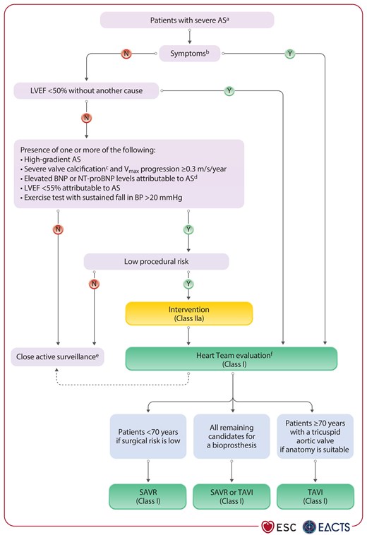
Management of patients with severe aortic stenosis.
Figure 7

Management of patients with severe aortic stenosis.

AS, aortic stenosis; AVCS, aortic valve calcium score; BNP, brain natriuretic peptide; BP, blood pressure; LVEF, left ventricular ejection fraction; NT-proBNP, N-terminal pro-B-type natriuretic peptide; SAVR, surgical aortic valve replacement; TAVI, transcatheter aortic valve implantation; Vmax, peak transvalvular velocity. aIntegrative imaging assessment of AS (Figure 6). bConfirmed by a normal exercise test, if feasible. cAVCS >2000 in men, >1200 in women. dMore than three times age- and sex-corrected normal range. eEducate patient and reassess at least every 6 months (or promptly if symptoms occur). fHeart Team assessment based upon individual patient factors (Figure 9; Recommendation Table 4). Dashed arrow only applies to asymptomatic patients.

Intervention is recommended in all eligible symptomatic patients with high-gradient severe AS. However, management of patients with low-gradient AS is more challenging:

  • Low-flow, low-gradient AS with reduced LVEF: reduced LV function usually improves after intervention if it is predominantly caused by excessive afterload.341–343 However, improvement is unlikely if the primary cause is fibrosis due to myocardial infarction or cardiomyopathy. Intervention is recommended when severe AS is confirmed by CCT (calcium scoring) or stress echocardiography,341 while patients with pseudo-severe AS should receive GDMT.339,344 Although the absence of flow reserve is associated with increased surgical and long-term mortality, both modes of intervention improved LVEF and clinical outcomes in observational studies.341,342,345–347

  • Low-flow, low-gradient AS with preserved LVEF: outcomes are improved with intervention (either TAVI or SAVR) compared with medical treatment alone in patients with low-flow, low-gradient AS and preserved LVEF.348,349 Intervention should therefore be considered in patients with symptoms after careful confirmation that AS is severe.

  • Normal-flow, low-gradient AS with preserved LVEF: prognosis of these patients is similar to that of moderate AS. Unless multimodality diagnostic evaluation clearly suggests severe AS, regular clinical and echocardiographic surveillance is recommended.293,294,350

8.4.2. Asymptomatic severe aortic stenosis

Up to 40% of patients with severe AS do not report symptoms at the time of diagnosis.351,352 In roughly one-third of these, exercise testing can uncover symptoms or reduced exercise capacity attributable to AS,102,103,318 and such patients should be treated as symptomatic. However, exercise testing is not always feasible because of frailty or impaired mobility.353 Intervention is recommended in asymptomatic patients with severe AS and an LVEF of <50% without another cause.14,102,318,319,354–360 For patients with severe high-gradient AS and no adverse prognostic features, close active clinical surveillance, so-called ‘watchful waiting’, has previously been the default management strategy. However, four RCTs comparing early AV intervention with clinical surveillance suggest that early intervention should be considered as an alternative in patients at low procedural risk.360–363 This approach is reinforced if additional adverse prognostic features (very high Vmax,14,353,364 elevated natriuretic peptides,97,317,365,366 severe valve calcification,303,364 rapid Vmax progression,353,364 or LVEF of <55%14,354,356–359) are present. Restricted local resources (that may impede close surveillance) or long waiting lists (that preclude prompt treatment when symptoms develop) are further arguments favouring an early intervention.

The Evaluation of TAVR Compared to Surveillance for Patients with Asymptomatic Severe Aortic Stenosis (EARLY TAVR) trial360 randomized 901 patients to early TAVI or clinical surveillance and demonstrated a reduction of 50% in the primary composite endpoint of all-cause mortality, stroke, or unplanned hospitalization for cardiovascular causes associated with pre-emptive intervention.360 The result was driven by 26.2% of the patients in the clinical surveillance group who converted to TAVI within 6 months of randomization due to the development of symptoms or adverse prognostic factors. There was no significant difference in strokes and all-cause mortality over 5-year follow-up.

In the Early Valve Replacement Guided by Biomarkers of LV Decompensation in Asymptomatic Patients with Severe AS (EVoLVeD) RCT, which included 224 asymptomatic patients with severe AS and myocardial fibrosis (late gadolinium enhancement on CMR), early valve intervention (with SAVR or TAVI) failed to reduce the incidence of all-cause death or unplanned AS-related hospitalization compared with clinical surveillance.361 However, the study was underpowered and the median time to intervention was prolonged to 5 months in the experimental arm.

Two previous smaller trials compared early SAVR with clinical surveillance. In the Randomized Comparison of Early Surgery vs Conventional Treatment in Very Severe Aortic Stenosis (RECOVERY) trial (145 patients), there was a reduction in all-cause mortality following early SAVR over a mean follow-up of 6.2 years.362 In the Aortic Valve Replacement Versus Conservative Treatment in Asymptomatic Severe Aortic Stenosis (AVATAR) trial (157 patients), a reduction in the composite primary endpoint was noted after a mean follow-up of 2.5 years with a significant reduction in HF hospitalizations and mortality at long-term follow-up.363,367 Limitations of these surgical studies included their small sample sizes, enrolment of selected young populations at low surgical risk (mean ages 64 and 67 years, respectively), and inclusion of patients with mostly very severe AS.

A meta-analysis of the four RCTs showed that early intervention is associated with a significant reduction in unplanned cardiovascular or HF hospitalization and stroke, but not all-cause or cardiovascular mortality. Limitations of this analysis include the heterogeneity of the pooled trials and lack of granularity with respect to specific events owing to the study-level analysis.368

Additional RCTs (NCT04204915 and NCT03972644) will further inform and refine the management of patients with severe asymptomatic AS.

8.4.3. Moderate aortic stenosis

Surgical intervention for moderate AS should only be performed in patients undergoing CABG,369 surgery of the ascending aorta or other valve disease (see Section 13). There is evidence of an association between moderate AS and adverse outcomes in patients with and without HF with reduced EF (HFrEF),370–372 but it is unknown whether this is causal or related to comorbidities. In an early terminated trial randomizing 178 patients with moderate AS and HFrEF to TAVI or clinical surveillance, no differences with respect to hard clinical endpoints were found, while patients undergoing TAVI had improved quality of life (change in KCCQ of 12.8 ± 21.9 points vs 3.2 ± 22.8 points; P = 0.018).373 Ongoing trials are expected to provide new insights (NCT04889872 and NCT05149755).

8.5. Treatment options

The mode of AV intervention depends on the estimated life expectancy, expected prosthesis durability, patient preference, and specific trade-offs associated with different treatment options (Figure 8). Most patients with AS undergoing valve intervention in Europe and North America receive a BHV (by either SAVR or TAVI).374 BHVs do not require long-term anticoagulation but have limited durability that varies between devices and is inversely associated with age.375 MHVs are durable but require long-term anticoagulation, with associated thromboembolic and bleeding risks.260,376 In general, an MHV should be preferred in patients aged <60 years and a BHV in patients aged >65 years in aortic position (see Section 14.1).
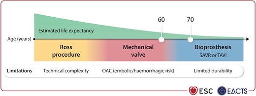
Aortic valve treatment options.
Figure 8

Aortic valve treatment options.

OAC, oral anticoagulation; SAVR, surgical aortic valve replacement; TAVI, transcatheter aortic valve implantation.

The Ross procedure (replacement of the AV with the patient's pulmonary autograft combined with homograft pulmonary valve replacement) is associated with excellent long-term survival when performed in selected patients at centres with high expertise.260,263,377,378 Although a valuable surgical option in young patients with prolonged life expectancy in whom anticoagulation is undesirable or contraindicated, it is associated with procedural complexity and the need for reintervention in ∼15% of patients within 15 years (Figure 8).263,377

Balloon aortic valvuloplasty may be rarely considered as a bridge to TAVI or SAVR in carefully selected patients with decompensated AS, and in those with severe AS who require urgent high-risk non-cardiac surgery (NCS) (see Section 15.2.1). The procedure carries significant risks of acute complications.379

A substantial increase in the number of patients undergoing AV intervention has been observed over the past decade as a consequence of the introduction of TAVI, improved diagnostic techniques, and evolving indications for intervention.374,380–383 Nevertheless, there is still wide variation in worldwide access to TAVI as a result of high device costs in comparison with surgical prostheses and variation in healthcare resources and reimbursement systems between countries.384–386 In addition, registries suggest that late referral and undertreatment remain frequent.12,387

8.5.1. The mode of intervention in candidates for a bioprosthesis

The two modes of BHV replacement, TAVI and SAVR, have been compared in RCTs across the spectrum of surgical risk in predominantly elderly patients with tricuspid AS.

In patients unsuitable for surgery, TAVI was superior to medical therapy with a number-needed-to-treat of five to prevent one death at 1-year follow-up.388 Subsequently, RCTs showed non-inferiority of TAVI compared with SAVR in patients at high,389–391 intermediate,3,392,393 and low surgical risk,1,394,395 with comparable longer-term outcomes demonstrated during follow-up periods ranging from 4 to 10 years.2,4,396–398 Of note, the majority of patients included in RCTs were male, while patients with low-flow low-gradient AS or adverse anatomical characteristics (including BAVs or complex CAD) were excluded per protocol.

Meta-analyses of RCTs show a risk reduction in all-cause death and disabling stroke with TAVI in low-risk patients at 1 year, but no differences to SAVR at longer-term follow-up or in patients at intermediate or high surgical risk.399,400 The early benefit of TAVI in low-risk patients has since been corroborated in the investigator-initiated Randomized, Multicenter, Event-Driven Trial of TAVI vs SAVR in Patients with Symptomatic Severe Aortic-Valve Stenosis (DEDICATE), which met its non-inferiority target with a composite of death and stroke rate at 1 year of 5.4% in the TAVI group vs 10.0% in the SAVR group [hazard ratio (HR), 0.53; 95% confidence interval (CI), 0.35–0.79] and all-cause death rates of 2.6% and 6.2% (HR 0.43; 95% CI, 0.24–0.73), respectively.395

Rates of vascular complications and paravalvular leak (PVL) are consistently higher after TAVI despite refined transcatheter heart valve (THV) designs, whereas severe bleeding, acute kidney injury, and new-onset AF are more frequent after SAVR.2,4,399,401 Even though PVL has been associated with adverse clinical outcomes,402,403 it does not seem to impact the comparison of clinical outcomes between TAVI and SAVR in RCTs.2,4,395

New pacemaker implantations are more frequent after TAVI, particularly when using self-expanding valves.3,394,404 Conflicting data exist regarding the long-term impact of new pacemaker implantation or new-onset left bundle branch block following AV intervention.405–407 Patients undergoing TAVI have quicker recoveries, shorter hospital stays, and more rapid improvements in quality of life compared with those who undergo SAVR.1,2,408,409

Available data do not suggest systematic differences in durability between the two treatment modalities. Randomized controlled trials and observational studies have reported comparable rates of BHV failure related to SVD with transcatheter and surgical valves up to 10 years.2,398,410 However, potential selection and survival bias, the use of variable definitions of SVD, limited follow-up durations, differential attrition rates,2,4,396,400 competing risk of death, and the use of multiple valve types in the surgical arms of RCTs may limit direct comparison.411

Although conclusive evidence is still lacking, concomitant non-complex CAD can be addressed either by CABG or PCI, while complex CAD favours CABG (see Section 6.1). In the only RCT comparing these two strategies, the transcatheter valve and vessels (TCW) trial, randomization of patients with severe AS and concomitant CAD to FFR-guided PCI plus TAVI resulted in fewer deaths and major bleeding events compared with combined SAVR plus CABG at 12 months.412 However, inferences from trial results are limited due to early termination, modest sample sizes, event rates deviating from those observed in registries and other RCTs, and a low prevalence of complex CAD.

8.5.1.1. Age and life expectancy

The relationship between estimated life expectancy and prosthetic heart valve durability determines the likelihood of a future reintervention. Although life expectancy may be a theoretically better guide than age alone for treatment decisions, it is difficult to estimate for an individual patient owing to large geographical (https://ghdx.healthdata.org/record/ihme-data/gbd-2019-life-tables-1950-2019) and interindividual variability.413 In combination with cardiac and extracardiac comorbidities, as well as anatomical factors, age contributes to risk estimation and represents a pragmatic surrogate for life expectancy. In addition, age thresholds characterize the populations enrolled in RCTs better than life expectancy. It is notable that younger patients with AS seem to have lower life expectancy than the general population despite valve replacement, whereas life expectancy almost normalizes after treatment in older cohorts.414,415 While several trials compared TAVI with SAVR in study populations aged 70–85 years accross the surgical risk spectrum, the representation of patients aged <70 years in RCTs is low and therefore evidence limited.1,394,395,416

8.5.1.2. Anatomical features

The advantages of TAVI demonstrated in RCTs are largely confined to patients treated via the transfemoral approach.401 While SAVR remains the preferred treatment option when iliofemoral artery disease precludes transfemoral TAVI, TAVI via a non-transfemoral access (transaxillary, transcarotid, transcaval, transinnominate, or transapical) constitutes an alternative supported by observational data in patients unsuitable for surgery.417–423

Other anatomical factors that favour SAVR or led to exclusion of patients from RCTs comparing TAVI with SAVR are (Figure 9):

  • Aortic annular dimensions that lie outside sizing recommendations for currently available transcatheter devices.

  • Excessive or bulky calcifications of the annulus or LVOT, which increase the risk of PVL and annular rupture.424,425

  • Increased risk of coronary obstruction with TAVI (cusp height greater than coronary height in combination with shallow sinuses of Valsalva, or high calcium burden of corresponding cusp).329
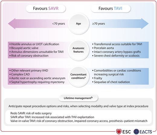
Factors to be considered when selecting the mode of intervention for aortic stenosis.
Figure 9

Factors to be considered when selecting the mode of intervention for aortic stenosis.

CAD, coronary artery disease; LV, left ventricular; LVOT, left ventricular outflow tract; SAVR, surgical aortic valve replacement; TAVI, transcatheter aortic valve implantation; THV, transcatheter heart valve; VHD, valvular heart disease. aLV thrombus and infective endocarditis are relative contraindications to TAVI and are therefore not listed. bParticularly relevant for patients in whom the anticipated life expectancy is thought to exceed valve durability.

In contrast, anatomical findings such as porcelain aorta, severe chest deformation, or intact grafts post-CABG favour TAVI. Right anterior thoracotomy or upper hemisternotomy are minimally invasive access alternatives to sternotomy for performing SAVR, which are being used with increasing frequency.426,427

In BAV patients, severe AS usually occurs earlier compared with tricuspid AV and is frequently associated with aortopathy.219,281,428 Prevalence of BAV anatomy sharply increases in younger AS patients.429 BAV anatomy adds complexity to TAVI because of asymmetric AV calcification and elliptical annular shape, as well as the lack of standardization of valve sizing.

Patients with BAV have been excluded from almost all landmark RCTs comparing TAVI with SAVR to date.429,430 In the NOTION 2 trial, the composite of all-cause death, stroke, or valve- or HF-related rehospitalization was numerically more frequent (seven vs two events) at 1 year in the underpowered subgroup of 100 patients with BAV (HR, 3.8; 95% CI, 0.8–18.5; P = 0.07).416 Whereas some observational studies report favourable outcomes with TAVI in selected BAV patients,430–432 others suggest higher stroke, annular rupture, and PVL rates as compared with TAVI in tricuspid AS.433 Heavy cusp calcification, particularly in conjunction with a calcified raphe, is associated with increased risk of aortic root injury, PVL, and mortality after TAVI.434 Data on TAVI in two-sinus BAV (Sievers type 0) are scarce.219,435

For the above-mentioned reasons, SAVR remains the primary mode of treatment for stenotic BAV, particularly if patients are young or have coexistent aortopathy or unfavourable valve morphology. TAVI may be considered in patients at increased surgical risk, if anatomy is suitable.

Patients with small annuli in relation to their body stature are at increased risk of prosthesis–patient mismatch (PPM) after valve replacement. Annular enlargement allows for implantation of larger BHVs with SAVR. Given its technical complexity it should be performed in experienced centres, and the benefit must be balanced against a possibly higher risk of operative mortality.436 Prostheses with supra-annular design reduce PPM risk with TAVI, although randomized long-term data evaluating the impact on clinical outcomes or valve durability are pending.394,437,438

8.5.1.3. Lifetime management

Decision-making concerning the mode of intervention and type of prosthesis needs to integrate expected valve durability, and the potential risks of future reinterventions (Figure 9). Surgical THV explantation followed by SAVR is a rare (<1% of all TAVI procedures) but technically challenging procedure.439–441 Although its incidence among patients undergoing TAVI is stable, absolute numbers are increasing due to the total increase in TAVI procedures performed and the peri-operative risk remains high (early mortality rates as high as 12%–17%).440–445 The majority of surgical THV explantations are performed in patients at high surgical risk with urgent or emergent, non-SVD-related indication for reintervention (frequently endocarditis) within 2 years after TAVI.440,442–444

Implanting a transcatheter aortic valve inside a surgical (TAV-in-SAV) or prior transcatheter valve (TAV-in-TAV) is associated with lower peri-procedural risk compared with redo SAVR.446–451 However, valve-in-valve implantation (particularly TAV-in-SAV) increases the risk of severe PPM,448 which has been linked to adverse outcomes in observational studies.452–455 In addition, valve-in-valve implantation immobilizes the leaflets of the failed prosthesis in an open position, creating a covered tube (or neo-skirt) that may cause direct coronary obstruction in patients with shallow sinuses of Valsalva or indirect coronary flow obstruction as a result of sinus sequestration if the ascending aorta is narrow and the neo-skirt reaches the sinotubular junction.456,457

The risk of coronary obstruction varies considerably depending on the index valve type, and is particularly increased if a stentless SAV or stented SAV with externally mounted leaflets is in place.457 The risk of sinus sequestration at the time of TAV-in-TAV implantation is particularly increased in supra-annular valves with a high neo-skirt.458,459 Even if coronary flow is preserved, coronary access may be difficult or impossible in a relevant proportion of patients after valve-in-valve implantation, especially after TAV-in-TAV.460–462

In patients who require a reintervention due to dysfunction of a surgical or transcatheter bioprosthesis, but are at increased risk of severe PPM or coronary obstruction, redo SAVR may be preferable despite the increased surgical risk. Fracture of surgical valves and leaflet modification techniques have been proposed for patients at high surgical risk and, if contemplated, should be performed in carefully selected patients at experienced centres (Table 6).463,464

The need for a meticulous CT-based anatomical analysis is paramount in patients with an estimated life expectancy exceeding the assumed valve durability to anticipate future risks at the time of the index valve intervention. Based on individual assessment, the following measures should be considered with respect to lifetime management:

  • Use of surgical and transcatheter valves with proven long-term durability to reduce the likelihood of reintervention.2,4,375,410,411,465–467

  • SAVR with aortic root enlargement or implantation of a supra-annular transcatheter valve in patients with a small annulus at risk of severe PPM based on the predicted effective orifice area (EOA).436,438,468,469

  • No implantation of stentless prostheses or prostheses with externally mounted leaflets in patients at risk of coronary obstruction during future TAV-in-SAV implantation.457

  • Anticipation of the feasibility and risks of a possible future TAV-in-TAV procedure considering related technical aspects at the index TAVI (device choice, neo-skirt height, commissural alignment, and implantation depth).456,470

Considering life-time management aspects and the scarcity of randomized data in patients younger than 70 years, SAVR remains the preferred treatment in patients <70 years of age if surgical risk is low. TAVI is recommended as the primary treatment modality in elderly patients ≥70 years of age with a tricuspid AV, if anatomy is suitable and transfemoral access is feasible, to reduce the risk of early adverse outcomes and accelerate recovery.

For all other candidates for a bioprosthesis, the most appropriate mode of intervention should be carefully selected by the Heart Team, taking into account procedural risk based on anatomical characteristics and comorbidities, expected outcomes, lifetime management considerations, and patient preference (Figure 9; Recommendation Table 4). Recommendations for concomitant valve replacement at the time of CABG or ascending aortic surgery are listed in Recommendation Table 5.

Recommendation Table 4

Recommendations on indications for intervention in symptomatic and asymptomatic severe aortic stenosis, and recommended mode of intervention (see also Supplementary data online, Evidence Tables 9–13)
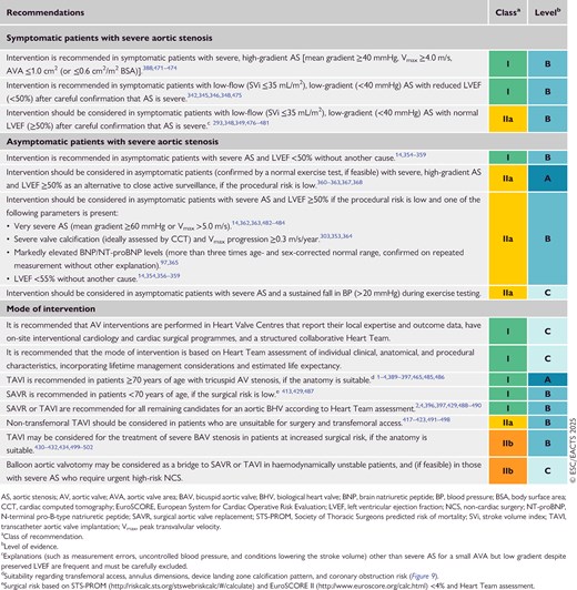
Recommendation Table 5

Recommendations on indications for concomitant aortic valve replacement at the time of coronary artery bypass grafting or ascending aorta surgery
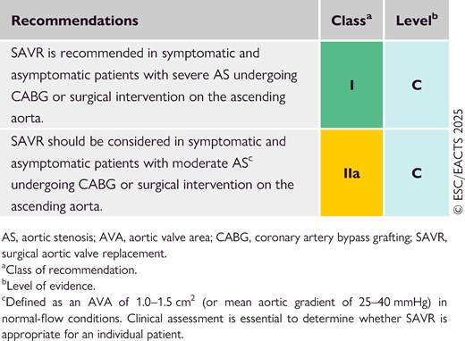
8.6. Follow-up

The rate of progression of AS varies widely and asymptomatic patients, their family, and medical caregivers need careful education, with particular emphasis on the importance of regular follow-up (ideally at a Heart Valve Centre)14 and prompt reporting of symptoms. Asymptomatic patients with severe AS should be followed up at least every 6 months to allow detection of early symptoms (using exercise testing if complaints are inconclusive) and any change in echocardiographic parameters (particularly LVEF). Serial measurements of natriuretic peptides can provide additional useful information regarding the timing of treatment.

Younger patients with mild AS and no significant leaflet calcification may be followed up every 2–3 years. With increasing stenosis severity, progression accelerates and follow-up intervals should be gradually reduced.283,503 Several studies suggest that the prognosis of moderate degenerative AS is worse than previously considered,504,505 particularly if there is significant valve calcification, and these patients should be re-evaluated at least annually.

Cardiac rehabilitation is frequently performed after AV intervention, especially after surgery and in elderly patients, and is associated with improved activities of daily living and 6-minute walking distance.506 After valve intervention, an early echocardiographic examination within the first weeks after valve replacement is recommended to document baseline prosthetic valve function. Cardiological evaluations and echocardiographic examinations are recommended annually in patients with a bioprosthesis, and whenever changes in clinical symptoms or signs suggestive of valve dysfunction are noted.

9. Mitral regurgitation

Chronic MR is one of the most common acquired valve pathologies,12,187 while acute MR is observed in the context of infective endocarditis, chordal rupture, or as a complication of myocardial infarction (papillary muscle rupture). MR either relates to anatomical changes of the MV apparatus (primary), or to LV or LA dilatation and dysfunction (secondary).193 Since natural history, prognosis, and management differ according to aetiologies, populations should be clearly distinguished in clinical practice and research.12,507

9.1. Primary mitral regurgitation

9.1.1. Prevalence and aetiology

Primary mitral regurgitation relates to an anatomical lesion of one or more of the three main components (not including the annulus) of the MV apparatus (valve leaflets, chordae tendineae, and papillary muscles). PMR is observed in 55% of patients with MR who require treatment.507 While degenerative disease related to fibroelastic deficiency or myxomatous alterations (in its most severe form, Barlow's disease) are the most common aetiologies in higher-income countries, RHD is most frequently found in the rest of the world. MV endocarditis is a separate entity of PMR caused by acute or chronic infectious conditions and is discussed in the corresponding Guidelines.5 In a small subgroup of patients, PMR is associated with a higher incidence of ventricular arrhythmias, and sudden cardiac death has been reported in individual cases, especially in patients with Barlow's disease.508 The arrhythmogenic burden is independent from MR severity and has been associated with mitral annular disjunction. Atrial displacement of the posterior MV leaflet hinge point is assumed to cause excessive mobility of the valvular apparatus and increases tension on the papillary muscles and the posterobasal myocardium, causing local fibrosis, which may lead to ventricular arrhythmias and sudden cardiac death.509

9.1.2. Evaluation

9.1.2.1. Echocardiography and right heart catheterization

Echocardiography is the diagnostic method of choice for the quantification of MR, determination of its aetiology, and identification of cardiac consequences (Figure 10). TTE is used for initial evaluation that includes: (i) assessment of valve morphology (presence and location of prolapse or flail, calcifications, and/or mitral annular disjunction); (ii) integrative severity grading; and (iii) quantification of LV and LA dimensions and function, as well as assessment of concomitant RV dysfunction.46 Quantitative parameters such as the effective regurgitant orifice area (EROA) have prognostic implications.510,511 Volumetric methods provide additional information on MR severity [RVol and regurgitant fraction (RF)].512 Accurate colour flow settings must be used to avoid overestimation of MR severity.
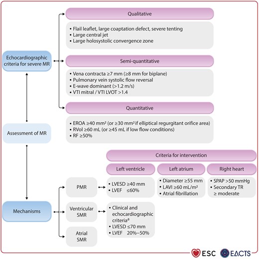
Echocardiographic assessment of patients with mitral regurgitation.
Figure 10

Echocardiographic assessment of patients with mitral regurgitation.

EROA, effective regurgitant orifice area; LAVI, left atrial volume index; LVEF, left ventricular ejection fraction; LVESD, left ventricular end-systolic diameter; LVOT, left ventricular outflow tract; MR, mitral regurgitation; PMR, primary mitral regurgitation; RF, regurgitant fraction; RVol, regurgitant volume; SMR, secondary mitral regurgitation; SPAP, systolic pulmonary artery pressure; TR, tricuspid regurgitation; VTI, velocity time integral. aSee Table 7 for criteria predicting outcome improvement.

Transoesophageal echocardiography (TOE) represents the method of choice to assess valve anatomy, leaflet quality, motion, and coaptation, as well as to confirm MR severity.513 Three-dimensional TOE provides an excellent morphological and functional view of the different valve segments, and should be used systematically when planning and performing surgical or transcatheter repair.514

Exercise echocardiography evaluates dynamic changes in regurgitant jet and pulmonary pressures during peak exercise, and might be helpful in patients with discordant symptoms and regurgitation severity at rest.515,516

In asymptomatic patients with severe PMR, increased LV or LA dimensions, as well as elevated pulmonary pressures (>50 mmHg at rest), moderate or more secondary TR, and AF are important markers of worse prognosis, and should be considered for intervention timing (Figure 11; Section 9.1.4).517,518
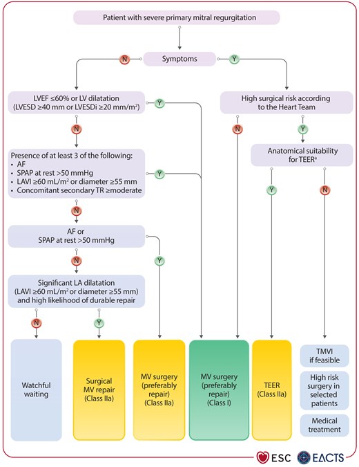
Management of patients with severe primary mitral regurgitation.
Figure 11

Management of patients with severe primary mitral regurgitation.

AF, atrial fibrillation; LA, left atrial; LAVI, left atrial volume index; LV, left ventricle/left ventricular; LVEF, left ventricular ejection fraction; LVESD, left ventricular end-systolic diameter; LVESDi, left ventricular end-systolic diameter indexed to BSA; MV, mitral valve; SPAP, systolic pulmonary artery pressure; TEER, transcatheter edge-to-edge repair; TMVI, transcatheter mitral valve implantation; TR, tricuspid regurgitation. aSee Supplementary data online, Table S2.

Right heart catheterization remains important in patients with PMR for confirmation of pulmonary artery pressures in case of discrepancy between echocardiographic MR severity and clinical symptoms, as well as in the presence of concomitant lung disease.519

9.1.2.2. Biomarkers

Cardiac biomarkers are recognized indicators of disease severity with prognostic implications, but may also be non-specific. NT-proBNP levels are directly related to the New York Heart Association (NYHA) functional class in PMR.520 In a multicentre registry including more than 1300 patients with PMR, increased BNP level was an independent predictor of long-term mortality under medical treatment. However, pre-operative BNP activation did not impact long-term mortality after surgical treatment.96

9.1.2.3. Cardiac magnetic resonance and computed tomography

Cardiac magnetic resonance imaging is an alternative to precisely quantify RVol and RF in cases of inconclusive or discordant measurements, and the gold standard to determine cardiac dimensions and chamber volumes.521,522 The combination of planimetered volumetric methods and phase contrast measurement of the MV inflow are used for this purpose.523 In patients with Barlow's disease and mitral annular disjunction, CMR-detected myocardial fibrosis524 has been associated with ventricular arrhythmias and sudden cardiac death.525

Cardiac computed tomography provides high-resolution anatomical details of the entire MV apparatus523,526 and increasingly plays a role in MV intervention planning, particularly in the assessment of arterial access for extracorporeal circulation and the presence of MV calcification in minimally invasive surgery,527 as well as the feasibility of transcatheter MV implantation (TMVI) based on annulus size and risk of LVOT obstruction.528 Due its high sensitivity for detection of calcifications, CCT is instrumental for procedural planning (surgical and transcatheter) in patients with MAC.529

9.1.2.4. Genetic evaluation

There is increasing evidence that specific mitral pathologies may be associated with genetic conditions. A meta-analysis of six genome-wide association studies identified 14 potential genetic loci associated with primary MR. By comprising epigenetic, transcriptional, and proteomic data, the following genes could be identified as potential genetic sources of the pathology: LMCD1, SPTBN1, LTBP2, TGFB2, NMB, and ALPK3.530 However, genetic testing is not recommended in routine practice.

9.1.3. Medical therapy

Medical therapy has a limited role in patients with PMR. Afterload reduction with sodium nitroprusside has been used as a bridge to an intervention in patients with acute severe PMR without signs of hypotension. Inotropic agents and diuretics are usually indicated to reduce filling pressures and control pulmonary congestion, while the implantation of an intra-aortic balloon pump helps to further reduce afterload in exceptional cases of acute PMR.

In chronic PMR without signs of LV dysfunction or criteria for an intervention, there is no evidence supporting prophylactic afterload reduction. Patients with PMR and impaired LV function should receive GDMT according to HF Guidelines.340

9.1.4. Indications for intervention

Urgent surgery or transcatheter treatment is indicated in patients with acute severe PMR because it is poorly haemodynamically tolerated. In patients with papillary muscle rupture and endocarditis, surgical valve replacement is generally required, while acute degenerative chordal rupture can be treated with surgical MV repair or TEER in high-risk patients.

Indications for surgery in patients with chronic asymptomatic and symptomatic PMR are summarized in Recommendation Table 6 and Figure 11. In cases of severe PMR, restoring the anatomy by surgical MV repair, including annuloplasty, is the treatment of choice in operable patients when an optimal and durable result is expected. According to contemporary data, the procedure can be performed with a low mortality risk (1.2%) in appropriately selected patients.531 Compared with replacement, MV repair has been associated with lower peri-operative mortality along with significantly better long-term survival and functional outcomes.532,533 Repair of more complex pathologies such as valves with annular or leaflet calcifications, as well as in cases of RHD, is challenging and should be attempted in experienced Heart Valve Centres.534–536 When MV repair is not feasible, valve replacement with preservation of the subvalvular apparatus should be performed.537

High-risk and elderly patients with chronic PMR, though uncommon, may benefit from a less-invasive M-TEER procedure.538 Peri-interventional and mid-term results with regard to residual MR and mean transmitral gradient are closely related to patient outcomes.538 The use of latest-generation devices along with a growing team experience have improved results, and allow for the successful treatment of more complex anatomical conditions (Supplementary data online, Table S2).539,540 The decision regarding the mode of intervention or conservative treatment should be made by the Heart Team, considering clinical and anatomical characteristics, as well as procedural risks and patient preference. The longer-term efficacy of TEER compared with surgery is still under investigation in high- (NCT03271762) and intermediate-risk PMR patients (NCT04198870), as well as in all-risk-category patients >60 years of age (NCT05051033).541 TMVI is very effective in abolishing MR in selected high-risk patients, particularly those with complex MV anatomy for TEER (Supplementary data online, Table S2).542,543 The major drawbacks of current TMVI systems include limited availability, high screening failure rate, as well as the risk of LVOT obstruction and valve thrombosis. Limited data exist for PMR patients and regarding mid-term prosthesis durability.

In patients with asymptomatic PMR with signs of LV dysfunction (i.e. LVEF ≤60%, LVESD ≥40 mm, or LVESDi ≥20 mm/m2) the benefit of early surgery is well established.544–546 Furthermore, there is increasing evidence that the presence of LA dilatation [LA volume index (LAVI) ≥60 mL/m2 or LA diameter ≥55 mm], AF, systolic pulmonary artery pressure (SPAP) at rest of >50 mmHg, and concomitant moderate or severe secondary TR are associated with worse long-term prognosis irrespective of LV function after corrective surgery, and should therefore prompt referral in low-risk patients, particularly if a high probability of MV repair is expected.517,518,547 A recent study showed that women have a higher risk of long-term mortality after MV repair than men, even at lower degrees of ventricular dilatation and dysfunction, suggesting the potential usefulness of sex-specific indexed thresholds.548 Ventricular arrhythmias in patients with MV prolapse have been linked to impaired prognosis and possibly sudden cardiac death, especially in the presence of mitral annular disjunction.549–551 Ring annuloplasty stabilizes the posterior annulus, reduces traction on the subvalvular apparatus,552 and may lower the risk of arrhythmias.553 Minimally invasive surgery via right mini-thoracotomy is used with increasing frequency in experienced centres.554–556 A recent RCT demonstrated similar safety and efficacy compared with conventional sternotomy. Mini-thoracotomy was associated with shorter hospital stay and improved physical activity within the first 6 weeks after surgery, a difference that disappeared at 12 weeks.557 The use of minimally invasive MV surgery may therefore be considered to reduce hospital stay and accelerate recovery in experienced centres. However, in a national registry, these benefits were less clear.558

Recommendation Table 6

Recommendations on indications for intervention in severe primary mitral regurgitation (see also Supplementary data online, Evidence Tables 14–16)
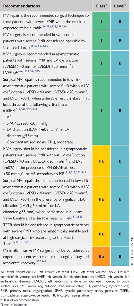
9.1.5. Follow-up

Asymptomatic patients with severe MR not fulfilling the criteria for an intervention, and with documented preserved exercise capacity, should undergo clinical and echocardiographic follow-up twice per year (watchful waiting strategy), ideally in the setting of a Heart Valve Clinic.568 Follow-up may also include serial measurements of BNP levels, electrocardiogram (ECG) and/or Holter monitoring, and (in selected cases) exercise echocardiography and CMR to confirm MR severity, and cardiac chamber function and dimensions.46 Asymptomatic patients with moderate MR and preserved LV function can be followed on a yearly basis with echocardiographic assessment every 1 or 2 years.

The frequency of follow-up after an intervention depends on the type of procedure performed. Very good long-term durability of MV repair in PMR due to valve prolapse with a low recurrence rate has been reported at experienced centres with freedom from moderate or severe MR of 87.5% at 20 years.533,559 Serial clinical and echocardiographic follow-up in patients without pre-operative LV dysfunction or rhythm abnormalities every 2–3 years thereafter is sufficient. Patients with atrial or ventricular arrhythmias possibly related to MV disease should be further evaluated using continuous ECG monitoring. Patients with recurrent MR after surgical ring annuloplasty frequently undergo repeat surgery (usually MV replacement), while transcatheter alternatives are reserved for selected high-risk patients because of the risk of increased gradients (M-TEER), PVL, and LVOT obstruction (mitral valve-in-ring procedures).569,570 In patients undergoing MV replacement, closer follow-up on a yearly basis is required due to the risk of prosthetic valve dysfunction or SVD (see Section 14.4). Following TEER, reported rates of residual MR and increased transmitral gradients are higher than after surgical repair, suggesting that yearly follow-up is appropriate.571 Although rare, the need for surgical treatment after failed TEER is associated with increased peri-operative mortality and low valve repair rates,541,572 while transcatheter solutions to detach TEER implants and replace the valve have been described in few cases.573,574

9.2. Secondary mitral regurgitation

9.2.1. Prevalence and aetiology

Secondary mitral regurgitation is present when the MV structure appears grossly normal but the MV is nonetheless incompetent, due to alterations in LV and LA geometry, dyssynchrony, and imbalances between MV closing and tethering forces.575 The prevalence of severe SMR in patients with chronic HF is ∼10% and higher in patients with reduced vs preserved LVEF (25% vs 4%).576 Secondary mitral regurgitation can be classified as atrial or ventricular with different pathophysiological and morphological characteristics, as well as contrasting prognostic and therapeutic implications.577 Ventricular SMR is more common and associated with worse long-term prognosis.513,578–580 Dilated or ischaemic cardiomyopathy are the most frequent causes of severe ventricular SMR. Acute HF exacerbation may occur in patients with chronic HF due to a renewed ischaemic event, arrhythmia, infection, or volume overload. Atrial SMR is due to pure mitral annular dilation and is observed in patients with long-standing AF and/or HFpEF.581 Factors predisposing to atrial SMR include age ≥65 years, female sex, LA dilatation, and diastolic dysfunction.582 From a morphological point of view, ventricular SMR is characterized by leaflet tethering and restricted motion combined with annular dilation, while annulus enlargement and flattening leading to planar coaptation are predominantly observed in atrial SMR. The prevalence of atrial SMR has been underestimated in the past and was occasionally misclassified as PMR due to pseudo-prolapse with leaflet tethering in advanced stages.

9.2.2. Evaluation

Echocardiographic criteria to define significant SMR according to aetiology are reported in Figure 10. Importantly, SMR assessment should be performed after optimization of medical therapy and in a euvolaemic and normotensive state. When quantifying EROA and RVol in SMR, lower thresholds may apply to define severe regurgitation because of the potential elliptical regurgitant orifice and/or the low-flow state. An EROA of ≥30 mm2 and/or an RVol of ≥45 mL has been identified as having a significant impact on outcomes,45 with prognosis improved after treatment.583,584 Cardiac magnetic resonance is used to confirm SMR severity and assess cardiac chamber function and dimensions. The extent of myocardial fibrosis, as assessed with CMR, has been associated with poor prognosis.585 Owing to the dynamic nature of SMR, exercise echocardiography may help to identify patients with severe SMR when values at rest are inconclusive.45

9.2.3. Definition of atrial secondary mitral regurgitation

Characteristics distinguishing between atrial and ventricular SMR are displayed in Figure 12.
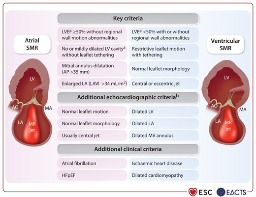
Most frequently used criteria for the diagnosis of atrial secondary mitral regurgitation.
Figure 12

Most frequently used criteria for the diagnosis of atrial secondary mitral regurgitation.

AO, aorta; AP, anteroposterior; HFpEF, heart failure with preserved ejection fraction; LA, left atrium/left atrial; LAVI, left atrial volume index; LV, left ventricle/left ventricular; LVEF, left ventricular ejection fraction; MA, mitral annulus; MR, mitral regurgitation; MV, mitral valve; SMR, secondary mitral regurgitation. aLV end-diastolic dimension of <56 mm in females and <63 mm in males; indexed LV end-diastolic volume <71 mL/m2 (in women) or <79 mL/m2 (in men). bAdditional echocardiographic criteria for atrial SMR may no longer be fulfilled in advanced stages.

Atrial SMR is most frequently defined by the presence of the following key criteria:513,578–580,586–590

  • preserved LVEF (≥50%) without regional wall motion abnormalities or leaflet tethering; AND

  • no or mildly dilated LV cavity [LV end-diastolic dimension of <56 mm in women and <63 mm in men; indexed LV end-diastolic volume of <71 mL/m2 (in women) or <79 mL/m2 (in men)]; AND

  • mitral annulus (MA) dilatation [anteroposterior (AP) diameter of >35 mm]; AND

  • enlarged LA (LAVI >34 mL/m2).

Echocardiography frequently reveals normal leaflet motion with planar coaptation, and normal leaflet morphology with a central MR jet in atrial SMR. However, at advanced stages, an overlap between atrial and ventricular SMR criteria can be observed in the case of late LV damage due to continuous volume overload.591,592 Clinical criteria (i.e. history of AF and/or diagnosis of HFpEF) are also useful and should be taken into consideration.

9.2.4. Management of ventricular secondary mitral regurgitation

9.2.4.1. Medical and device therapy

In patients with ventricular SMR, GDMT for the treatment of HF is recommended prior to any MV intervention.339,340 The combination of ACE-Is/ARBs or angiotensin receptor/neprilysin inhibitors, beta-blockers, mineralocorticoid receptor antagonists, and sodium–glucose co-transporter 2 inhibitors (SGLT2is) at the maximum tolerated doses is recommended according to the HF Guidelines.340 Initiation and up-titration of neurohormonal drugs need to be tailored according to the patient profile, mainly based on BP, heart rate, potassium levels, and renal function.593 Notably, GDMT up-titration must be rapid (within 6 weeks) and take place in the context of close follow-up visits, especially in the case of a recent hospitalization for acute HF.594 About 40% of patients with ventricular SMR experience improvement of SMR severity after 1–3 months of optimized GDMT.595,596 Cardiac resynchronization therapy (CRT) should also be considered as part of HF management before an MV intervention according to HF guideline criteria (LVEF ≤35% and wide QRS).597,598 Although no dedicated RCTs exist, SMR reduction by at least one grade has been described in 40%–60% of patients and is associated with reverse LV remodelling and improved clinical outcomes.597,599,600

9.2.4.2. Indications for intervention

The management of patients with ventricular SMR should be discussed by a multidisciplinary Heart Team including HF specialists. The indication for intervention is based on the persistence of symptoms (i.e. NYHA class II–IV) despite adequate GDMT and CRT, if indicated (Figure 13). GDMT is the only option for very frail patients or those with limited life expectancy.
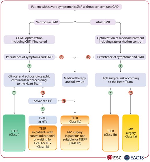
Treatment of severe secondary mitral regurgitation without concomitant coronary artery disease.
Figure 13

Treatment of severe secondary mitral regurgitation without concomitant coronary artery disease.

CAD, coronary artery disease; CRT, cardiac resynchronization therapy; GDMT, guideline-directed medical therapy; HF, heart failure; HTx, heart transplantation; LVAD, left ventricular assist device; MV, mitral valve; SMR, secondary mitral regurgitation; TEER, transcatheter edge-to-edge repair. aSee Table 7. bSee Supplementary data online, Table S2.

In the Cardiovascular Outcomes Assessment of the MitraClip Percutaneous Therapy for Heart Failure Patients with Functional Mitral Regurgitation (COAPT) trial, M-TEER was shown to be safe and reduced recurrent HF hospitalization and all-cause mortality at 2 and 5 years of follow-up, compared with optimized GDMT in patients with ventricular SMR without relevant CAD.583,584 In a second study (Percutaneous Repair with the MitraClip Device for Severe Functional/Secondary Mitral Regurgitation, MITRA-FR), no differences were demonstrated for the combined primary endpoint of all-cause mortality or HF hospitalization at 1 and 2 years.601,602 These diverging results might be explained by effect sizes of the trials, differences in trial design, patient selection and follow-up, echocardiographic assessment of SMR severity, use of GDMT, and technical factors.603–605 A third RCT, the Randomized Investigation of the MitraClip Device in Heart Failure: 2nd Trial in Patients with Clinically Significant Functional Mitral Regurgitation (RESHAPE-HF-2) trial showed a 36% reduction in the rates of HF hospitalization or cardiovascular death at 2 years in the intervention arm driven by reduction of first or recurrent HF hospitalization. When considered in isolation, cardiovascular mortality was not significantly reduced during the observation period. In addition, there was a significant improvement in quality of life as measured by the KCCQ overall score (mean difference between study groups, 10.9 points; 95% CI, 6.8–15.0; P <0.001).606 A recent study-level meta-analysis of these three trials showed a significant reduction of HF rehospitalization at 24 months (HR, 0.63, 95% CI, 0.50–0.80) and the composite of death and all-cause hospitalizations (HR, 0.72, 95% CI, 0.51–0.999).607 However, there was no statistically significant difference in death from any cause or cardiovascular death at 24 months. Transcatheter edge-to-edge repair is therefore recommended to reduce HF hospitalizations, and improve quality of life, in symptomatic patients with persisting severe SMR despite optimized GDMT fulfilling specific clinical and echocardiographic criteria (Table 7). Although less challenging than in PMR, anatomical suitability for TEER needs to be assessed upfront (Supplementary data online, Table S2). There is also increasing observational evidence supporting the use of TEER for the improvement of symptoms, functional capacity, and quality of life in patients with ventricular SMR not fulfilling the clinical and echocardiographic criteria for outcome improvement.608–612 This particularly applies to patients not tolerating GDMT in whom TEER may help up-titration, as well as those with recent myocardial infarction and persistent severe ventricular SMR.203,613

Table 7

Clinical and echocardiographic criteria predicting outcome improvement in patients with severe ventricular secondary mitral regurgitation undergoing mitral transcatheter edge-to-edge repair

Anatomy deemed suitable for M-TEER
NYHA class ≥II
LVEF 20%–50%
LVESD ≤70 mm
At least one HF hospitalization within the previous year or increased natriuretic peptide levels (BNP ≥300 pg/mL or NT-proBNP ≥1000 pg/mL)
SPAP ≤70 mmHg
No severe RV dysfunction
No Stage D or advanced HF
No CAD requiring revascularization
No severe AV and/or TV disease
No hypertrophic, restrictive, or infiltrative cardiomyopathies

AV, aortic valve; BNP, brain natriuretic peptide; CAD, coronary artery disease; HF, heart failure; LVEF, left ventricular ejection fraction; LVESD, left ventricular end-systolic diameter; M-TEER, mitral transcatheter edge-to-edge repair; NT-proBNP, N-terminal pro-B-type natriuretic protein; NYHA, New York Heart Association; RV, right ventricle/right ventricular; SPAP, systolic pulmonary artery pressure; TV, tricuspid valve.

© ESC/EACTS 2025

Transcatheter implantation of an indirect annuloplasty device into the coronary sinus has been proposed as an alternative that preserves valve integrity. Despite rather modest SMR reduction (22.4% decrease in mitral RVol) and no significant effect on quality of life in a small sham-controlled RCT, symptomatic improvement and reverse remodelling were observed in registries at 1 year.614,615 Heart transplantation or left ventricular assist device (LVAD) implantation should be considered in selected patients with severe ventricular SMR and advanced HF.

In patients with ventricular SMR without relevant CAD, indications for isolated MV surgery are restrictive owing to procedural risks and the absence of proven mortality benefit.616 The Multicenter, Randomized, Controlled Study to Assess Mitral Valve Reconstruction for Advanced Insufficiency of Functional or Ischemic Origin (MATTERHORN) trial, which included a mixed population mainly composed of patients with ventricular SMR (84%), demonstrated that TEER is non-inferior to surgical repair or replacement with regard to a composite endpoint of death, HF hospitalization, MV reintervention, implantation of an assist device, or stroke within 1 year after the procedure, and showed a better safety profile.617 In patients with severe ischaemic ventricular SMR and concomitant CAD requiring coronary revascularization, MV surgery at the time of CABG is recommended, unless the patient is at high surgical risk and/or the coronary anatomy is suitable for PCI. Although isolated undersized mitral annuloplasty is the most commonly performed MV repair procedure, recurrent MR rates were high with this technique in an RCT618 and reverse LV remodelling is limited, especially in patients with an increased tenting area (>1.35 cm2/m2 BSA),619 in whom MV replacement is usually required.620 The addition of subvalvular modification in patients with LV dilatation and pronounced MV leaflet tenting may improve results for MV repair,621 but durability and impact on HF symptoms with this technique require further investigation.

The treatment of moderate ischaemic SMR in patients undergoing CABG remains controversial. Meta-analyses, including four RCTs comparing CABG with concomitant MV surgery vs CABG alone, have shown lower rates of recurrent MR but no benefit in terms of mortality and clinical outcomes.622–624 Therefore, clinical decision-making should weigh peri-operative risks of more complex surgery against the long-term risk of MR progression.

9.2.4.3. Follow-up

Patients with ventricular SMR need to be followed up carefully after intervention by an HF specialist, because they remain at increased risk of events despite intervention. The 5-year cumulative incidence of all-cause death or HF hospitalization was 73.6% in the device arm of the COAPT study.584 Clinical, laboratory, and echocardiographic evaluation every 3 or 6 months, according to the HF stage, is recommended. Durability of the procedural result, as well as congestion status and the need for further GDMT optimization facilitated by SMR reduction,613 need to be assessed. Patients and families should be trained in monitoring vital signs, body weight, and HF symptoms to avoid late hospital admissions and facilitate management of possible decompensation. Also, patients must be educated on the importance of not discontinuing medical therapies after intervention since the two treatments (devices and drugs) are complementary.

Patients with ventricular SMR, who are asymptomatic and/or have moderate or dynamic MR, should undergo clinical and echocardiographic follow-up at least twice per year.

9.2.5. Management of atrial secondary mitral regurgitation

9.2.5.1. Medical therapy and rhythm management

In patients with atrial SMR, underlying causes need to be recognized and treated. Associated HFpEF and AF should be managed according to the relevant Guidelines.7,340 The use of SGLT2is should be encouraged in patients with HFpEF due to their proven efficacy in reducing cardiovascular death and HF hospitalization.625 Limited data show that rhythm control may contribute to reduce atrial SMR severity and reverse LA dilatation.626

9.2.5.2. Indications for intervention

Registry data demonstrate that patients with atrial SMR are typically elderly with associated AF. Mitral valve surgery has been recently associated with lower rates of HF hospitalizations and mortality compared with GDMT in a matched population, despite a higher risk profile in the surgical arm at baseline.627 Data from several observational studies also suggest that surgical annuloplasty is effective and durable in patients with atrial SMR, because it counteracts the main mechanism of MR progression.628–630 Its combination with surgical AF ablation (Maze procedure) and concomitant LAAO may have further advantages,630,631 while the frequently associated relevant TR can also be addressed during the same procedure.627 Transcatheter edge-to-edge repair may also be considered because observational studies have demonstrated high safety and procedural success,588–590,632,633 as well as compared with surgery in a small subgroup (n = 34) of the MATTERHORN RCT.634 However, the risk of increased gradient due to planar leaflet coaptation, large regurgitant jet, and limited MV area (MVA) needs to be taken into consideration.592,635 Further studies are warranted to investigate the treatment modalities of patients with atrial SMR.

9.2.5.3. Follow-up

Patients with atrial SMR undergoing surgical or transcatheter intervention should be followed up on a yearly basis, including clinical and echocardiographic evaluation. In cases of HFpEF, as an underlying cause of atrial SMR, consultation with an HF specialist is necessary.

Asymptomatic patients with severe atrial SMR not fulfilling the criteria for an intervention should undergo clinical and echocardiographic follow-up at least once per year.

Recommendation Table 7

Recommendations on indications for intervention in secondary mitral regurgitation (see also Supplementary data online, Evidence Tables 17–20)
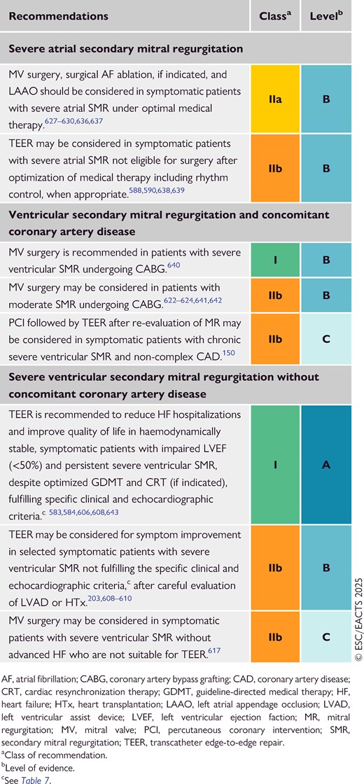
10. Mitral stenosis

10.1. Prevalence and aetiology

The aetiology of MS is most frequently rheumatic or degenerative, while rare forms can be drug-induced, inflammatory, or carcinoid-related. Rheumatic fever is the most common cause of MS and death due to VHD worldwide. Its prevalence has decreased in high- and middle-income countries, but remains a major healthcare problem in low-income countries, where it predominantly affects young patients.12,187,644 Degenerative MS related to MAC is a distinct age-dependent pathology requiring different treatment strategies.645–647 Both aetiologies are more frequent in females.648

10.2. Rheumatic mitral stenosis

10.2.1. Evaluation

Echocardiography is the preferred method for screening in endemic regions and the assessment of the severity, extent of anatomical lesions, and haemodynamic consequences of MS. Involvement of other valves, particularly secondary TR, should be identified. Mitral valve area using 2D planimetry is the most commonly used measurement to assess stenosis severity, but 3D TTE and TOE have additional diagnostic value.649 An MVA of ≤1.5 cm2 in conjunction with clinical factors (symptoms, high risk of thromboembolism, or haemodynamic decompensation) is indicative of clinically severe MS. Mean transvalvular gradient and pulmonary pressures reflect its consequences and have prognostic value.649,650 Leaflet thickening and fibrosis, along with commissural fusion and shortening of the subvalvular apparatus, are the most important pathomechanisms of stenosis associated with RHD. The presence and extent of leaflet and subvalvular calcifications influence treatment decisions. Scoring systems have been developed to assess the suitability of patients for percutaneous mitral commissurotomy (PMC) (see Supplementary data online, Table S3).651–653

Exercise testing is indicated in asymptomatic patients or patients with symptoms that are equivocal or discordant with the severity of stenosis. Exercise echocardiography provides additional information on exercise capacity and related changes in mitral gradient and pulmonary artery pressure, and is preferred over DSE, especially when there are contraindications to dobutamine.654 Transoesophageal echocardiography should be systematically performed in PMC candidates to exclude LA thrombus or after an embolic episode, and may play an essential role for procedural guidance.52,655

10.2.2. Medical therapy

Diuretics, beta-blockers, digoxin, non-dihydropyridine calcium channel blockers, and ivabradine can improve symptoms by controlling volume overload and heart rate. Anticoagulation with a VKA with a target international normalized ratio (INR) between 2 and 3 is indicated in patients with AF, and DOACs should be avoided in patients with an MVA of ≤2.0 cm2 according to current evidence.165,656,657 Interventions to restore sinus rhythm (cardioversion or catheter pulmonary vein isolation) are unlikely to be successful in patients with untreated severe MS. If AF is of recent onset and the LA moderately enlarged, cardioversion can be attempted soon after successful intervention or in patients with moderate MS combined with amiodarone treatment.658,659 In patients in sinus rhythm, OAC is indicated after systemic embolism or if a thrombus is present in the LA, and should also be considered when TOE shows dense spontaneous echocardiographic contrast or an enlarged LA (M-mode diameter >50 mm or LA volume >60 mL/m2).660 Prophylaxis of infective endocarditis is indicated as appropriate.5

10.2.3. Indications for intervention

The type (PMC or surgery) and timing of treatment should be decided based on clinical characteristics, the anatomy of the valve and subvalvular apparatus, and local expertise.661–663 The management of clinically severe rheumatic MS is summarized in Figure 14 and Recommendation Table 8.
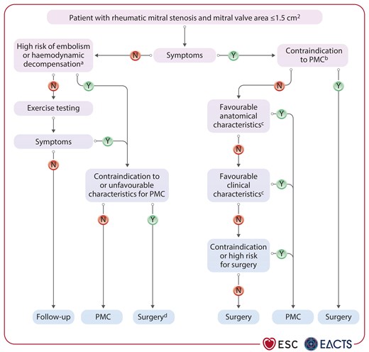
Management of clinically severe rheumatic mitral stenosis (mitral valve area ≤1.5 cm2).
Figure 14

Management of clinically severe rheumatic mitral stenosis (mitral valve area ≤1.5 cm2).

AF, atrial fibrillation; LA, left atrium; MS, mitral stenosis; MV, mitral valve; MVA, mitral valve area; NCS, non-cardiac surgery; NYHA, New York Heart Association; PH, pulmonary hypertension; PMC, percutaneous mitral commissurotomy; SPAP, systolic pulmonary artery pressure; TR, tricuspid regurgitation. aHigh thromboembolic risk: history of systemic embolism, dense spontaneous contrast in the LA, new-onset AF. High-risk of haemodynamic decompensation: SPAP >50 mmHg at rest, need for major NCS, desire for pregnancy or pregnant. bSee Table 8. cFavourable = absence of unfavourable characteristics for PMC defined by unfavourable anatomical characteristics [echocardiographic score >8, Cormier score 3 (calcification of MV of any extent as assessed by fluoroscopy), severe TR] or unfavourable clinical characteristics (old age, history of commissurotomy, NYHA class IV, permanent AF, severe PH) (for the definition of scores see Supplementary data online, Table S3). dIf operative risk is low.

In general, indication for intervention should be limited to patients with clinically severe rheumatic MS (MVA <1.5 cm2) in whom PMC is expected to have a significant impact on clinical outcome. In higher-income countries, where the incidence of rheumatic fever and the number of PMCs performed is low, this treatment should be restricted to expert operators in specialized centres to improve safety and procedural success rate.661,664 Efforts should be made to increase the availability of PMC in lower-income countries, where access to treatment is limited for economic reasons.665,666 Percutaneous mitral commissurotomy may be also considered in symptomatic patients with an MVA of >1.5 cm2, if symptoms cannot be explained by another cause and if the anatomy is favourable (see Table 8). Percutaneous mitral commissurotomy should be considered as a first-line treatment for patients with anatomically suitable rheumatic MS and mild-to-moderate calcification without severe subvalvular impairment. Selected patients with unfavourable anatomical and clinical characteristics can still benefit from PMC, particularly if they are at increased surgical risk. When symptomatic restenosis occurs after surgical commissurotomy or PMC, reintervention in most cases requires surgical valve replacement, but redo PMC can be proposed in selected candidates with favourable characteristics, if the predominant mechanism is commissural refusion. Long-term follow-up has shown favourable results following PMC despite a growing number of elderly patients with suboptimal clinical and anatomical characteristics.663–665

Table 8

Contraindications for percutaneous mitral commissurotomy in rheumatic mitral stenosis

Contraindications
MVA >1.5 cm2  a
LA thrombusb
More than mild MR
Severe or bi-commissural calcification
Absence of commissural fusion
Severe concomitant AV disease, or severe combined tricuspid stenosis and regurgitation requiring surgery
Concomitant CAD requiring bypass surgery

AV, aortic valve; CAD, coronary artery disease; LA, left atrium/left atrial; MR, mitral regurgitation; MVA, mitral valve area; OAC, oral anticoagulation; PMC, percutaneous mitral commissurotomy; TOE, transoesophageal echocardiography.

aPMC may be considered in patients with MVA of >1.5 cm2 with symptoms that cannot be explained by another cause and if the anatomy is favourable.

bWhen the thrombus is located in the LA appendage, PMC may be considered in patients with contraindications to surgery or those without urgent need for intervention, in whom OAC can be safely given for 1–3 months, provided repeat TOE confirms resolution of thrombus.

© ESC/EACTS 2025

For patients in whom PMC is contraindicated (Table 8), surgical MV repair or, more frequently, replacement are good alternatives. Although repair is much more challenging than for PMR, it can be attempted at experienced centres.663

For patients with multiple VHD including MS, a comprehensive evaluation by the Heart Team and an individualized approach is necessary. Surgery is preferable to PMC in patients with severe MS and severe AV disease, unless the surgical risk is high. In selected cases with severe MS and moderate AV disease, PMC can be performed to postpone surgical treatment of both valves.

In high-risk cases with concomitant severe TR, PMC may be considered in selected patients with sinus rhythm, moderate atrial enlargement, and secondary TR due to post-capillary PH. In non-high-risk cases, surgery on both valves is preferred.651,652,662,667 Treatment of patients with low-gradient severe MS (MVA <1.5 cm2, mean gradient <10 mmHg) is challenging, because these patients are often older and have unfavourable anatomy.668,669

10.2.4. Follow-up

Asymptomatic patients with clinically severe MS who have not undergone intervention should be followed up yearly by TTE, and at longer intervals (2–3 years) in cases of moderate stenosis. After PMC, the post-procedural MVA and mean mitral gradient are important parameters that influence long-term clinical outcomes. Follow-up of patients after successful PMC is necessary because asymptomatic restenosis may occur. Progressive rheumatic involvement of other valves should be periodically assessed, irrespective of the therapy modality. Finally, education and the engagement of the family is key in patients with rheumatic MS, since it usually affects young individuals and women of childbearing age.

10.3. Degenerative mitral stenosis with mitral annular calcification

Patients presenting with MAC are elderly and have significant comorbidities, including disease of other valves. Mitral annular calcification is also an indicator of cardiovascular disease severity and is associated with an increased risk of AF, stroke, and death.670,671 The incidence of MAC varies substantially, depending on the age of the studied population and the imaging modality used. It can be a consequence of many different pathological processes and, depending on the underlying disease, can be accompanied by stenosis, regurgitation, or both. However, most patients with MAC do not have significant valvular dysfunction.647,672

Generally, MS occurs due to calcific extension into the MV leaflets or subvalvular apparatus, and in some patients it is associated with combined MR.673 Treatment options (including transcatheter and surgical approaches) are high-risk procedures and evidence from RCTs is lacking.

10.3.1. Evaluation

Echocardiography is used for initial evaluation, but is frequently limited by acoustic shadowing due to severe calcification. Evaluation of MVA by planimetry is less reliable than in rheumatic MS, and TOE should therefore be used liberally. Degenerative MS can coexist with varying degrees of MR. Mean transmitral gradient has been shown to be associated with increased mortality irrespective of MR severity.674 Electrocardiogram-gated CCT is necessary to assess the degree and locations of calcifications, especially if an intervention is planned.647,674–676 Calcifications are usually more prominent at the posterior aspect of the annulus.

10.3.2. Indications for intervention

Intervention is recommended in symptomatic patients who are not responsive to medical therapy, weighing the potential benefits of the procedure against its associated risks. In elderly patients with degenerative MS and MAC, surgery is technically challenging and high risk. However, surgical MV repair or replacement with extensive decalcification and patch reconstruction of the annulus can be performed in selected patients at experienced centres (e.g. young patients post-chest radiation), where mortality rates of <5% have been reported.677–679

Degenerative MS is not amenable to PMC because commissural fusion is absent. In symptomatic high-risk patients with suitable anatomy, transcatheter implantation of a TAVI prosthesis in the mitral position is feasible but associated with frequent complications, including LVOT obstruction, valve embolization, stroke, and haemolysis due to PVL. Open surgical valve replacement via the LA with a TAVI device is an alternative that allows complete anterior leaflet removal;680 however, mortality remains high. The use of dedicated TMVI devices is therefore encouraged because it appears to be safer.542,681 Heart Team evaluation should guide the choice of treatment avoiding futility, because mortality remains high, even after successful treatment (10%–30% within 1 year).

Recommendation Table 8

Recommendations on indications for percutaneous mitral commissurotomy, mitral valve surgery, and transcatheter intervention in clinically severe rheumatic and degenerative mitral stenosis (see also Supplementary data online, Evidence Table 21)
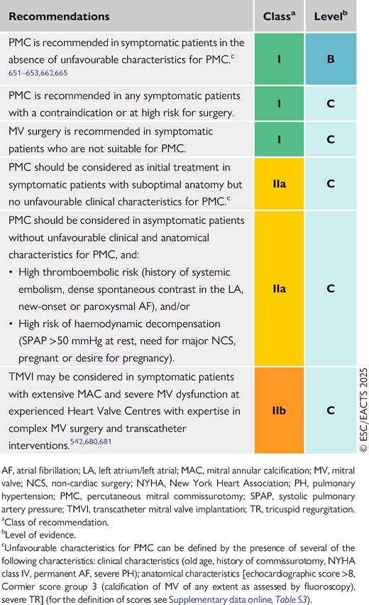
11. Tricuspid regurgitation

11.1. Prevalence and aetiology

TR is a common echocardiographic finding in the general population, with higher prevalence in women and older patients. Trivial or mild TR is mostly a benign condition. Significant TR (≥ moderate) has a reported age- and sex-adjusted prevalence of 0.55% (4% in people aged ≥75 years).682 Severe TR is associated with increased risk of death and HF, independent of comorbidities, ventricular function, and pulmonary pressures.683–686

Only 8%–10% of patients with TR present with clear anatomical abnormalities of the TV apparatus (primary TR), which can be due to infective endocarditis, RHD, carcinoid syndrome, congenital abnormalities (e.g. Ebstein's anomaly), chest radiation, or myxomatous disease, as well as trauma or iatrogenic valve damage (e.g. after endomyocardial biopsy).682 Cardiac implantable electronic device (CIED)-related TR represents a separate entity requiring a specific diagnostic approach and management.687 In patients with a CIED, diagnostic efforts should be made to clarify if the lead is the cause of TR (CIED-related TR) or incidental (CIED-associated TR).688

In patients with secondary TR, TV leaflets are structurally normal and regurgitation is caused by annular dilatation and/or leaflet tethering due to RA dilatation, and/or RV dilation and dysfunction. Based on the main morphological and haemodynamic characteristics, two phenotypes of secondary TR have been proposed:689 (i) atrial secondary TR, mainly due to AF and characterized by the absence of significant leaflet tethering, but with marked RA and annular dilatation along with preserved RV size/function, pulmonary pressure, and LV function; and (ii) ventricular secondary TR, due to annular dilatation and leaflet tethering as a consequence of left-sided ventricular or valvular disease (post-capillary PH), pre-capillary PH, or primary RV cardiomyopathy/ischaemia (also after left-sided valve surgery).690 At an advanced disease stage, these two phenotypes may no longer be distinguishable, and therefore early characterization is key to determine outcome.691,692 Evidence of an impact on patient management is currently lacking; therefore, current recommendations for intervention consider mainly primary vs secondary TR.

11.2. Evaluation

Echocardiography is recommended to assess patients with TR45 and should include evaluation of severity and aetiology (including characterization of left-sided heart disease and, if applicable, CIED lead location and interaction with the valve apparatus), the impact of TR on the right-sided chambers (RV and RA size and function), and assessment of central venous (inferior vena cava) and pulmonary pressures. Transthoracic echocardiography provides sufficient diagnostic information in most patients. Transoesophageal echocardiography is necessary for when visualization of the TV apparatus. In candidates for an intervention, advanced techniques, such as strain analysis and 3D echocardiography, should also be applied when available.

Assessment of TR severity should be ideally performed in euvolaemic status, with optimized pulmonary and systemic pressures, and based on an integrative approach considering multiple qualitative and quantitative parameters (Figure 15).45,693 A grading scheme extending beyond severe, including ‘massive’ and ‘torrential’ grades, has been proposed to refine TR reduction assessment after transcatheter interventions and has been used in several studies.694,695 Although this five-grade scale may be associated with a proportional increase in symptoms and event risk,694,695 an intervention should be considered without delay, as soon as TR is severe, with the aim of reducing TR to moderate or less.696,697
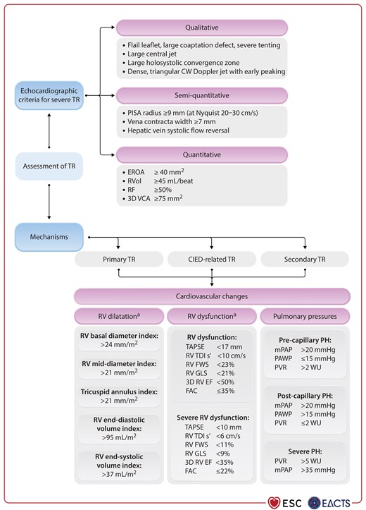
Echocardiographic and invasive assessment of tricuspid regurgitation.
Figure 15

Echocardiographic and invasive assessment of tricuspid regurgitation.

3D, three-dimensional; CIED, cardiac implantable electronic device; CW, continuous-wave; EROA, effective regurgitant orifice area; FAC, fractional area change; FWS, free wall strain; GLS, global longitudinal strain; mPAP, mean pulmonary artery pressure; PAWP, pulmonary artery wedge pressure; PH, pulmonary hypertension; PISA, proximal isovelocity surface area; PVR, pulmonary vascular resistance; RF, regurgitant fraction; RV, right ventricle/ventricular; RVEF, right ventricular ejection fraction; RVol, regurgitant volume; TAPSE, tricuspid annular plane systolic excursion; TDI, tissue Doppler imaging; TR, tricuspid regurgitation; VCA, vena contracta area; WU, wood unit. aRV apical focused view.

Echocardiographic assessment of the RV is challenging due to its complex geometry, imaging constraints, and the high dependency on loading conditions. When accurate measurements of RV size and function, as well as RV volume, are necessary for decision-making, CMR should be used because of its high accuracy and reproducibility.698,699 In Figure 15, upper limits of normal for different RV size parameters are provided to guide definition of RV dilatation and remodelling. In the setting of severe TR, RV function is often overestimated and therefore the most conservative/cautious thresholds are suggested for the currently used echocardiographic parameters to identify RV dysfunction at the earliest stage possible. Cut-off values for severe RV dysfunction are also provided to indicate high-risk or possibly futile interventions. Although robust validation is lacking, all these reference values are chosen based on large multicentre reports of normative data and outcomes.60,697,700–704

Echocardiography often underestimates pulmonary pressures in cases of severe TR.705 Right heart catheterization is therefore recommended in all candidates for an intervention to assess the haemodynamic consequences of TR on the RA and venous circulation (e.g. ventricularization of the RA pressure curves), measure end-diastolic RV pressure, and document volume overload. The assessment of pulmonary pressures and vascular resistance are key to exclude masked severe pre-capillary PH.706

RV–pulmonary artery coupling refers to the ability of the RV systolic performance to match a given pulmonary afterload maintaining adequate cardiac output, and can be measured invasively or approximated using echocardiography [i.e. tricuspid annular plane systolic excursion (TAPSE)/SPAP].707,708 RV–pulmonary artery uncoupling (low TAPSE/SPAP) occurs when sustained increases in afterload cannot be matched by RV contractile reserve and has been associated with poor prognosis in different HF conditions, including severe TR. Although not yet prospectively validated, this index may improve risk stratification.

Electrocardiogram-gated CCT with dedicated protocols ensuring sufficient contrast enhancement of the right heart cavities provides detailed characterization of the RA, RV, and vena cava anatomy, the location of the right coronary artery, and is crucial to assess suitability and device sizing for several transcatheter interventions.689,697

Before any intervention is considered, careful evaluation of TR aetiology, disease stage (TR severity, RV and LV dysfunction, and PH), patient operative risk, and likelihood of recovery by a dedicated collaborative Heart Team is recommended (Figure 16). Dedicated clinical risk scores for TR patients have been described recently. The TRI-SCORE709 and the STS isolated TV risk calculator710 take into consideration clinical and echocardiographic signs of RV dysfunction, and secondary organ (particularly hepatic and renal) impairment. They both allow improved estimates of peri-procedural risk in patients with severe TR undergoing surgery and possibly help to avoid futile interventions.84,711 The importance of risk stratification was demonstrated in a recent registry analysis (n = 2413) comparing any interventions with conservative management. Early TV intervention (transcatheter valve repair or surgery) was associated with improved mid-term survival in patients with a low or intermediate TRI-SCORE (up to 5 points), while patients with a high TRI-SCORE (≥6) did not derive any benefit compared with conservative management.696 Moreover, isolated TV surgery (repair or replacement) improved survival at 10 years in patients with a low TRI-SCORE (≤3). The same benefit was observed in patients with an intermediate TRI-SCORE (4–5) after successful TV repair only.689,697,709 Patients with moderate or severe TR should be regularly followed up clinically and by echocardiography at least every 6 months.
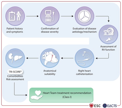
Stepwise evaluation of patients with tricuspid regurgitation.
Figure 16

Stepwise evaluation of patients with tricuspid regurgitation.

RV, right ventricle/right ventricular. aSee Supplementary data online, Table S4.

11.3. Medical therapy

Patients with relevant TR should be first treated according to the assumed aetiology, including optimal HF treatment, pulmonary vasodilators for PH, and rhythm control for AF.339,693

In the case of HF symptoms, diuretics should be initiated in a stepwise approach,339 beginning with loop diuretics eventually combined with aldosterone antagonists, thiazide diuretics, and/or SGLT2is.712 However, according to current knowledge, medical therapy has very limited effect on the evolution of TR severity and none of these measures should delay evaluation of an intervention at an expert centre.713

11.4. Indications for intervention

11.4.1. Surgery

Patients are often referred too late for surgery when significant RV and other organ failure have occurred. Isolated TV surgery has therefore been considered to be generally high risk, with in-hospital mortality rates of 8%–10% in several reports,714,715 but contemporary cohorts have demonstrated improved outcomes when patients are referred earlier and more effective techniques are used.716 Valve repair using an annuloplasty ring is preferred over replacement, whenever technically feasible, especially in low-risk patients with suitable anatomy.709 However, TV replacement may be necessary in cases of advanced disease with marked annular dilatation and leaflet tethering.688,717 For CIED-related TR, preparation of any entrapped tricuspid leaflet and possibly lead extraction with implantation of an epicardial system has been associated with improved TV function.688,717

11.4.1.1. Patients without indication for left-sided valve surgery

In patients with severe TR but without the need for left-sided valve surgery, surgical intervention is recommended in operable symptomatic patients with primary TR (Figure 17; Recommendation Table 9).718 Furthermore, it should be considered in symptomatic patients with secondary TR, or in asymptomatic patients with primary or secondary TR and signs of RV dilatation or RV function deterioration.719 However, patients with severe LV/RV dysfunction or PH do not qualify due to high operative risk.84,686,720
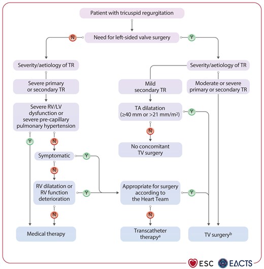
Management of patients with tricuspid regurgitation.
Figure 17

Management of patients with tricuspid regurgitation.

LV, left ventricle/left ventricular; RV, right ventricle/right ventricular; TA, tricuspid annulus; TR, tricuspid regurgitation; TV, tricuspid valve. aThe Heart Team with expertise in the treatment of TV disease evaluates anatomical eligibility for transcatheter therapy including jet location, coaptation gap, leaflet tethering, and potential interference with pacing lead. bRepair whenever possible, particularly in cases of moderate TR or mild TR with significant TA dilatation.

11.4.1.2. Patients with indication for left-sided valve surgery

Severe primary or secondary TR is unlikely to improve after isolated surgical treatment of left-sided valve disease, and reoperation for TR treatment is associated with high peri-operative mortality.721 Therefore, TV surgery is recommended at the time of the index procedure.

Mild TR with associated significant annular dilatation or moderate TR, if left uncorrected at the time of left-sided valve surgery, will progress in approximately one-quarter of patients and is associated with worse outcome.690,722 In patients with moderate TR, TV repair annuloplasty during MV surgery should be considered, because large retrospective studies723,724 and two recent RCTs have shown beneficial effects on TR progression and RV remodelling over time.725,726 However, no effects on mortality, HF events, or reoperation were observed in the repair group.725,727 Concomitant TV repair has also been associated with a higher risk of conduction disturbances requiring pacemaker implantation (up to 14%),725,728 with potential negative impacts on longer-term outcomes.729,730 In patients with mild TR and annular dilatation (≥40 mm or >21 mm/m2) undergoing left-sided valve surgery, previous observational studies have demonstrated a benefit of concomitant TV repair in terms of TR progression723,724 and RV function,731 and a trend towards improved long-term survival.724 However, a subanalysis of a recent RCT did not identify any difference in the progression of TR or other outcomes in this category of patients during a 2-year follow-up period.725 In turn, the need of pacemaker implantation after surgery led to a subsequent increase in HF hospitalizations, endocarditis, and mortality.729 Annuloplasty may therefore be considered after careful evaluation of the risk factors for progressive annular dilatation and TR (AF, RA size, pulmonary pressures, etc.), balancing against the risk of possible pacemaker implantation (Supplementary data online, Table S5).732

11.4.2. Transcatheter techniques

Several transcatheter approaches for the treatment of TR have been developed, including TEER, direct annuloplasty, and orthotopic and heterotopic TV replacement. Data from large multicentre registries, single-arm clinical trials, and two recent RCTs in patients with severe TR at intermediate and high risk for surgery have shown the safety of transcatheter repair, as well as the ability to reduce TR to moderate or less in more than 80% of cases when anatomical suitability was confirmed.713,733–735 The Clinical Trial to Evaluate Cardiovascular Outcomes in Patients Treated With the Tricuspid Valve Repair System (TRILUMINATE) Pivotal Trial showed also a lower incidence of the composite endpoint of death from any cause or TV surgery, HF hospitalization, and improvement in quality of life as measured with the KCCQ score after tricuspid TEER compared with medical treatment that was exclusively driven by improved quality of life [11.7 points (95% CI, 6.8–16.6); P <0.001].713 At 2 years, a lower incidence of HF hospitalizations has been observed in the intervention group, despite a high rate of crossovers (59%).736

Another investigator-initiated RCT (the Tri.Fr trial) demonstrated the benefit of tricuspid TEER in combination with GDMT over medical therapy alone, for a composite score driven by improved PROMs.737 Another recent RCT (the Edwards EVOQUE Transcatheter Tricuspid Valve Replacement: Pivotal Clinical Investigation of Safety and Clinical Efficacy using a Novel Device (TRISCEND) II trial), comparing transcatheter TV replacement with optimized medical therapy in patients with symptomatic severe TR, showed similar results with a win ratio favouring TV replacement mainly explained by symptom and quality-of-life improvement. In these studies, reverse RV remodelling was also observed. However, the safety profile of transcatheter TV replacement was less favourable, including higher risk for major bleeding (15%) and post-procedural pacemaker implantation in about one-quarter of the pacemaker-naïve patients after 12 months of follow-up.738 Based on these data, transcatheter treatment should be considered to improve quality of life and RV remodelling in high-risk patients with symptomatic severe TR, despite optimal medical therapy, but without severe RV dysfunction or pre-capillary PH.

Transvenous CIED lead repositioning or extraction can be considered in selected patients to improve TR or avoid lead jailing before any tricuspid interventions, although the efficacy of this procedure is uncertain and the risk of damaging the TV not negligible.688,717,739

Recurrent TR after previous tricuspid annuloplasty usually requires cardiac reoperation for surgical TV replacement. Transcatheter valve-in-ring implantation is an off-label procedure to treat residual TR in high-risk patients. Challenges are the non-circular shape and the open form of the surgical annuloplasty ring.740 However, transcatheter tricuspid valve-in-valve procedures have been performed with satisfactory results.741

Transcatheter TV procedures should be performed at an experienced Heart Valve Centre with expertise in the treatment of TV disease (Table 6). Careful evaluation of clinical and anatomical suitability is key for appropriate patient and device selection to achieve optimal TR reduction and symptomatic response to the therapy.

Recommendation Table 9

Recommendations on indications for intervention in tricuspid regurgitation (see also Supplementary data online, Evidence Tables 22 and 23)
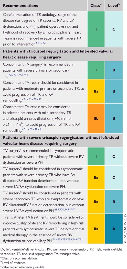
12. Tricuspid stenosis

12.1. Prevalence and aetiology

Tricuspid stenosis (TS) is a relatively rare disease that is most commonly associated with congenital conditions or enzymatic disorders, such as Whipple's or Fabry's disease. It can also be acquired as an isolated manifestation of RHD or occur in combination with aortic and/or MV involvement. Moreover, TS can be the consequence of carcinoid disease due to serotonin-mediated proliferation, causing apposition of fibroblasts and extracellular matrix on the valve leaflets and the subvalvular apparatus. Rare causes include medications (e.g. fenfluramine or methysergide) or inflow obstruction due to CIED-associated thrombus formation or endocarditis with large vegetations.

12.2. Evaluation

Valve evaluation and diagnosis of TS is based on echocardiography, and consists of the anatomical assessment of the leaflet tissue and the subvalvular apparatus. Leaflet thickening with or without calcifications and commissural fusions are pathognomonic findings of rheumatic involvement. A mean diastolic transvalvular gradient of >5 mmHg at a normal heart rate indicates severe TS.649

12.3. Medical therapy

Medical therapy is a bridge to surgical or transcatheter intervention. Intensive sodium restriction and concomitant diuretic therapy can lead to symptom improvement and diminish hepatic congestion.

12.4. Indications for intervention

Although valve repair is preferred in younger patients,752 valve replacement is frequently required (see Recommendation Table 10). Biological heart valves have demonstrated adequate mid- and long-term results, and are preferred over MHVs because of high thrombogenicity in the low-pressure system.753 In the case of BHV degeneration, transcatheter valve-in-valve procedures are good alternatives to re-replacement.754 Transcatheter TV implantation is an emerging field, although limited experience is available for the treatment of TS.749,755 In patients with carcinoid disease, a stable oncological situation is a prerequisite for any valve intervention to maximize survival and valve durability.756

Recommendation Table 10

Recommendations on indications for intervention in tricuspid stenosis
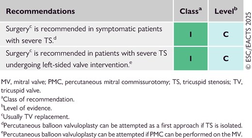
Although data are very limited, TV balloon valvuloplasty can be an option in selected patients with TS (and no relevant TR), as well as in those with concomitant mitral and tricuspid rheumatic pathology.757 However, in contrast to rheumatic MS, tricuspid rheumatic disease more frequently presents as combined stenosis and regurgitation, which limits the applicability of balloon valvuloplasty.

13. Multiple and mixed valvular heart disease

13.1. Prevalence and undertreatment

Patients frequently present with disease of more than one native heart valve [multiple VHD (MVHD)], or coexisting stenosis and regurgitation of the same valve (mixed VHD).758 While the main cause of MVHD or mixed VHD has shifted to degeneration in high-income countries, the leading aetiology in low- and middle-income countries remains RHD.758,759 Regurgitation of atrioventricular valves secondary to cardiomyopathy or long-standing primary valve disease, and late effects of radiation therapy, are further causes of MVHD.758,760 Challenges in diagnostic evaluation coupled with limited data to guide clinical decisions contribute to late referral and undertreatment of patients with MVHD.758

13.2. Evaluation and diagnostic pitfalls

In view of the complex haemodynamic interplay of multiple and mixed valve lesions, assessment by a Heart Team at a Heart Valve Centre and use of an integrative multimodality approach is key to gain diagnostic certainty, detect cardiac damage, and evaluate therapeutic options.761,762 In the light of paucity of data on diagnostic and prognostic parameters in patients with MVHD, assessment largely focuses on pathophysiological considerations and evidence derived from isolated valve lesions.

Echocardiography is the main tool to diagnose MVHD, assess mechanism, severity, and associated cardiac damage, and monitor disease progression.761 Haemodynamic interdependence between multiple valve defects alters the loading and flow conditions, thereby limiting the diagnostic validity of measures established to grade single valve defects (Table 9). In the presence of MVHD, low-flow states are frequent. The continuity equation becomes erroneous if transvalvular flows are unequal, and pressure half-time (PHT)-derived methods are inaccurate if the ventricular compliance or filling is altered.761 In this context, TOE can provide important detailed anatomical and mechanistic flow-independent information.46,761

Table 9

Echocardiographic pitfalls, robust measures, and complementary multimodality imaging parameters in multiple or mixed valvular heart disease
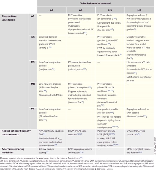
If symptoms or echocardiographic findings are equivocal, multimodality diagnostics should be considered on an individual basis to assess the cumulative repercussions of MVHD.

Measures obtained during cardiopulmonary exercise testing reflect the effect of MVHD on functional capacity.52,105,654,761,779 Levels of natriuretic peptides such as NT-proBNP correlate with functional and echocardiographic parameters, and provide incremental prognostic value in patients with mixed aortic disease and MVHD.98,780 AV calcium scoring confirms the diagnosis of true severe AS under low-flow conditions, as described in Section 8.2.777 Cardiac magnetic resonance imaging enables the independent assessment of valvular regurgitation using volumetric methods or direct flow quantification.45,46,522,762,781 Importantly, invasive cardiac output-derived measures based on thermodilution or the Fick equation using estimated oxygen uptake are inaccurate in low-flow conditions or severe TR, commonly present in MVHD.782

13.3. Indications for intervention

Given the heterogeneity of clinical scenarios and the lack of evidence on optimal treatment pathways, it is recommended that patients with MVHD are evaluated for intervention by a collaborative Heart Team at a Heart Valve Centre with experience in multimodality imaging and treatment of complex VHD.16,764

Patients presenting with a lesion fulfilling criteria for an intervention based on recommendations for single VHD should be treated accordingly. In the remaining patients, assessment of symptoms and functional status, as well as cardiac damage (which may be masked by the consequences of concomitant lesions and occur before symptoms manifest), is required. The risk–benefit ratio of intervention needs to take into account diagnostic (un)certainty, the mechanisms and severity of MVHD, and patient-specific factors, as well as procedural options and risks to determine the mode, timing, and sequence of valve treatment.

13.3.1. Multiple valvular heart disease

Multiple VHD with primary (as opposed to secondary) valvular disease usually requires surgical treatment of all relevant valvular lesions. Simultaneous treatment of concomitant severe valve defects is recommended and treatment of concomitant moderate AS, or moderate TR, should be considered (Recommendation Table 11).

Recommendation Table 11

Recommendations on indications for surgery of concomitant left-sided valvular heart diseasea
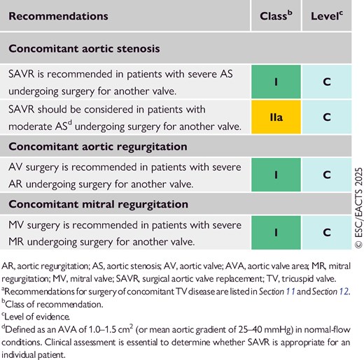
Transcatheter treatment options are established for severe AS (TAVI), rheumatic MS (PMC), and primary MR and TR (TEER or transcatheter replacement) in patients with MVHD at high surgical risk, if anatomy is suitable.401,783 In the context of a transcatheter strategy, a staged approach—typically beginning with the downstream lesion (aortic, followed by mitral and tricuspid)—is generally preferred to avoid potential haemodynamic deterioration.784,785

MVHD with severe secondary regurgitation of both atrioventricular valves is usually the consequence of either HFrEF with ventricular SMR and secondary TR, or atrial dilatation leading to both atrial MR and TR (often TR > MR). While surgery aims to address all relevant valve lesions in a single procedure, a stepwise transcatheter strategy offers the possibility of reassessing the upstream valve(s) under altered loading conditions, usually 3 months after the initial intervention. In very selected cases simultaneous transcatheter valve treatment may be considered.788,789 However, evidence is limited and primarily derived from modestly sized observational studies.786–789

13.3.2. Mixed aortic valve disease

The severity of mixed AV disease is often underestimated and patients with balanced moderate AR and AS show adverse event rates comparable to patients with severe isolated AS.790,791

Transvalvular gradients measured by Doppler reflect the overall haemodynamic burden of both regurgitation and stenosis, and are strongly associated with adverse outcomes.765,791–793 Therefore, the presence of high transvalvular gradients justifies valve intervention in patients with moderate mixed AV disease, even if regurgitation is graded as moderate and the calculated or planimetric AVA is >1 cm2 (Recommendation Table 12). Patients presenting with mixed AV disease, but with gradients below thresholds for intervention, should undergo careful multimodality diagnostics including assessment of cardiac damage to inform individual treatment strategies. Global longitudinal strain and natriuretic peptides have shown incremental prognostic value beyond symptom status and single lesion severity in patients with preserved LVEF.762,794–796

Recommendation Table 12

Recommendations on indications for intervention in patients with mixed moderate aortic stenosis and moderate aortic regurgitation (see also Supplementary data online, Evidence Table 24)
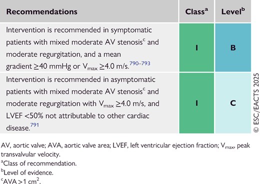
13.3.3. Mixed mitral valve disease

Mixed MV disease is usually present in patients with rheumatic valve disease or MAC. If MVA is ≤1.5 cm2, recommendations for isolated MS apply. However, patients with an MVA of >1.5 cm2 and moderate MR may be evaluated for valve replacement based on symptoms, anatomical characteristics, transmitral gradient, and signs of cardiac damage such as LA dilatation, AF, or PH.529,674,776,797

13.4. Follow-up

Due to the cumulative haemodynamic impact of MVHD or mixed VHD, progression of its severity and the development of cardiac damage may be faster than in single VHD.761 Therefore, follow-up intervals should be adjusted according to individual patient characteristics.

14. Management of patients with prosthetic valves or valve repair

14.1. Choice of prosthetic valve

When choosing between an MHV and a BHV prosthesis for an individual patient, age, life expectancy, lifestyle, bleeding and thromboembolic risks, possibility of pregnancy, and patient preference should be considered. Life expectancy is estimated according to age, sex, comorbidities, ethnicity, and geographical area.798 An MHV is preferred in younger patients with longer life expectancy and in those with a pre-existing indication for long-term OAC (Recommendation Table 13). Generally, a BHV is implanted in patients with shorter life expectancy, increased bleeding risk due to frailty or comorbidities, in women contemplating pregnancy, and in patients in whom stable INRs with adequate times in therapeutic range are unlikely.799 Importantly, the performance of different BHV prostheses can vary considerably.800 The impact of the aetiology of the native valve disease, if any, on the choice between an MHV and a BHV remains unexplored.

Recommendation Table 13

Recommendations for prosthetic valve selection
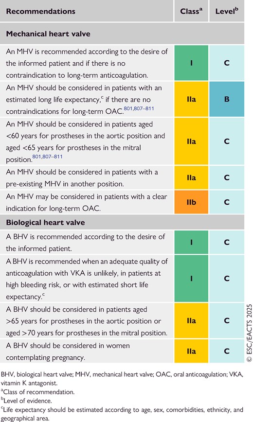
Several large observational studies, smaller RCTs, and meta-analyses have compared long-term mortality with BHV and MHV prostheses in patients aged 50–70 years.376,801–805 Some of the studies showed lower mortality with an MHV in AV patients <60 years and MV patients <65 years old,801,804 while others failed to show any differences.376,802,803 Most of these studies were limited by their observational nature and missing information on the type of prostheses implanted. RCTs with sufficient statistical power comparing biological and mechanical prostheses are warranted.

Replacement of the AV using an autograft (Ross procedure) is an alternative to an MHV in young patients that should be performed at experienced centres by operators with dedicated expertise (see also Section 8).806 General recommendations are summarized in Recommendation Table 13.

14.2. Follow-up of patients with prosthetic valves

All patients with prosthetic valves require lifelong clinical and echocardiographic follow-up to detect deterioration of prosthetic function, associated cardiac damage, or progressive disease of another valve. Serial TTE measurements of transprosthetic gradients, calculation of the effective valve area, and evaluation of leaflet motion and morphology should be performed in patients receiving a BHV within 3 months after valve implantation, again at 1 year, and annually thereafter, or sooner if new cardiovascular symptoms occur.812 TOE is recommended in all cases of suspected prosthetic valve dysfunction or endocarditis. In the latter case, CCT and PET-CT are also recommended, if the diagnosis is unclear, and to identify primary or secondary infection foci.5

Cinefluoroscopy for MHVs and CCT provide useful additional information, if valve thrombus or pannus is suspected.812 Imaging should be repeated in the case of thrombolytic and antithrombotic treatment of MVH thrombosis, even if gradients are normalized.813

14.3. Antithrombotic therapy in patients with treated valvular heart disease

14.3.1. Mechanical heart valves

14.3.1.1. Post-operative anticoagulation and therapeutic targets

Mechanical heart valves require lifelong treatment with a VKA guided by the INR. Bridging with either therapeutic unfractionated heparin (UFH) or low-molecular-weight heparin (LMWH) and VKA should be initiated within 24 h after MHV implantation, or as soon as considered safe. Heparin can be stopped when the INR is documented for 2 consecutive days within the therapeutic range (Figure 18).814 Two meta-analyses and a prospective study have suggested slightly lower bleeding rates using UFH bridging compared with LMWH after MHV replacement or cardiac surgery.814–816 However, RCTs comparing the timing and dosage of each bridging strategy are lacking.
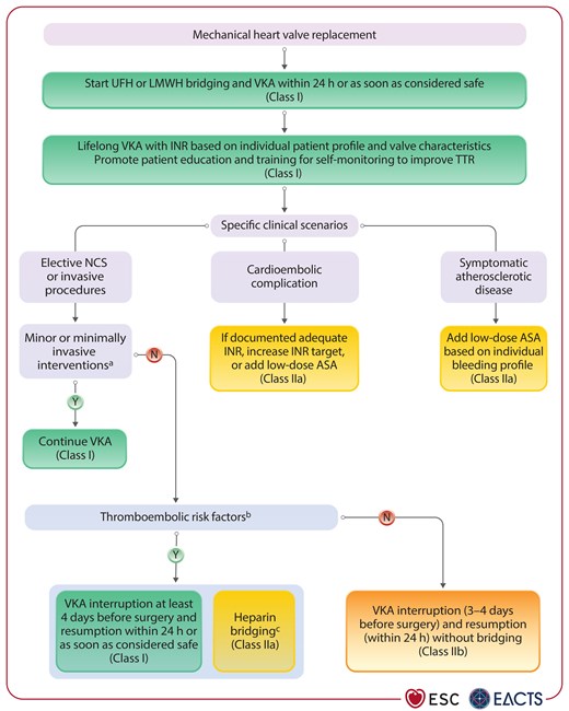
Antithrombotic therapy following mechanical heart valve implantation.
Figure 18

Antithrombotic therapy following mechanical heart valve implantation.

AF, atrial fibrillation; ASA, acetylsalicylic acid; INR, international normalized ratio; LMWH, low-molecular-weight heparin; LV, left ventricle/left ventricular; LVEF, left ventricular ejection fraction; MHV, mechanical heart valve; MS, mitral stenosis; N, no; NCS, non-cardiac surgery; TTR, time in therapeutic range; UFH: unfractionated heparin; VKA, vitamin K antagonist; Y, yes. aSkin; minor eye surgery including cataract; dental cleaning, treatment of caries, and dental extractions; pacemaker or device implantation; and diagnostic cardiac catheterization. bMHV in mitral or tricuspid position, older MHV generations in any position, inherited or acquired hypercoagulable state, LV dysfunction (LVEF <35%), AF with significant MS, recent (<12 months) major thrombotic event (i.e. cardioembolic stroke, deep vein thrombosis, pulmonary embolism). cBridging needs to be started as soon as INR reaches a sub-therapeutic value and on the first postoperative day or as soon as considered safe.

For all patients with an MHV, lifelong VKA is recommended to avoid major thrombotic complications, since cardioembolic or valve thrombosis rates without anticoagulation are substantial (12% per year and 22% per year for first-generation aortic and mitral MHVs, respectively).817 The INR target and range should be chosen considering the type, position, and number of valves, the patient's thrombotic risk, and comorbidities (see Table 10 and Recommendation Table 14). In patients with an MHV developing a major thromboembolic complication despite adequate INR and time in therapeutic range (TTR, usually defined as >60%),820 either increased VKA intensity (e.g. INR target and range increased by 0.5 units) or the addition of low-dose acetylsalicylic acid (ASA) (75–100 mg/day) should be considered.818 Direct oral anticoagulants or dual antiplatelet therapy (DAPT) are contraindicated to prevent thromboembolism in patients with an MHV.821–824

Table 10

International normalized ratio targets and therapeutic ranges for patients with a mechanical heart valve

MHV type and positionAdditional pro-thrombotic factorsaINR target and (range)
First-line treatment with VKA only
Ball-in cage, tilting disc valve in any position, all MHV in mitral/tricuspid positionNo3 (2.5–3.5)
Yes3.5 (3–4)b
Bileaflet, current-generation single-tilting aortic MHVNo2.5 (2–3)c
Yes3 (2.5–3.5)

AF, atrial fibrillation; ASA, acetylsalicylic acid; INR, international normalized ratio; LV, left ventricle/left ventricular; LVEF, left ventricular ejection fraction; MHV, mechanical heart valve; MS, mitral stenosis; VKA, vitamin K antagonist.

aInherited or acquired hypercoagulable state, LV dysfunction (LVEF <35%), AF with significant MS, recent (<12 months) major thrombotic event (i.e. cardioembolic stroke, deep vein thrombosis, pulmonary embolism).

bIn patients at very high thrombotic risk, low-dose ASA may be added instead.818

cIn patients at high bleeding risk, INR target could be maintained at a lower interval: 2 (1.5–2.5).819

© ESC/EACTS 2025
Recommendation Table 14

Recommendations for the management of antithrombotic therapy in patients with a mechanical heart valve
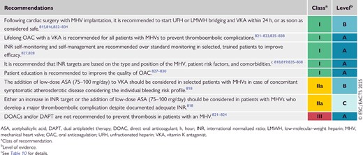
Given the complexity of lifelong VKA therapy due to high intra- and interpatient variability, the need for monitoring, drug and food interactions,825,826 the narrow therapeutic window, influence of comorbidities, and non-modifiable characteristics (e.g. ageing, genetics, and ethnicity), RCTs have shown that patient's education, as well as disease and treatment awareness, significantly improves anticoagulation quality and adherence.827–830 International normalized ratio self-monitoring and/or self-management increase efficacy, but not safety, as compared with a standard approach; INR self-monitoring can be used by motivated patients after adequate training.827–830

The indication for VKA and DAPT combination in MHV patients presenting with acute coronary syndrome is described in the corresponding ESC Guidelines.151,831 In patients with an MHV and an indication for single antiplatelet therapy (SAPT) due to symptomatic major atherosclerotic diseases and low bleeding risk, low-dose ASA in combination with a VKA should be considered, because this strategy has been shown to significantly lower the incidence of major adverse cardiovascular events.818 However, the combination of antiplatelet agents (single or dual) and VKA increases the risk of bleeding,825 and therefore their use should be carefully weighted. In a large meta-analysis that included MHV patients, the combination of VKA with either ASA or clopidogrel increased clinically relevant bleeding compared with VKA alone (clopidogrel odds ratio, 3.55; 95% CI, 2.78–4.54; ASA odds ratio, 1.50; 95% CI, 1.29–1.74), which may be aggravated because of potential pharmacokinetic drug interactions on the cytochrome P (CYP)450s 2C19 and 3A4.825

14.3.1.2. Prevention and management of bleeding

Since patients with MHVs receive lifelong VKA, strategies to prevent bleeding need to be implemented. Proton pump inhibitors reduce the risk of upper gastrointestinal bleeding by approximately 40% in patients taking VKAs825,839,840 and should therefore be prescribed to patients with MHVs, particularly in those with additional bleeding risk factors (e.g. elderly, antiplatelet agent co-administration, chronic use of non-steroidal anti-inflammatory drugs, high INR, or alcohol abuse). Randomized controlled trials and a meta-analysis did not demonstrate a benefit of oral vitamin K1 supplementation in addition to temporary VKA cessation in non-bleeding patients with a supratherapeutic INR (4.5–10.0), with a trend toward reduced safety, which may be relevant for MHV patients.841,842 For non-bleeding patients with an INR of >10.0, oral vitamin K1 (2.0–3.0 mg) should be administered, avoiding overcorrection.841,843

In cases of uncontrolled life-threatening or other major bleeding,844 VKA use must be interrupted and reversal with non-activated four-factor prothrombin complex concentrates is preferred over fresh frozen plasma, because of higher safety and effectiveness.845–848 A reduced starting dose (12.5 rather than 25 IU/kg) in patients with more thrombogenic MHVs may be considered.849 In addition, vitamin K1 should be administered to reverse the effect of VKA. Intravenous route corrects INR ∼4 h faster than oral administration,850 with no differences at 24 h and an unknown clinical impact. VKA should be restarted as soon as major bleeding is controlled.

14.3.1.3. Management of anticoagulation therapy before and after non-cardiac invasive procedures

In patients with an MHV, VKA treatment should not be interrupted for: minor or minimally invasive procedures on the skin or eyes (including cataract with topical anaesthesia); dental cleaning, treatment of caries, and dental extractions; pacemaker implantation; cardiac catheterization; and endoscopic procedures (Recommendation Table 15; Table 11).851–856 Topical antifibrinolytic or haemostatic agents may improve local haemostasis.853

Table 11

Peri-operative management of antithrombotic treatment in patients with a mechanical heart valve undergoing non-cardiac surgery based on type of procedure and underlying risk
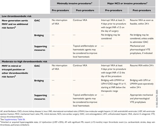
Recommendation Table 15

Recommendations for the management of antithrombotic therapy in patients with a mechanical heart valve undergoing elective non-cardiac surgery or invasive procedures
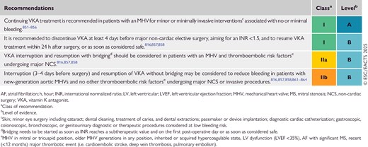
In patients with an MHV and high thromboembolic risk (Recommendation Table 16; Table 11) undergoing non-cardiac, elective major invasive procedures with high risk of bleeding (see Supplementary data online, Table S6), VKA treatment must be interrupted at least 4 days before the procedure, bridging with LMWH should be started as soon as the INR reaches a subtherapeutic value, and the INR should be <1.5 on the day of surgery.816,857,858 Measuring anti-Xa activity at peak and trough may be appropriate to manage LMWH dosing in selected patients, such as those with severe obesity or underweight.859 In cases of urgent, major invasive procedures, four-factor prothrombin complex concentrate should be administrated to timely correct the INR for the intervention, if needed. For the management of antithrombotic drugs in patients undergoing major cardiac surgery, please refer to recent EACTS Guidelines.860

For patients with an MHV and low cardioembolic risk (e.g. a new-generation MHV in the aortic position without additional risk factors) undergoing elective major NCS or invasive procedures, VKA interruption (3–4 days before surgery)816,857,858,861–864 and resumption861–863 within 24 h may be performed without bridging to reduce post-surgical bleeding without increasing the risk of thrombosis (see Figure 18).

14.3.2. Biological heart valves

The management of antithrombotic treatment after BHV implantation or valve repair is summarized in Recommendation Table 16 and Figure 19.
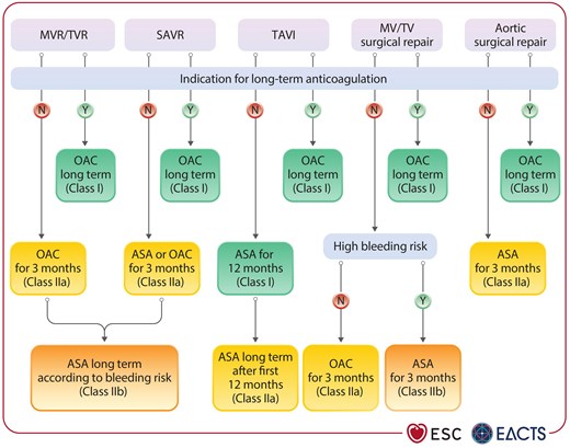
Antithrombotic therapy following biological heart valve implantation or surgical valve repair.
Figure 19

Antithrombotic therapy following biological heart valve implantation or surgical valve repair.

ASA, acetylsalicylic acid; MV, mitral valve; MVR, mitral valve replacement; OAC, oral anticoagulation; SAVR, surgical aortic valve replacement; TAVI, transcatheter aortic valve implantation; TV, tricuspid valve; TVR, tricuspid valve replacement.

Recommendation Table 16

Recommendations for the management of antithrombotic therapy in patients with a biological heart valve or valve repair
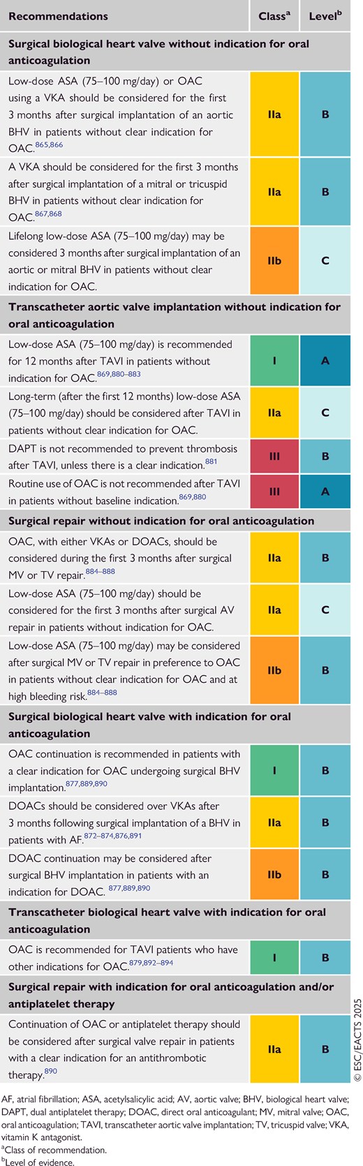
14.3.2.1. Patients with a surgical biological heart valve and no indication for oral anticoagulation

The optimal antithrombotic strategy early after surgical implantation of an aortic BHV remains controversial due to the lack of high-quality evidence. Multiple observational studies support the short-term use of VKAs to reduce the risk of thromboembolism,865,866 while data on DOACs are missing. A small RCT and a meta-analysis found that VKA treatment for 3 months significantly increased major bleeding compared with low-dose ASA, without reducing mortality or thromboembolic events, but statistical power was low.867,868 Therefore, both strategies (OAC or ASA) are reasonable within 3 months of surgical aortic BHV implantation. In the absence of randomized evidence, patients undergoing mitral or tricuspid BHV implantation should receive OAC for at least 3 months due to the increased risk of AF and thromboembolisms.

14.3.2.2. Patients with a transcatheter heart valve and no indication for oral anticoagulation

Based on evidence from RCTs, lifelong low-dose ASA is the recommended antithrombotic treatment after TAVI in patients without OAC indication. In the Antiplatelet Therapy for Patients Undergoing Transcatheter Aortic Valve Implantation (POPular TAVI) trial (cohort A), the incidence of bleeding and the composite of bleeding or thromboembolic events were both reduced with ASA compared with DAPT at 1 year.869 One RCT [Global multicenter, open-label, randomized, event-driven, active-controlled study comparing a rivAroxaban-based antithrombotic strategy to an antipLatelet-based strategy after transcatheter aortIc vaLve rEplacement (TAVR) to Optimize clinical outcomes, GALILEO], which investigated a systematic antithrombotic strategy of rivaroxaban (10 mg/day) in combination with low-dose ASA compared with DAPT with clopidogrel for the first 3 months, was halted prematurely due to an increased risk of death or thromboembolic complications, and an increased risk of bleeding in the rivaroxaban/ASA group.870 The systematic use of DOACs after TAVI in patients without indication for OAC is therefore not recommended. Data on antithrombotic management after implantation of transcatheter mitral BHVs or tricuspid BHVs are limited. Vitamin K antagonist treatment for ≥3 months is commonly prescribed, while DOACs may represent an alternative allowing for earlier discharge and a lower risk of short-term bleeding complications (median follow-up, 4.7 months).871

For patients without baseline OAC undergoing mitral or tricuspid transcatheter valve implantation, OAC (either with DOAC or VKA) is initiated and usually pursued for at least 6 months or indefinitely (in particular after valve implantation in the tricuspid position), while patients undergoing TEER usually receive single long-term antiplatelet therapy with ASA.

14.3.2.3. Patients with a surgical biological heart valve and an indication for oral anticoagulation

Lifelong OAC is recommended for patients with surgical BHVs with other indications for OAC. The evidence supports the use of DOACs in preference to VKAs in patients with AF, even during the early post-operative period.872–877 A previously existing therapy with a DOAC may be continued after BHV implantation and should be restarted, as soon as considered surgically safe, usually within 2–3 days of surgery.860

14.3.2.4. Patients with a transcatheter biological heart valve and an indication for oral anticoagulation

In the POPular TAVI trial (cohort B), the incidence of bleeding over a period of 1 month or 1 year was lower with OAC than with OAC plus clopidogrel.878 Oral anticoagulation alone was non-inferior to OAC plus clopidogrel with respect to ischaemic events, but the non-inferiority margin was large. In Edoxaban vs Standard of Care and Their Effects on Clinical Outcomes in Patients Having Undergone Transcatheter Aortic Valve Implantation–Atrial Fibrillation (ENVISAGE-TAVI-AF),879 edoxaban was non-inferior to VKA regarding the composite primary endpoint (death from any cause, myocardial infarction, ischaemic stroke, systemic thromboembolic event, valve thrombosis, or major bleeding), but the incidence of major bleeding was higher with edoxaban than with VKA. The Anti-Thrombotic Strategy to Lower All Cardiovascular and Neurologic Ischemic and Hemorrhagic Events after Trans-Aortic Valve Implantation for Aortic Stenosis (ATLANTIS) trial880 showed that apixaban was not superior to VKA after TAVI when there was a pre-existing indication for OAC, and there was a signal of higher non-cardiovascular mortality with apixaban. Therefore, no definitive recommendation can be made concerning the use of VKAs vs DOACs in patients who have undergone TAVI with a pre-existing indication for OAC.

Antithrombotic therapy after transcatheter MV or TV implantation remains empirical because data are limited. A high proportion of patients are already under OAC because of AF (almost 50% of the population with MR and 80%–90% of the candidates for TR treatment). Common practice is to continue the pre-existing anticoagulation regimen. Vitamin K antagonists and DOACs have been used in this setting with or without combination with ASA.871 However, the high bleeding risk of this usually elderly population needs to be taken into consideration.

14.4. Management of prosthetic valve dysfunction and complications

Prosthetic valve dysfunction can occur due to intrinsic permanent changes to the prosthetic valve (defined as SVD) or non-structural valve dysfunction resulting from any abnormality not intrinsic to the prosthetic valve itself. Valve thrombosis and endocarditis are considered separate entities due to their specific presentation and management, but may both result in SVD.

14.4.1. Structural valve deterioration

Structural valve deterioration has been defined by several consensus documents812,895,896 and includes wear and tear, leaflet disruption, leaflet fibrosis or calcification, and stent or strut fracture or deformation. BHV (transcatheter or surgical) SVD is more frequent than MHV SVD. The incidence of SVD may be underestimated by the simple analysis of patients with valve-related deaths or those undergoing reintervention. To ensure timely diagnosis, serial measurement should be performed and compared with the TTE performed at discharge, or within 1–3 months after valve implantation.

The criteria for haemodynamic deterioration associated with aortic and mitral SVD can be found in Table 12.895,896 The diagnosis of moderate or severe haemodynamic deterioration should prompt referral to an experienced Heart Valve Centre for evaluation and treatment, and to exclude all causes of non-structural valve dysfunction, particularly PVL or PPM, as well as thrombosis and endocarditis. This step requires the use of advanced imaging techniques (TOE, CCT, and/or PET-CT) to document SVD-related morphological changes and elucidate its mechanism. Structural valve deterioration associated with corresponding clinical criteria (e.g. new onset or worsening of symptoms, LV or RV dilation/dysfunction, or PH) indicates BHV failure with potential need for reintervention. Decisions about the treatment modality (redo surgery or transcatheter valve-in-valve implantation) should be made within the interdisciplinary Heart Team, depending on reoperation risk and anatomical considerations,445,897 including the risk of coronary obstruction,144,442,446,461 as well as prosthesis type and size.898–900 When considering a valve-in-valve procedure for a degenerated aortic BHV, the possibility of creating a PPM in small valves should be anticipated and may impact intervention or valve selection.448

Table 12

Criteria for the diagnosis of moderate or severe aortic and mitral haemodynamic valve deterioration

ModerateSevere
Aortic BHV  
SVD or non-structural valve dysfunction (except PVL or PPM),a thrombosis, or endocarditis
Increase in mean transvalvular gradient ≥10 mmHg resulting in mean gradient ≥20 mmHgIncrease in mean transvalvular gradient ≥20 mmHg resulting in mean gradient ≥30 mmHg
ANDAND
Decrease in EOA ≥0.3 cm2 or ≥25%, and/or decrease in DVI ≥0.1 or ≥20%, compared with echocardiographic assessment performed 1–3 months post-procedureDecrease in EOA ≥0.6 cm2 or ≥50%, and/or decrease in DVI ≥0.2 or ≥40%, compared with echocardiographic assessment performed 1–3 months post-procedure
OROR
New occurrence or increase of ≥1 grade of intraprosthetic AR resulting in ≥ moderate ARNew occurrence or increase of ≥2 grades of intraprosthetic AR resulting in ≥ moderate-to-severe AR
Mitral BHV  
SVD or non-structural valve dysfunction (except PVL or PPM),b thrombosis, or endocarditis
Increase in DVI ≥0.4 or ≥20%, resulting in DVI ≥2.2, or decrease in EOA ≥0.5 cm2 or ≥25%, resulting in EOA <1.5 cm2, usually associated with increase of transmitral gradient ≥5 mmHgIncrease in DVI ≥0.8 or ≥40%, resulting in DVI ≥2.7, or decrease in EOA ≥1.0 cm2 or ≥50%, resulting in EOA <1 cm2, usually associated with increase of transmitral gradient ≥10 mmHg
OROR
New occurrence or increase of ≥1 grade of intraprosthetic MR resulting in ≥moderate MRNew occurrence or increase of ≥2 grades of intraprosthetic MR resulting in ≥moderate-to-severe MR

Adapted from 895,896.

AR, aortic regurgitation; AV, aortic valve; BHV, biological heart valve; DVI, Doppler velocity index; EOA, effective orifice area; MR, mitral regurgitation; PPM, prosthesis–patient mismatch; PVL, paravalvular leak; SVD, structural valve deterioration.

aObstruction by pannus; dilatation of the aortic root after stentless BHV; or AV-sparing operations.

bLeaflet entrapment by pannus, chordae, or suture.

© ESC/EACTS 2025

Given the larger sizes of BHVs in mitral or tricuspid positions, transfemoral/transseptal valve-in-valve implantation represents an attractive alternative to redo open surgery.570,900–903 In the case of mitral valve-in-valve implantation the risk of LVOT obstruction, although infrequent, should be carefully ruled out, especially in patients with small and hypertrophic ventricles.904

14.4.2. Non-structural valve dysfunction

14.4.2.1. Prosthesis–patient mismatch

Prosthesis–patient mismatch should be prevented whenever possible after either transcatheter or surgical valve-replacement procedures (see Section 8.5.1.2). When occurring in the aortic position, severe PPM is associated with decreased quality of life, increased rate of rehospitalization and reintervention, and possible reduction in long-term survival, although findings are not consistent throughout all studies.454,905–909 Moderate PPM is more common, but seems to have a limited impact on outcomes. The projected indexed EOA may be predicted prior to valve implantation to avoid severe PPM,908 although this concept has been challenged.910 Less is known about the prevalence and consequences of PPM in the mitral and tricuspid positions, and established definitions are lacking.

Prosthesis–patient mismatch is an infrequent indication for reintervention. However, reoperation should be considered in symptomatic patients with severe PPM, particularly if the patient is low risk.394,437,438

14.4.2.2. Paravalvular leak and haemolysis

The diagnosis of PVL requires systematic TOE, because TTE may be inconclusive. Haemolytic anaemia can often be detected in patients with prosthetic valves and is best assessed by measuring lactate dehydrogenase and haptoglobin serum levels, but rarely leads to symptoms. Intervention is needed if a PVL causes haemolysis requiring blood transfusions or symptoms, or if secondary to valve endocarditis. Transcatheter PVL closure is a valid alternative to surgery in the case of significant regurgitation or haemolysis, if feasible depending on the size and location of the leak, but requires specific expertise and planning. Care must be taken not to interfere with mechanical leaflet motion in MHV patients. Reported results for transcatheter PVL closure are inconsistent, with several patients returning with recurrent PVL and/or haemolysis.911 When surgery or transcatheter intervention are contraindicated, medical therapy aims to counteract the effect of haemolysis (iron supplementation and erythropoietin) or reduce it (beta-blockers).912

14.4.3. Endocarditis

Antibiotic prophylaxis is recommended in all patients with a prosthetic valve (including transcatheter valve prostheses) and after valve repair using prosthetic material or with previous episode(s) of infective endocarditis, and should also be considered after transcatheter MV or TV repair.5 It is recommended in such patients when undergoing dental extractions, oral surgery, or other procedures requiring manipulation of the gingival or peri-apical region of the teeth. Particular attention to dental and cutaneous hygiene, and strict aseptic measures during any invasive procedure, are also advised in this population. Additional details on endocarditis prophylaxis are mentioned in the 2023 ESC Guidelines for the management of endocarditis.5

14.4.4. Valve thrombosis

Valve thrombosis occurs mainly in MHVs,913,914 but can also be observed in BHVs.915,916 The spectrum of BHV thrombosis ranges from incidental CCT findings, such as hypo-attenuated leaflet thickening (HALT) with or without reduced leaflet motion but normal gradients, to clinically apparent presentations with elevated gradients, symptoms of valve obstruction, or thromboembolic events.917–919

14.4.4.1. Hypo-attenuated leaflet thickening

Hypo-attenuated leaflet thickening is detected by CCT in 10%–30% of aortic BHVs depending on antithrombotic management, the definition of HALT, the timepoint of assessment, and the type of valve.917,919–921 The significance and clinical implications of these findings with respect to thromboembolic risk and valve durability remain uncertain. Accordingly, routine use of CCT to detect HALT is not indicated.918–920 In patients with relevant increases in gradients, in whom HALT and restricted leaflet motion is confirmed by CCT, elective use of DOACs or VKAs should be considered to resolve leaflet thrombosis.918,921,922

14.4.4.2. Clinically significant valve thrombosis

Obstructive valve thrombosis should be suspected in any patient with any type of prosthetic valve who presents with new-onset dyspnoea or HF symptoms, an embolic event, or an unexpected increase in transvalvular gradients. If TTE findings are uncertain, the diagnosis should be confirmed by TOE and/or CCT to distinguish between thrombus, pannus, and degeneration.923–925 Cinefluoroscopy can detect impaired MHV leaflet motion and reduced opening angles.

Adequate anticoagulation must be promptly restored in all patients with MHV thrombosis and subtherapeutic INR. Although surgery remains the first-line option in critically ill patients, emergency valve replacement is associated with increased risk, whereas bleeding and systemic embolism are increased with fibrinolysis.926–928 Slow, low-dose infusion appears to lower complication rates, while preserving thrombolytic success rates.928–931a It is recommended that the decision between surgery and fibrinolysis is taken within the Heart Team, and individualized by weighing clinical factors and local expertise (Figure 20).
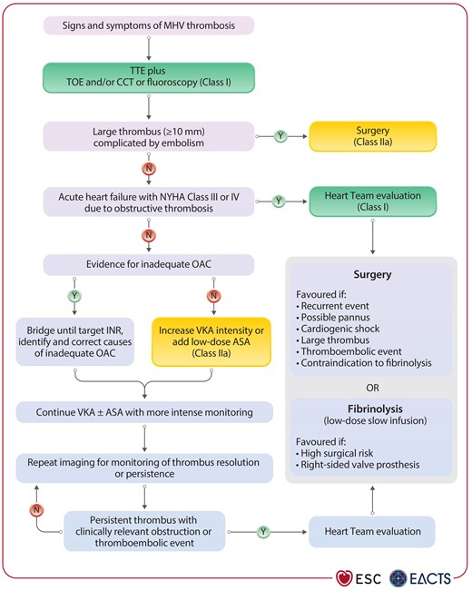
Management of left-sided obstructive and non-obstructive mechanical heart valve thrombosis.
Figure 20

Management of left-sided obstructive and non-obstructive mechanical heart valve thrombosis.

ASA, acetylsalicylic acid; CCT, cardiac computed tomography; INR, international normalized ratio; MHV, mechanical heart valve; NYHA, New York Heart Association; OAC, oral anticoagulation; TOE, transoesophageal echocardiography; TTE, transthoracic echocardiography; VKA, vitamin K antagonist.

Management of non-obstructive thrombosis or obstructive thrombosis of an MHV without pronounced HF symptoms depends mainly on the occurrence of a thromboembolic event and the size of the thrombus. Surgery should be considered for a large (>10 mm) non-obstructive prosthetic valve thrombus that is complicated by embolism or persists despite optimal OAC.913,932,933

Anticoagulation using a VKA is the first-line treatment for clinically relevant BHV thrombosis, unless urgent reintervention or fibrinolysis is required due to progressive acute HF or haemodynamic instability.934–938 Because clinically relevant BHV thrombosis is associated with recurrence and may contribute to prosthetic degeneration, indefinite anticoagulation may be considered after a confirmed episode, but this strategy must be balanced against the risk of bleeding.936,939

Recommendation Table 17

Recommendations for the management of prosthetic valve dysfunction (see also Supplementary data online, Evidence Table 25)
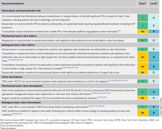
15. Management during non-cardiac surgery

In patients with significant VHD who undergo NCS, the risk of peri-operative cardiovascular complications is increased and related to both the timing of the procedure (i.e. urgent vs non-urgent) and type of surgery (low, intermediate, or high risk), as well as patient-specific factors (type and severity of VHD, LV function, etc.).194,957–959 Detailed recommendations related to NCS are available in the 2022 ESC Guidelines on cardiovascular assessment and management of patients undergoing non-cardiac surgery.960

15.1. Pre-operative evaluation

Echocardiography should be performed in all patients with VHD requiring NCS. Patient- and surgery-specific factors, along with risk calculators, can be used to guide the treatment strategy. Determination of functional capacity is a pivotal step for pre-operative risk assessment, measured either by the ability to perform activities in daily life or by exercise testing. Screening for frailty using validated tools is advisable.90 Decisions on pre- and peri-operative management, surveillance, and continuation of chronic cardiovascular medical treatment should be taken after multidisciplinary discussion involving cardiologists, surgeons, and cardiac anaesthesiologists, as well as the team who will be in charge of the NCS. Patients receiving OAC treatment should be managed as described in Section 14.

15.2. Specific valve lesions

15.2.1. Aortic stenosis

In patients with severe symptomatic AS, the treatment depends on the urgency and risk of NCS. If life-saving time-sensitive NCS is needed, it should be performed under careful haemodynamic monitoring avoiding rapid changes of volume status, with prompt treatment of arrhythmia regardless of AS severity. In cases of urgent high-risk NCS, TAVI or balloon aortic valvuloplasty should be considered in patients with critical AS prior to surgery, considering the risk of developing severe acute AR after balloon aortic valvuloplasty.194,961 Urgent low- and intermediate-risk NCS can be performed relatively safely in patients with severe AS.962

Patients in whom NCS can be deferred (i.e. non-urgent NCS) should undergo pre-operative Heart Team evaluation to determine whether SAVR or TAVI is preferable. TAVI may be preferred to SAVR if faster recovery plays a role, particularly in elderly patients in whom complex or high-risk NCS is planned.963 The treatment of asymptomatic patients with severe AS should be individualized (Figure 21).964
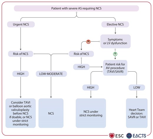
Management of non-cardiac surgery in patients with severe aortic stenosis.
Figure 21

Management of non-cardiac surgery in patients with severe aortic stenosis.

AS, aortic stenosis; AV, aortic valve; LV, left ventricular; NCS, non-cardiac surgery; SAVR, surgical aortic valve replacement; TAVI, transcatheter aortic valve implantation.

15.2.2. Mitral stenosis

Heart rate and fluid balance should be controlled to prevent pulmonary oedema during NCS and arterial vasodilators should be avoided. Non-cardiac surgery is safe in patients with an MVA of >1.5 cm2, and in asymptomatic patients with MVA ≤1.5 cm2 and SPAP of <50 mmHg. Symptomatic patients or those with SPAP of >50 mmHg should undergo PMC or other appropriate valve intervention before high-risk NCS, if possible.965,966 Asymptomatic patients with MVA ≤1.5 cm2 can undergo low-to-moderate-risk NCS under careful monitoring, especially if PMC is unsuitable. Multidisciplinary management is advised for patients with significant MS who are ineligible for valve intervention.

15.2.3. Aortic and mitral regurgitation

Non-cardiac surgery can usually be performed safely in asymptomatic patients with severe MR or AR and preserved LV function. If NCS is urgent, patients should undergo surgery under strict haemodynamic monitoring, regardless of symptom status. In cases of elective (non-urgent) NCS in patients with severe ventricular SMR, medical therapy should be optimized. If symptoms persist and NCS is intermediate or high risk, TEER should be considered after Heart Team discussion based on clinical and anatomical selection criteria.194,584 Valve treatment should be performed for patients with AR meeting the criteria for valve intervention before any elective intermediate- or high-risk NCS.966,967

15.3. Peri-operative monitoring

Heart rate control (particularly in MS) and careful fluid management (particularly in AS and MR with reduced LVEF) are needed throughout the procedure. The involvement of specialized cardiovascular anaesthesiologists should be considered in complex situations because TOE monitoring may be considered. Pulmonary artery catheterization is not routinely used.

16. Management of valvular heart disease during pregnancy

Specific ESC Guidelines on this topic are available (the 2025 ESC Guidelines on cardiovascular disease during pregnancy) and should be consulted for further details.968 Pregnancies in patients with VHD should be considered high risk and managed under the close supervision of a cardiologist and a multidisciplinary Pregnancy Heart Team. The importance of midwives and other specialized nursing personnel is increasingly recognized for high-quality direct patient interactions. Shared decision-making is especially important when addressing the cardiovascular risk of pregnancy and the benefit–risk ratios of therapeutic options and modes of delivery (Figure 22).
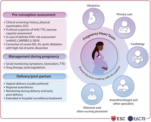
The Pregnancy Heart Team model of care.
Figure 22

The Pregnancy Heart Team model of care.

AS, aortic stenosis; ECG, electrocardiogram; MS, mitral stenosis; TTE, transthoracic echocardiography; VHD, valvular heart disease.

16.1. Management before pregnancy

Ideally, women should undergo a thorough physical examination by their general practitioner for VHD screening prior to pregnancy. A specialized evaluation including TTE should be performed by a cardiologist in the case of clinical suspicion.968,969

If VHD is diagnosed, pre-pregnancy counselling is imperative. Unplanned pregnancies in patients with VHD should be discouraged. Contraception methods should be recommended after discussion with the patient in the presence of a significant maternal and/or foetal risk. In patients contemplating pregnancy, the maternal risk should be assessed using the modified WHO classification and other scores such as the CARPREG or DEVI risk score (see Supplementary data online, Table S7).970

Before recommending pregnancy, the following conditions need to be corrected:

  • MS with MVA <1.5 cm2, even when asymptomatic.971

  • Severe AS with symptoms, or abnormal exercise test, or LV systolic dysfunction.972

  • Heritable aortic disorders and high risk of aortic dissection: prophylactic aortic repair is recommended prior to pregnancy in women with Marfan syndrome and an aortic diameter of >45 mm, and may be considered in women with an aortic diameter between 40 and 45 mm when risk factors for dissection exist.973 Aneurysmal dilation in women with BAV should be corrected when the aortic diameter is ≥50 mm.974 Close follow-up in dedicated units and beta-blocker therapy during pregnancy and post-partum are recommended unless contraindicated, although strong evidence is only available for Marfan syndrome.

Valvular regurgitant lesions are generally well tolerated during pregnancy. Prophylactic intervention is therefore not recommended in the absence of class I or IIa indications.

The first therapeutic option for MS in a woman considering pregnancy should be PMC. When implantation of a prosthetic valve is necessary, BHVs are recommended, although early SVD remains a serious concern.975 Mechanical heart valves must be avoided due to the high risk of maternal and foetal complications linked to the potential teratogenic effects of VKAs, as well as the increased risk of bleeding.976 The Ross procedure may be considered for the treatment of AV disease at centres with expertise.

16.2. Management during pregnancy

16.2.1. Patients with native valve disease

Pregnancy can worsen the clinical course of left-sided stenotic valvular lesions because increased cardiac output causes an increment of transvalvular gradient of ∼50%, mainly between the first and second trimesters. Regurgitant lesions are less likely to cause complications, except in high-risk cases (LV systolic dysfunction, PH, and cardiac events before pregnancy).977

Mild MS is generally well tolerated.978 Heart failure occurs in one-third of pregnant women with an MVA of <1.5 cm2 and in one-half of those with an MVA of <1.0 cm2, most often during the second trimester.971 Percutaneous mitral commissurotomy should be considered if symptoms or SPAP of >50 mmHg persist despite optimal medical therapy (diuretics and beta-1-selective beta-blockers), preferably after the 20th week of pregnancy.

Regarding AS, pregnancy is generally well tolerated if prior exercise tolerance was normal, even in severe AS, while HF has been reported to occur in up to 25% of symptomatic patients.972 In symptomatic patients despite medical therapy (i.e. diuretics), TAVI seems to be the preferred option in very selected patients, although evidence is lacking. Percutaneous balloon valvuloplasty may be an alternative option. Procedures should be performed in an experienced valve centre.972,979,980

Surgery under cardiopulmonary bypass is associated with a high rate of foetal loss and should be restricted to conditions that threaten the mother's life if transcatheter intervention is not possible or has failed.

Vaginal delivery is the first choice for the majority of patients. Caesarean section is preferred if there is an obstetric indication, and in the case of severe MS or AS, ascending aortic diameter of >45 mm, severe PH, or if delivery starts while the patient is being treated with OAC.

16.2.2. Patients with prosthetic valves

Pregnant patients with MHVs should be followed in a centre with corresponding expertise. In a cohort of 212 women with MHVs, valve thrombosis occurred in 10 (4.7%) pregnancies and haemorrhagic events occurred in 49 (23.1%).976 Therapeutic OAC during pregnancy is essential to avoid thrombosis. The higher efficacy of VKAs compared with LMWH to prevent thrombosis must be balanced against increased foetal risks.981

In patients requiring ≤5 mg/day of warfarin, OAC with warfarin throughout pregnancy, changing to UFH before delivery, is advocated, in order to reduce the risk of thrombosis. In patients requiring higher doses, switching to dose-adjusted LMWH at least twice per day with strict anti-Xa monitoring during the first trimester is recommended to avoid the teratogenic effect of VKAs (for corresponding flowcharts see the 2025 ESC Guidelines for the management of cardiovascular diseases during pregnancy968).

Lifetime management of women with a BHV considering pregnancy is central and transcatheter valve-in-valve implantation may be considered as a bridge to MHV implantation.982

17. Sex-specific considerations in patients with valvular heart disease

Although men and women are equally likely to experience VHD, sex-specific prevalence exists according to valve type and disease pathophysiology.644,648,983 Women more frequently suffer from MV disease such as prolapse, RHD or TR, while men more often present with AS or AR, especially associated with BAV.984 Men also suffer from endocarditis of any valve more frequently.985

Female sex is associated with higher mortality in the presence of various VHDs,986 including early post-treatment,987 and is included as a risk factor in surgical risk prediction tools such as the STS and EuroSCORE calculators. However, risk scores are often derived from populations including a majority of men.988 In addition, risk prediction scores are susceptible to referral bias that results in a well-established higher risk of undertreatment or delayed treatment of VHD in women.983,984,986

17.1. Aortic valve disease

Female patients with severe AS present more often with shortness of breath, whereas males more frequently have angina, presumably due to the higher incidence of CAD.989 The pathophysiology of AS seems to differ according to sex, with women having less calcium and more fibrosis.990 Concentric ventricular hypertrophy and remodelling are also more frequently observed in women than in men, resulting in higher LVEF but smaller LV cavity and stroke volume.991 Consequently, paradoxical low-flow, low-gradient constellations are frequent and may both contribute to the underdiagnosis of severe AS in women and delay an intervention.992 The use of sex-specific thresholds to define flow limitation (<40 mL/m2 for men and <32 mL/m2 for women) has therefore been suggested,292 and the use of CCT to quantify the calcium score should be performed in women with discordant echocardiographic parameters.777

Women are less likely to be referred to a cardiologist and undergo investigations. Surgical aortic valve replacement is performed less frequently in women, especially if echocardiographic parameters are discordant.993 In patients presenting with severe AS and Class I indications for SAVR, a significantly higher proportion of men than women are referred for evaluation by a cardiac surgeon.994,995

Male patients derive a clearer benefit from SAVR compared with women, who have higher in-hospital and long-term mortality and morbidity,987,996 possibly explained by more severe concentric hypertrophy991 and a smaller aortic annulus predisposing to PPM.997 Conversely, in a randomized trial (RHEIA) that compared TAVI with SAVR in 443 women with a mean age of 73 years, TAVI was superior in reducing the primary endpoint composed of death, stroke, or rehospitalization at 1 year, predominantly driven by a reduction in rehospitalization for valve- or procedure-related symptoms or worsening HF.998 Several observational studies suggest that elderly women have lower mortality after TAVI,999 even if the rates of major vascular complications and bleeding tend to be higher.997

Indexed cut-offs to indicate treatment of AR are validated, but only partially account for sex differences. Newer studies using echocardiographic volumes1000 and CMR1001 suggest that women may experience higher event rates at lower cut-offs compared with men, but this requires further investigation.

17.2. Mitral valve disease

Patients with RHD are often young women and have a high prevalence of major cardiovascular complications with far-reaching impacts on reproductive health and access to care.282 Although women experience favourable outcomes compared with men when treated by percutaneous balloon valvuloplasty, access to these procedures is still limited in low-income countries.662

Fibroelastic disease with MV prolapse is also more frequent in women, and may be accompanied by morphological abnormalities of the MA associated with fibrosis of the papillary muscles or the inferobasal LV that may act as the substrate for sudden cardiac death, even in the absence of severe MR.1002 Recent evidence has supported considering lower women-specific cut-offs for intervention with regard to LVESD (36 mm; indexed LVESD, 1.8 cm/m2), while the LVEF cut-off was similar to men (58%), albeit with higher mortality.548

Ventricular SMR associated with low LVEF is more frequent in men with HF,1003 while atrial SMR due to chronic AF and/or HFpEF affects more women (58% vs 42% male).579

Female sex is a risk factor for the development of MAC and was reported to account for 68% of the patients included in a large-scale transcatheter MV replacement in MAC registry.1004 Furthermore, women present with a faster disease progression.1005

17.3. Tricuspid valve disease

Tricuspid regurgitation is more prevalent in women,208 and female sex is associated with accelerated disease progression.1006–1008 Hypotheses to explain these observations include the overall higher prevalence of HFpEF and AF in women.1009 Women are usually diagnosed at an older age and the cause of TR is most frequently a consequence of left valvular disease or annular dilatation attributable to RA dilatation.1010 No differences have been detected to date in terms of adverse events after surgery1011 or transcatheter TV interventions.1012,1013

18. Key messages

Heart Team and Heart Valve Centre

  • An integrated regional Heart Valve Network incorporating outpatient Heart Valve Clinics and specialist Heart Valve Centres allows optimal patient care.

  • Heart Valve Centres should fulfil institutional and local statutory requirements, and strive for high procedural volume and excellent clinical outcomes.

  • Heart Team recommendations should be based upon these guideline recommendations, relevant updated evidence, key medical considerations, and patient preferences.

  • Core members of the Heart Team include the primary clinical cardiologist, cardiologists with subspecialty expertise in VHD, specialists in advanced cardiovascular imaging and peri-procedural imaging guidance, and surgeons and interventional cardiologists with training and expertise in valve interventions.

  • A network approach that distinguishes between higher- and lower-volume centres is appropriate, with more complex procedures focused in the most experienced (i.e. upper quartile) centres. Information on the network organization should be communicated to patients, as well as referring cardiologists and general practitioners.

Aortic regurgitation

  • Assessment of AR severity with TTE remains challenging and current cut-offs for intervention are mostly based on 2D measurements, although 3D echocardiography and CMR allow more accurate evaluation of LV volumes and LVEF.

  • Mechanisms of AR may be closely related to the aortic diameters that should be measured accurately at all levels of the aortic root (annulus, sinuses, and sinotubular junction).

  • Indication for operation is based on symptoms, LV volumes, LVEF, and aortic diameters. Although valve replacement remains the standard treatment, AV repair (or AV sparing when associated with root aneurysm) is being increasingly used to avoid prosthesis-related complications, especially in experienced centres.

  • Current transcatheter options for AR are limited and applicable only in patients who are ineligible for surgery.

Aortic stenosis

  • Diagnosis of severe AS requires integrative evaluation of pressure gradients (the most robust measurements), AVA, flow conditions, the extent of valve calcification, and LV function.

  • Selection of the most appropriate mode of intervention should take into account clinical characteristics (age and estimated life expectancy, concomitant conditions), access and valve anatomy (particularly the feasibility of transfemoral TAVI and calcification patterns), and surgical risk, as well as repeat procedure options and risks (lifetime management).

Mitral regurgitation

  • The echocardiographic diagnostic workup of patients with MR includes multiparametric assessment of MR severity, evaluation of MV anatomy (often with 3D TOE), identification of the mechanism (PMR, ventricular SMR, or atrial SMR), and evaluation of cardiac damage.

  • Surgical MV repair is the preferred method of treatment in severe PMR. Transcatheter edge-to-edge repair is recommended in patients who are inoperable or high risk according to the Heart Team.

  • Surgical MV repair is the procedure of choice for asymptomatic patients with primary MR and signs of cardiac damage, including moderate or more TR.

  • In patients with ventricular SMR, GDMT (including CRT if indicated) is the initial and essential treatment step. In symptomatic patients without CAD needing revascularization, M-TEER is recommended. In patients with concomitant complex CAD and those not suitable for TEER, mitral surgery may be considered.

  • In patients with atrial SMR, MV surgery, AF ablation if indicated, and LAAO should be considered after optimization of medical therapy. Transcatheter edge-to-edge repair may be considered in patients at high surgical risk.

Mitral stenosis

  • Most patients with severe rheumatic MS and favourable valve anatomy should undergo PMC, which is the standard of care. Surgery is recommended for symptomatic patients with contraindications or unfavourable anatomical and clinical characteristics for PMC.

  • Decision-making in patients with unfavourable anatomy should take into account local PMC experience.

  • In selected patients with clinically severe degenerative MS and MAC, transcatheter intervention or surgery may improve symptoms.

Tricuspid regurgitation

  • Concomitant TV repair is the preferred method for patients with left-sided valve pathology and associated moderate or severe TR.

  • The use of risk scores for the assessment of RV and secondary organ dysfunction should be strongly encouraged in patients with isolated severe TV disease.

  • In isolated severe TR without severe RV dysfunction, surgery should be performed at an early stage in patients at low operative risk.

  • In isolated severe TR patients at increased surgical risk, tricuspid TEER or transcatheter replacement should be considered to improve quality of life and RV remodelling, in the absence of severe RV dysfunction or pre-capillary PH.

Tricuspid stenosis

  • TS is a very rare manifestation of acquired VHD in high-income countries.

  • TS is mainly associated with rheumatic valve disease, carcinoid syndrome, or enzymatic disorders such as Fabry's or Whipple's disease.

  • Treatment of symptomatic TS mainly involves surgical TV replacement.

Multiple and mixed valvular heart disease

  • Transvalvular gradients and velocities reflect the combined burden of regurgitation and stenosis in mixed aortic and mitral disease.

  • Treatment decisions should be based on the assessment of symptom and functional status, cardiac damage, anatomical suitability, and the risk–benefit ratio of intervention and lifetime management considerations.

  • Patients with mixed moderate AS and AR have similar detrimental outcomes compared with those with severe isolated AS.

  • In transcatheter procedures, which allow a sequential approach, downstream lesions should be treated first to prevent potential haemodynamic deterioration and allow improvement of upstream lesions due to changing loading conditions and reverse remodelling.

Antithrombotic treatment in patients with a mechanical heart valve

  • International normalized ratio therapeutic range should be balanced to the type and anatomical site of MHV, as well to the thrombotic risk profile of the individual patient.

  • Patient training, self-monitoring, and education can increase INR stability and TTR.

  • Minor or minimally invasive NCS procedures do not require VKA interruption in patients with an MHV.

  • In patients with an MHV undergoing elective major NCS, bridging may be omitted if the thromboembolic risk is low.

Non-cardiac surgery

  • The risk of peri-operative cardiovascular complications related to surgery and to patient-specific factors should be evaluated and communicated to the patient and surgical team.

  • In patients with symptomatic severe AS requiring urgent high-risk NCS, BAV or TAVI should be considered prior to surgery. In patients planned for elective NCS, AV intervention is recommended prior to NCS.

Pregnancy

  • In women with VHD, decisions regarding management before and during pregnancy should be taken after discussion by the multidisciplinary Pregnancy Heart Team. Unplanned pregnancies should be discouraged.

  • The following conditions should be corrected prior to considering pregnancy:

    • –

      clinically severe MS (MVA <1.5 cm2), even when asymptomatic

    • –

      severe symptomatic AS, or asymptomatic patients with impaired LV function or a pathological exercise test

    • –

      heritable aortic disorders and high risk of aortic dissection.

  • Vaginal delivery is the first choice for the majority of patients. Indications for Caesarean section include pre-term labour in patients on OAC, severe MS or AS, aggressive aortic pathology, acute intractable HF, and severe PH.

  • Women with MHVs should be managed in expert centres.

19. Gaps in evidence

General aspects

  • Patient-reported outcome measures are infrequently reported in VHD studies. Patient-reported outcome measure-oriented studies are required to improve quality of life and patient satisfaction.

  • Methods to address underdiagnosis and undertreatment of VHD need to be identified and implemented.

Heart Team and Heart Valve Centre

  • Structured research is required to investigate the relationship between procedural volume and clinical outcomes, in order to define minimum annual thresholds for individual operators and institutions undertaking surgical and transcatheter valve interventions.

  • There is a pressing need to ensure higher dispersion and adoption of interventions for VHD, especially in middle- and low-income countries.

Conditions associated with valvular heart disease

  • CAD:

    • –

      The prognostic value of functional assessment of stable, moderate coronary stenosis in VHD patients remains to be determined.

    • –

      The optimal strategy (invasive vs non-invasive) for CAD assessment in specific VHD populations remains to be elucidated.

    • –

      The optimal timing of PCI in patients with CAD undergoing TAVI is yet to be determined.

    • –

      The benefit of complete coronary revascularization with CABG in patients with combined VHD and CAD requires further research.

  • AF:

    • –

      It is unclear which patients with chronic persistent AF and concomitant VHD are deemed to be suitable for rhythm control therapy.

    • –

      The protective effect against stroke of OAC with VKA or DOACs in patients after surgical or transcatheter LAAO remains to be determined.

  • Cardiogenic shock and acute HF:

    • –

      The optimal treatment strategy in VHD patients presenting with cardiogenic shock and acute HF is unknown.

Aortic regurgitation

  • Impact of early LV remodelling on prognosis in asymptomatic AR patients is unknown.

  • Prognostic value of CMR-derived indices in asymptomatic patients needs to be determined.

  • More data are required on long-term results of surgical AV repair for AR.

  • More evidence is required on transcatheter treatment options for AR, in particular using dedicated devices.

Aortic stenosis

  • Better understanding of the pathophysiology of AS is needed to propose innovative medical therapy.

  • Further research is required on:

    • –

      Refined prognostic markers to guide timing of intervention in asymptomatic patients.

    • –

      The role of revascularization in patients with severe AS and asymptomatic concomitant CAD.

    • –

      Further data on the long-term durability of transcatheter valves in comparison with surgical BHVs in younger patients.

    • –

      The role of TAVI in patients with BAV AS and patients <70 years of age.

    • –

      Results of intervention (valve or coronary) after TAVI or SAVR.

    • –

      Determining the optimal lifetime management strategy for AS patients.

Mitral regurgitation

  • The association between primary MR and ventricular arrhythmias requires more investigation, including the impact of intervention on ventricular arrhythmias.

  • More data are required on the role of TEER in patients with advanced HF.

  • Long-term results of TEER need to be further assessed, including the clinical relevance of transmitral gradients after treatment of both primary and secondary MR.

  • Results of ongoing trials comparing MV surgery with TEER in non-high risk primary MR patients are awaited.

  • Data on the mid- and long-term clinical impacts of transcatheter MV replacement are required.

  • More data on the clinical impacts of surgical and transcatheter treatment of atrial SMR are required.

Mitral stenosis

  • The potential role of TMVI using dedicated devices in high-risk patients is to be determined, particularly those with severe MAC.

Tricuspid regurgitation

  • The long-term risks and benefits of concomitant TV surgery in patients with less than moderate TR and annular dilatation undergoing left-sided valve surgery need to be determined.

  • Further investigations are required on the outcomes of TV intervention in asymptomatic patients with severe TR and RV dysfunction or significant dilation.

  • The importance of addressing concomitant AF in patients with TR needs to be investigated.

  • More data are required on the indications, timing, and long-term outcomes of TV repair and replacement for TV disease.

  • Better understanding is required of the respective role of surgery vs transcatheter TV therapy for TR treatment.

Tricuspid stenosis

  • The role of transcatheter TV replacement remains unexplored in patients with TS. The most efficient way to achieve ventricular pacing in patients after TV replacement needs to be investigated.

Multiple and mixed valvular heart disease

  • Further evaluation of the impact on outcomes and indication for intervention, as well as timing and modalities of intervention, is required.

Prosthetic valves

  • Further development of current prosthetic valve devices is required to address their main complications (e.g. improved tissue processing to reduce degeneration of bioprostheses or new mechanical valve designs to reduce risk of thrombosis).

  • Antithrombotic drugs in MHV patients:

    • –

      Whether UFH or LMWH should be preferred as bridging therapy after MHV implantation, as well as their timing and dosage, remains to be established.

    • –

      For patients with MHV undergoing major NCS, the optimal post-operative management and bridging of VKA needs further investigation.

    • –

      The role of pharmacogenomics for VKORC1, CYP2C9, and CYP4F2 in patients with highly variable INR, and low TTR or major vascular complications despite good adherence, should be further investigated.

    • –

      More data on the risks and benefits of slow thrombolysis for valve thrombosis are required.

Pregnancy

  • More data are required on optimal management of anticoagulation in pregnant women with MHVs. Prospective studies comparing different antithrombotic regimens are lacking.

Non-cardiac surgery

  • Clinical utility of scales for peri-operative risk evaluation needs to be determined.

Sex-specific considerations

  • The development of sex-adjusted surgical risk prediction tools is required.

  • Additional data are needed to validate sex-specific cut-offs indicating interventions.

  • Further research is needed to investigate sex-related differences in the prognosis and treatment of specific valve diseases, especially TR.

20. ‘What to do’ and ‘What not to do’ messages from the Guidelines

Class I and Class III recommendations from throughout the guideline document are summarized in Table 13.

Table 13

‘What to do’ and ‘what not to do’

21. Evidence tables

Evidence tables are available at European Heart Journal online.

22. Data availability statement

No new data were generated or analysed in support of this research.

23. Author information

Jonas Lanz, Department of Cardiology, Bern University Hospital, Bern, Switzerland; Mateo Marin-Cuartas, University Department of Cardiac Surgery, Leipzig Heart Center, Leipzig, Germany; Ana Abreu, Faculdade de Medicina da Universidade de Lisboa (FMUL), ISAMB, IMPSP, CCUL, CAML, Lisboa, Portugal; Serviço Cardiologia, Unidade de Reabilitação Cardiovascular, ULS Santa Maria/FMUL, Lisboa, Portugal; Marianna Adamo, Institute of Cardiology, ASST Spedali Civili, Brescia, Italy; Department of medical and surgical specialties, radiological sciences, and public health, University of Brescia, Italy; Nina Ajmone Marsan, Department of Cardiology, Leiden University Medical Center, Leiden, Netherlands; Fabio Barili, Department of Biomedical and Clinical Sciences, Università Degli Studi Di Milano, Milan, Italy; University Cardiac Surgery Unit, IRCCS Ospedale Galeazzi - Sant’Ambrogio, Milan, Italy; Department of Epidemiology, Harvard T.H. Chan School of Public Health Boston, MA, United States of America; Nikolaos Bonaros, Department of Cardiac Surgery, Medical University of Innsbruck, Innsbruck, Austria; University Hospital Innsbruck, Innsbruck, Austria; Bernard Cosyns, Cardiology department, Universitair Ziekenhuis Brussel, Brussels, Belgium; Ruggero De Paulis, Department of cardiac surgery, European Hospital, Rome, Italy; Unicamillus University, Rome, Italy; Habib Gamra, Cardiology University of Monastir, Monastir, Tunisia; Marjan Jahangiri, Cardiac Surgery, St George's Hospital, London, United Kingdom; Anders Jeppsson, Cardiothoracic surgery, Sahlgrenska University Hospital, Gothenburg, Sweden; Molecular and clinical medicine, Institute of medicine, Sahlgrenska Academy, University of Gothenburg, Gothenburg, Sweden; Robert J.M. Klautz, Department of Cardiothoracic Surgery, Leiden University Medical Center, Leiden, Netherlands; Department of Cardiothoracic Surgery, Amsterdam University Medical Center, Amsterdam, Netherlands; Benoit Mores, (Belgium), ESC Patient Forum, Sophia Antipolis, France; Esther Pérez-David, Cardiology Department, Hospital Universitario La Paz, IdiPaz (Hospital la Paz Research Institute), Facultad de Medicina Universidad Europea de Madrid, Madrid, Spain; Janine Pöss, Department of internal medicine/cardiology, Heart Centre Leipzig at University of Leipzig, Leipzig, Germany; Bernard D. Prendergast, Heart Vascular & Thoracic Institute, Cleveland Clinic London, London, United Kingdom; Department of Cardiology, St Thomas' Hospital, London, United Kingdom; Bianca Rocca, Department of Medicine and Surgery, LUM University Casamassima (Ba), Italy; Xavier Rossello, Cardiology Department, Hospital Universitari Son Espases, Palma de Mallorca, Spain; Health Research Institute of the Balearic Islands (IdISBa), Universitat de les Illes Balears (UIB), Palma de Mallorca, Spain; Mikio Suzuki, (Serbia), ESC Patient Forum, Sophia Antipolis, France; Holger Thiele, Department of Internal Medicine/Cardiology, Heart Centre Leipzig at Leipzig University, Leipzig, Germany; Leipzig Heart Science, Leipzig, Germany; Christophe Michel Tribouilloy, Cardiology department, Amiens University Hospital, Amiens, France; and Wojtek Wojakowski, Division of Cardiology and Structural Heart Diseases, Medical University of Silesia, Katowice, Poland.

24. Appendix

ESC/EACTS Scientific Document Group

Includes Document Reviewers, ESC National Cardiac Societies and EACTS Network of National Cardiac Surgery Societies

Document Reviewers: Alec Vahanian (ESC Review Co-ordinator) (France), Carlos-A. Mestres (EACTS Review Co-ordinator) (South Africa), Leila Abid (Tunisia), Suleman Aktaa (United Kingdom), Elena Arbelo (Spain), Folkert W. Asselbergs (Netherlands), Emanuele Barbato (Italy), Giuseppe Boriani (Italy), Margarita Brida (Croatia), Sergio Buccheri (Sweden), Robert Byrne (Republic of Ireland), Ovidiu Chioncel (Romania), Lenard Conradi (Germany), Michele De Bonis (Italy), Victoria Delgado (Spain), Enoch F. Akowuah (United Kingdom), Anna Franzone (Italy), Kristina Hermann Haugaa (Norway), Bettina Heidecker (Germany), Borja Ibanez (Spain), Bernard Iung (France), Stefan James (Sweden), Lars Køber (Denmark), Konstantinos C. Koskinas (Switzerland), Ulf Landmesser (Germany), Gregory Y.H. Lip (United Kingdom), John William McEvoy (Ireland), Gil Meltzer (Israel), David Messika-Zeitoun (Canada), Borislava Mihaylova (United Kingdom), Richard Mindham (United Kingdom), Inge Moelgaard (Denmark), Jens Cosedis Nielsen (Denmark), Gareth Owens (United Kingdom), Agnes A. Pasquet (Belgium), Thomas Pilgrim (Switzerland), Eva Prescott (Denmark), Eduard Quintana (Spain), Volker Rudolph (Germany), Rafael Sadaba (Spain), Anna Sannino (Germany), Felix C. Tanner (Switzerland), Marina Urena (France), Ilonca Vaartjes (Netherlands), Christiaan Vrints (Belgium), Alexander Wahba (Norway), Thomas Walther (Germany), Adam Witkowski (Poland), and Katja Zeppenfeld (Netherlands)

Contributor either withdrew or was engaged in only a part of the review process: Davide Capodanno

ESC National Cardiac Societies actively involved in the review process of the 2025 ESC/EACTS Guidelines for the management for valvular heart disease:

Albania: Albanian Society of Cardiology, Naltin Shuka; Algeria: Algerian Society of Cardiology, Brahim Kichou; Armenia: Armenian Cardiologists Association, Aram L. Chilingaryan; Austria: Austrian Society of Cardiology, Philipp Emanuel Bartko; Azerbaijan: Azerbaijan Society of Cardiology, Fuad Samadov; Belgium: Belgian Society of Cardiology, Caroline M. Van de Heyning; Bosnia and Herzegovina: Association of Cardiologists of Bosnia and Herzegovina, Zumreta Kusljugic; Bulgaria: Bulgarian Society of Cardiology, Elena Kinova; Croatia: Croatian Cardiac Society, Josko Bulum; Cyprus: Cyprus Society of Cardiology, Christos Eftychiou; Czechia: Czech Society of Cardiology, Hana Linkova; Denmark: Danish Society of Cardiology, Emil Fosbøl; Egypt: Egyptian Society of Cardiology, Hesham Bahaa; Estonia: Estonian Society of Cardiology, Jaagup Truusalu; Finland: Finnish Cardiac Society, Jarkko Piuhola; France: French Society of Cardiology, Erwan Donal; Georgia: Georgian Society of Cardiology, Shalva Petriashvili; Germany: German Cardiac Society, Tanja Katharina Rudolph; Greece: Hellenic Society of Cardiology, Maria Drakopoulou; Hungary: Hungarian Society of Cardiology, Attila Kertész; Iceland: Icelandic Society of Cardiology, Hjalti Guðmundsson; Ireland: Irish Cardiac Society, Ben Cole; Israel: Israel Heart Society, Shemy Carasso; Italy: Italian Federation of Cardiology, Alessandro Navazio; Kazakhstan: Association of Cardiologists of Kazakhstan, Madina Sugralimova; Kosovo (Republic of): Kosovo Society of Cardiology, Gani Bajraktari; Kyrgyzstan: Kyrgyz Society of Cardiology, Alina Kerimkulova; Libya: Libyan Cardiac Society, Elham Omran Elgdhafi; Lithuania: Lithuanian Society of Cardiology, Sigita Glaveckaite; Luxembourg: Luxembourg Society of Cardiology, Frederic Lebrun; Malta: Maltese Cardiac Society, Daniela Cassar Demarco; Moldova (Republic of): Moldavian Society of Cardiology, Dorin Mihail Lisii; Netherlands: Netherlands Society of Cardiology, Sebastian Streukens; North Macedonia: National Society of Cardiology of North Macedonia, Emilija Antova; Norway: Norwegian Society of Cardiology, Håvard Dalen; Poland: Polish Cardiac Society, Tomasz Kukulski; Portugal: Portuguese Society of Cardiology, Cristina Gavina; Romania: Romanian Society of Cardiology, Bogdan A. Popescu; San Marino: San Marino Society of Cardiology, Roberto Bini; Serbia: Cardiology Society of Serbia, Igor Ivanov; Slovakia: Slovak Society of Cardiology, Martin Hudec; Slovenia: Slovenian Society of Cardiology, Matjaž Bunc; Spain: Spanish Society of Cardiology, Thomas Javier Bermejo; Sweden: Swedish Society of Cardiology, Carl Johan Cronstedt Meurling; Switzerland: Swiss Society of Cardiology, Raban Jeger; Tunisia: Tunisian Society of Cardiology and Cardio-Vascular Surgery, Leila Abid; Türkiye: Turkish Society of Cardiology, Muzaffer M. Degertekin; Ukraine: Ukrainian Association of Cardiology, Elena G. Nesukay; United Kingdom of Great Britain and Northern Ireland: British Cardiovascular Society, Madalina Garbi; Uzbekistan: Association of Cardiologists of Uzbekistan, Guzal Mullabayeva.

EACTS Network of National Cardiac Surgery Societies actively involved in the review process of the 2025 ESC/EACTS Guidelines for the management for valvular heart disease:

Austria: Austrian Society of Cardiac and Thoracic Vascular Surgery, Martin Grabenwoeger; Belgium: Belgian Association for Cardio-Thoracic Surgery, Frederic Vanden Eynden; Czechia: The Czech Society for Cardiovascular Surgery, Jan Vojacek; France: The French Society for Thoracic and Cardiovascular Surgery; André Vincentelli; Germany: German Society for Thoracic and Cardiovascular Surgery, Volkmar Falk; Greece: Hellenic Society of Thoracic and Cardiovascular Surgeons, Panagiotis Dedeilias; Italy: Italian Society for Cardiac Surgery, Alessandro Parolari; Netherlands: Netherlands Association for Cardio-thoracic surgery, Jerry Braun; North Macedonia: North Macedonian Society of Cardiac Surgery, Aleksandar Nikolic; Norway: Norwegian Association for Cardiothoracic Surgery, Vegard S. Ellensen; Portugal: Sociedade Portuguesa de Cirurgia Cardíaca Torácica e Vascular, Miguel Sousa-Uva; Serbia: Serbian Society for Cardiovascular Surgery, Slobodan Micovic; Spain: Sociedad Española de Cirugía Cardiovascular y Endovascular, Juan J. Legarra; Switzerland: Swiss Society for Thoracic and Cardiovascular Surgery, Enrico Ferrari; United Kingdom: The Society for Cardiothoracic Surgery in Great Britain and Ireland, Narain Moorjani

ESC Clinical Practice Guidelines (CPG) Committee: Ulf Landmesser (Chairperson) (Germany), Stefan James (Co-Chairperson) (Sweden), Marianna Adamo (Italy), Suleman Aktaa (United Kingdom), Folkert W. Asselbergs (Netherlands), Colin Baigent (United Kingdom), Michael A. Borger (Germany), Giuseppe Boriani (Italy), Margarita Brida (Croatia), Robert A. Byrne (Ireland), Estelle Gandjbakhch (France), Bettina Heidecker (Germany), Anja Hennemuth (Germany), Borja Ibanez (Spain), Peter Jüni (United Kingdom), Gregory Y.H. Lip (United Kingdom), John William McEvoy (Ireland), Borislava Mihaylova (United Kingdom), Inge Moelgaard (Denmark), Lis Neubeck (United Kingdom), Eva Prescott (Denmark), Bianca Rocca (Italy), Xavier Rossello (Spain), Anna Sannino (Germany), Felix C. Tanner (Switzerland), Wojtek Wojakowski (Poland), and Katja Zeppenfeld (Netherlands)

EACTS Council: Volkmar Falk (President) (Germany), Patrick Myers (Secretary General) (Switzerland), Joseph Bavaria (United States of America), Korkut Bostanci (Türkiye), Filip Casselman (Belgium), Mario Gaudino (United States of America), Jurgen Hoerer (Germany), Nabil Hussein (United Kingdom), Virginia Litle (United States of America), Franca Melfi (Italy), Rafael Sadaba (Spain), Florian Schoenhoff (Switzerland), Matthias Siepe (Switzerland), and Tanya Warburton (United Kingdom)

25. References

1

Mack
 
MJ
,
Leon
 
MB
,
Thourani
 
VH
,
Makkar
 
R
,
Kodali
 
SK
,
Russo
 
M
, et al.  
Transcatheter aortic-valve replacement with a balloon-expandable valve in low-risk patients
.
N Engl J Med
 
2019
;
380
:
1695
–
705
.

2

Mack
 
MJ
,
Leon
 
MB
,
Thourani
 
VH
,
Pibarot
 
P
,
Hahn
 
RT
,
Genereux
 
P
, et al.  
Transcatheter aortic-valve replacement in low-risk patients at five years
.
N Engl J Med
 
2023
;
389
:
1949
–
960
.

3

Reardon
 
MJ
,
Van Mieghem
 
NM
,
Popma
 
JJ
,
Kleiman
 
NS
,
Sondergaard
 
L
,
Mumtaz
 
M
, et al.  
Surgical or transcatheter aortic-valve replacement in intermediate-risk patients
.
N Engl J Med
 
2017
;
376
:
1321
–
31
.

4

Forrest
 
JK
,
Deeb
 
GM
,
Yakubov
 
SJ
,
Gada
 
H
,
Mumtaz
 
MA
,
Ramlawi
 
B
, et al.  
4-Year outcomes of patients with aortic stenosis in the Evolut low risk trial
.
J Am Coll Cardiol
 
2023
;
82
:
2163
–
65
.

5

Delgado
 
V
,
Ajmone Marsan
 
N
,
de Waha
 
S
,
Bonaros
 
N
,
Brida
 
M
,
Burri
 
H
, et al.  
2023 ESC Guidelines for the management of endocarditis
.
Eur Heart J
 
2023
;
44
:
3948
–
4042
.

6

Vrints
 
C
,
Andreotti
 
F
,
Koskinas
 
KC
,
Rossello
 
X
,
Adamo
 
M
,
Ainslie
 
J
, et al.  
2024 ESC Guidelines for the management of chronic coronary syndromes: developed by the task force for the management of chronic coronary syndromes of the European Society of Cardiology (ESC) Endorsed by the European Association for Cardio-Thoracic Surgery (EACTS)
.
Eur Heart J
 
2024
;
45
:
3415
–
537
.

7

Van Gelder
 
IC
,
Rienstra
 
M
,
Bunting
 
KV
,
Casado-Arroyo
 
R
,
Caso
 
V
,
Crijns
 
H
, et al.  
2024 ESC Guidelines for the management of atrial fibrillation developed in collaboration with the European Association for Cardio-Thoracic Surgery (EACTS)
.
Eur Heart J
 
2024
;
45
:
3314
–
414
.

8

Mazzolai
 
L
,
Teixido-Tura
 
G
,
Lanzi
 
S
,
Boc
 
V
,
Bossone
 
E
,
Brodmann
 
M
, et al.  
2024 ESC Guidelines for the management of peripheral arterial and aortic diseases
.
Eur Heart J
 
2024
;
45
:
3538
–
700
.

9

Baumgartner
 
H
,
De Backer
 
J
,
Babu-Narayan
 
SV
,
Budts
 
W
,
Chessa
 
M
,
Diller
 
G-P
, et al.  
2020 ESC Guidelines for the management of adult congenital heart disease: the Task Force for the management of adult congenital heart disease of the European Society of Cardiology (ESC). Endorsed by: Association for European Paediatric and Congenital Cardiology (AEPC), International Society for Adult Congenital Heart Disease (ISACHD)
.
Eur Heart J
 
2020
;
42
:
563
–
645
.

10

Généreux
 
P
,
Sharma
 
RP
,
Cubeddu
 
RJ
,
Aaron
 
L
,
Abdelfattah
 
OM
,
Koulogiannis
 
KP
, et al.  
The mortality burden of untreated aortic stenosis
.
J Am Coll Cardiol
 
2023
;
82
:
2101
–
9
.

11

d’Arcy
 
JL
,
Coffey
 
S
,
Loudon
 
MA
,
Kennedy
 
A
,
Pearson-Stuttard
 
J
,
Birks
 
J
, et al.  
Large-scale community echocardiographic screening reveals a major burden of undiagnosed valvular heart disease in older people: the OxVALVE Population Cohort Study
.
Eur Heart J
 
2016
;
37
:
3515
–
22
.

12

Iung
 
B
,
Delgado
 
V
,
Rosenhek
 
R
,
Price
 
S
,
Prendergast
 
B
,
Wendler
 
O
, et al.  
Contemporary presentation and management of valvular heart disease: the EURObservational research programme valvular heart disease II survey
.
Circulation
 
2019
;
140
:
1156
–
69
.

13

Gaede
 
L
,
Di Bartolomeo
 
R
,
van der Kley
 
F
,
Elsässer
 
A
,
Iung
 
B
,
Möllmann
 
H
.
Aortic valve stenosis: what do people know? A heart valve disease awareness survey of over 8,800 people aged 60 or over
.
EuroIntervention
 
2016
;
12
:
883
–
9
.

14

Lancellotti
 
P
,
Magne
 
J
,
Dulgheru
 
R
,
Clavel
 
MA
,
Donal
 
E
,
Vannan
 
MA
, et al.  
Outcomes of patients with asymptomatic aortic stenosis followed up in heart valve clinics
.
JAMA Cardiol
 
2018
;
3
:
1060
–
8
.

15

Lancellotti
 
P
,
Rosenhek
 
R
,
Pibarot
 
P
,
Iung
 
B
,
Otto
 
CM
,
Tornos
 
P
, et al.  
ESC Working Group on Valvular Heart Disease Position Paper—heart valve clinics: organization, structure, and experiences
.
Eur Heart J
 
2013
;
34
:
1597
–
606
.

16

Chambers
 
JB
,
Prendergast
 
B
,
Iung
 
B
,
Rosenhek
 
R
,
Zamorano
 
JL
,
Pierard
 
LA
, et al.  
Standards defining a ‘Heart Valve Centre’: ESC Working Group on Valvular Heart Disease and European Association for Cardiothoracic Surgery Viewpoint
.
Eur Heart J
 
2017
;
38
:
2177
–
83
.

17

Nishimura
 
RA
,
O’Gara
 
PT
,
Bavaria
 
JE
,
Brindis
 
RG
,
Carroll
 
JD
,
Kavinsky
 
CJ
, et al.  
2019 AATS/ACC/ASE/SCAI/STS Expert Consensus Systems of Care document: a proposal to optimize care for patients with valvular heart disease: a joint report of the American Association for Thoracic Surgery, American College of Cardiology, American Society of Echocardiography, Society for Cardiovascular Angiography and Interventions, and Society of Thoracic Surgeons
.
J Am Coll Cardiol
 
2019
;
73
:
2609
–
35
.

18

Dreyfus
 
G
,
Windecker
 
S
.
How to shape the future of cardiology and cardiac surgery?
 
Eur Heart J
 
2020
;
41
:
3693
–
701
.

19

Vahanian
 
A
,
Beyersdorf
 
F
,
Praz
 
F
,
Milojevic
 
M
,
Baldus
 
S
,
Bauersachs
 
J
, et al.  
2021 ESC/EACTS Guidelines for the management of valvular heart disease
.
Eur Heart J
 
2022
;
43
:
561
–
632
.

20

Leonardi
 
S
,
Capodanno
 
D
,
Sousa-Uva
 
M
,
Vrints
 
C
,
Rex
 
S
,
Guarracino
 
F
, et al.  
Composition, structure, and function of heart teams: a joint position paper of the ACVC, EAPCI, EACTS, and EACTA focused on the management of patients with complex coronary artery disease requiring myocardial revascularization
.
Eur Heart J Acute Cardiovasc Care
 
2021
;
10
:
83
–
93
.

21

Agricola
 
E
,
Ancona
 
F
,
Brochet
 
E
,
Donal
 
E
,
Dweck
 
M
,
Faletra
 
F
, et al.  
The structural heart disease interventional imager rationale, skills and training: a position paper of the European Association of Cardiovascular Imaging
.
Eur Heart J Cardiovasc Imaging
 
2021
;
22
:
471
–
9
.

22

Hahn
 
RT
,
Mahmood
 
F
,
Kodali
 
S
,
Lang
 
R
,
Monaghan
 
M
,
Gillam
 
LD
, et al.  
Core competencies in echocardiography for imaging structural heart disease interventions: an expert consensus statement
.
JACC Cardiovasc Imaging
 
2019
;
12
:
2560
–
70
.

23

Birkmeyer
 
JD
,
Stukel
 
TA
,
Siewers
 
AE
,
Goodney
 
PP
,
Wennberg
 
DE
,
Lucas
 
FL
.
Surgeon volume and operative mortality in the United States
.
N Engl J Med
 
2003
;
349
:
2117
–
27
.

24

Khera
 
R
,
Pandey
 
A
,
Koshy
 
T
,
Ayers
 
C
,
Nallamothu
 
BK
,
Das
 
SR
, et al.  
Role of hospital volumes in identifying low-performing and high-performing aortic and mitral valve surgical centers in the United States
.
JAMA Cardiol
 
2017
;
2
:
1322
–
31
.

25

Gammie
 
JS
,
O’Brien
 
SM
,
Griffith
 
BP
,
Ferguson
 
TB
,
Peterson
 
ED
.
Influence of hospital procedural volume on care process and mortality for patients undergoing elective surgery for mitral regurgitation
.
Circulation
 
2007
;
115
:
881
–
7
.

26

Chikwe
 
J
,
Toyoda
 
N
,
Anyanwu
 
AC
,
Itagaki
 
S
,
Egorova
 
NN
,
Boateng
 
P
, et al.  
Relation of mitral valve surgery volume to repair rate, durability, and survival
.
J Am Coll Cardiol
 
2017
;
69
:
2397
–
406
.

27

Chhatriwalla
 
AK
,
Vemulapalli
 
S
,
Szerlip
 
M
,
Kodali
 
S
,
Hahn
 
RT
,
Saxon
 
JT
, et al.  
Operator experience and outcomes of transcatheter mitral valve repair in the United States
.
J Am Coll Cardiol
 
2019
;
74
:
2955
–
65
.

28

Bonow
 
RO
,
O’Gara
 
PT
,
Adams
 
DH
,
Badhwar
 
V
,
Bavaria
 
JE
,
Elmariah
 
S
, et al.  
2019 AATS/ACC/SCAI/STS Expert Consensus Systems of Care document: operator and institutional recommendations and requirements for transcatheter mitral valve intervention: a joint report of the American Association for Thoracic Surgery, the American College of Cardiology, the Society for Cardiovascular Angiography and Interventions, and The Society of Thoracic Surgeons
.
J Am Coll Cardiol
 
2020
;
76
:
96
–
117
.

29

Grayburn
 
PA
,
Mack
 
MJ
,
Manandhar
 
P
,
Kosinski
 
AS
,
Sannino
 
A
,
Smith
 
RL
 2nd
, et al.  
Comparison of transcatheter edge-to-edge mitral valve repair for primary mitral regurgitation outcomes to hospital volumes of surgical mitral valve repair
.
Circ Cardiovasc Interv
 
2024
;
17
:
e013581
.

30

Wild
 
MG
,
Stolz
 
L
,
Rosch
 
S
,
Rudolph
 
F
,
Goebel
 
B
,
Köll
 
B
, et al.  
Transcatheter valve repair for tricuspid regurgitation: 1-year results from a large European real-world registry
.
J Am Coll Cardiol
 
2025
;
85
:
220
–
31
.

31

Carroll
 
JD
,
Vemulapalli
 
S
,
Dai
 
D
,
Matsouaka
 
R
,
Blackstone
 
E
,
Edwards
 
F
, et al.  
Procedural experience for transcatheter aortic valve replacement and relation to outcomes: the STS/ACC TVT registry
.
J Am Coll Cardiol
 
2017
;
70
:
29
–
41
.

32

Mao
 
J
,
Redberg
 
RF
,
Carroll
 
JD
,
Marinac-Dabic
 
D
,
Laschinger
 
J
,
Thourani
 
V
, et al.  
Association between hospital surgical aortic valve replacement volume and transcatheter aortic valve replacement outcomes
.
JAMA Cardiol
 
2018
;
3
:
1070
–
8
.

33

Vemulapalli
 
S
,
Carroll
 
JD
,
Mack
 
MJ
,
Li
 
Z
,
Dai
 
D
,
Kosinski
 
AS
, et al.  
Procedural volume and outcomes for transcatheter aortic-valve replacement
.
N Engl J Med
 
2019
;
380
:
2541
–
50
.

34

Edwards
 
FH
,
Ferraris
 
VA
,
Kurlansky
 
PA
,
Lobdell
 
KW
,
He
 
X
,
O’Brien
 
SM
, et al.  
Failure to rescue rates after coronary artery bypass grafting: an analysis from the Society of Thoracic Surgeons adult cardiac surgery database
.
Ann Thorac Surg
 
2016
;
102
:
458
–
64
.

35

Wallen
 
T
,
Habertheuer
 
A
,
Bavaria
 
JE
,
Hughes
 
GC
,
Badhwar
 
V
,
Jacobs
 
JP
, et al.  
Elective aortic root replacement in North America: analysis of STS adult cardiac surgery database
.
Ann Thorac Surg
 
2019
;
107
:
1307
–
12
.

36

Geirsson
 
A
,
Ahlsson
 
A
,
Franco-Cereceda
 
A
,
Fuglsang
 
S
,
Gunn
 
J
,
Hansson
 
EC
, et al.  
Hospital volumes and later year of operation correlates with better outcomes in acute Type A aortic dissection
.
Eur J Cardiothorac Surg
 
2018
;
53
:
276
–
81
.

37

Gonzalez
 
AA
,
Dimick
 
JB
,
Birkmeyer
 
JD
,
Ghaferi
 
AA
.
Understanding the volume-outcome effect in cardiovascular surgery: the role of failure to rescue
.
JAMA Surg
 
2014
;
149
:
119
–
23
.

38

Wakeam
 
E
,
Asafu-Adjei
 
D
,
Ashley
 
SW
,
Cooper
 
Z
,
Weissman
 
JS
.
The association of intensivists with failure-to-rescue rates in outlier hospitals: results of a national survey of intensive care unit organizational characteristics
.
J Crit Care
 
2014
;
29
:
930
–
5
.

39

Ward
 
ST
,
Dimick
 
JB
,
Zhang
 
W
,
Campbell
 
DA
,
Ghaferi
 
AA
.
Association between hospital staffing models and failure to rescue
.
Ann Surg
 
2019
;
270
:
91
–
4
.

40

Timmis
 
A
,
Townsend
 
N
,
Gale
 
CP
,
Torbica
 
A
,
Lettino
 
M
,
Petersen
 
SE
, et al.  
European Society of Cardiology: cardiovascular disease statistics 2019
.
Eur Heart J
 
2020
;
41
:
12
–
85
.

41

Ali
 
N
,
Aktaa
 
S
,
Younsi
 
T
,
Beska
 
B
,
Batra
 
G
,
Blackman
 
DJ
, et al.  
European Society of Cardiology quality indicators for the care and outcomes of adults undergoing transcatheter aortic valve implantation
.
Eur Heart J Qual Care Clin Outcomes
 
2024
;
10
:
723
–
36
.

42

Messika-Zeitoun
 
D
,
Baumgartner
 
H
,
Burwash
 
IG
,
Vahanian
 
A
,
Bax
 
J
,
Pibarot
 
P
, et al.  
Unmet needs in valvular heart disease
.
Eur Heart J
 
2023
;
44
:
1862
–
73
.

43

van Dijk
 
WB
,
Schuit
 
E
,
van der Graaf
 
R
,
Groenwold
 
RHH
,
Laurijssen
 
S
,
Casadei
 
B
, et al.  
Applicability of European Society of Cardiology guidelines according to gross national income
.
Eur Heart J
 
2022
;
44
:
598
–
607
.

44

Sengupta
 
PP
,
Kluin
 
J
,
Lee
 
SP
,
Oh
 
JK
,
Smits
 
A
.
The future of valvular heart disease assessment and therapy
.
Lancet
 
2024
;
403
:
1590
–
602
.

45

Lancellotti
 
P
,
Pibarot
 
P
,
Chambers
 
J
,
La Canna
 
G
,
Pepi
 
M
,
Dulgheru
 
R
, et al.  
Multi-modality imaging assessment of native valvular regurgitation: an EACVI and ESC council of valvular heart disease position paper
.
Eur Heart J Cardiovasc Imaging
 
2022
;
23
:
e171
–
232
.

46

Zoghbi
 
WA
,
Adams
 
D
,
Bonow
 
RO
,
Enriquez-Sarano
 
M
,
Foster
 
E
,
Grayburn
 
PA
, et al.  
Recommendations for noninvasive evaluation of native valvular regurgitation: a report from the American Society of Echocardiography developed in collaboration with the Society for Cardiovascular Magnetic Resonance
.
J Am Soc Echocardiogr
 
2017
;
30
:
303
–
71
.

47

Popescu
 
BA
,
Andrade
 
MJ
,
Badano
 
LP
,
Fox
 
KF
,
Flachskampf
 
FA
,
Lancellotti
 
P
, et al.  
European Association of Echocardiography recommendations for training, competence, and quality improvement in echocardiography
.
Eur J Echocardiogr
 
2009
;
10
:
893
–
905
.

48

Chambers
 
JB
,
Garbi
 
M
,
Nieman
 
K
,
Myerson
 
S
,
Pierard
 
LA
,
Habib
 
G
, et al.  
Appropriateness criteria for the use of cardiovascular imaging in heart valve disease in adults: a European Association of Cardiovascular Imaging report of literature review and current practice
.
Eur Heart J Cardiovasc Imaging
 
2017
;
18
:
489
–
98
.

49

Agricola
 
E
,
Ancona
 
F
,
Bartel
 
T
,
Brochet
 
E
,
Dweck
 
M
,
Faletra
 
F
, et al.  
Multimodality imaging for patient selection, procedural guidance, and follow-up of transcatheter interventions for structural heart disease: a consensus document of the EACVI Task Force on Interventional Cardiovascular Imaging: part 1: access routes, transcatheter aortic valve implantation, and transcatheter mitral valve interventions
.
Eur Heart J Cardiovasc Imaging
 
2023
;
24
:
e209
–
68
.

50

Zoghbi
 
WA
,
Jone
 
PN
,
Chamsi-Pasha
 
MA
,
Chen
 
T
,
Collins
 
KA
,
Desai
 
MY
, et al.  
Guidelines for the evaluation of prosthetic valve function with cardiovascular imaging: a report from the American Society of Echocardiography developed in collaboration with the Society for Cardiovascular Magnetic Resonance and the Society of Cardiovascular Computed Tomography
.
J Am Soc Echocardiogr
 
2024
;
37
:
2
–
63
.

51

Faletra
 
FF
,
Agricola
 
E
,
Flachskampf
 
FA
,
Hahn
 
R
,
Pepi
 
M
,
Ajmone Marsan
 
N
, et al.  
Three-dimensional transoesophageal echocardiography: how to use and when to use—a clinical consensus statement from the European Association of Cardiovascular Imaging of the European Society of Cardiology
.
Eur Heart J Cardiovasc Imaging
 
2023
;
24
:
e119
–
97
.

52

Picano
 
E
,
Pibarot
 
P
,
Lancellotti
 
P
,
Monin
 
JL
,
Bonow
 
RO
.
The emerging role of exercise testing and stress echocardiography in valvular heart disease
.
J Am Coll Cardiol
 
2009
;
54
:
2251
–
60
.

53

Genereux
 
P
,
Pibarot
 
P
,
Redfors
 
B
,
Mack
 
MJ
,
Makkar
 
RR
,
Jaber
 
WA
, et al.  
Staging classification of aortic stenosis based on the extent of cardiac damage
.
Eur Heart J
 
2017
;
38
:
3351
–
8
.

54

Stolz
 
L
,
Doldi
 
PM
,
Orban
 
M
,
Karam
 
N
,
Puscas
 
T
,
Wild
 
MG
, et al.  
Staging heart failure patients with secondary mitral regurgitation undergoing transcatheter edge-to-edge repair
.
JACC Cardiovasc Interv
 
2023
;
16
:
140
–
51
.

55

Dietz
 
MF
,
Prihadi
 
EA
,
van der Bijl
 
P
,
Ajmone Marsan
 
N
,
Delgado
 
V
,
Bax
 
JJ
.
Prognostic implications of staging right heart failure in patients with significant secondary tricuspid regurgitation
.
JACC Heart Fail
 
2020
;
8
:
627
–
36
.

56

Généreux
 
P
,
Pibarot
 
P
,
Redfors
 
B
,
Bax
 
JJ
,
Zhao
 
Y
,
Makkar
 
RR
, et al.  
Evolution and prognostic impact of cardiac damage after aortic valve replacement
.
J Am Coll Cardiol
 
2022
;
80
:
783
–
800
.

57

Genereux
 
P
,
Cohen
 
DJ
,
Pibarot
 
P
,
Redfors
 
B
,
Bax
 
JJ
,
Zhao
 
Y
, et al.  
Cardiac damage and quality of life after aortic valve replacement in the PARTNER trials
.
J Am Coll Cardiol
 
2023
;
81
:
743
–
52
.

58

Kwak
 
S
,
Everett
 
RJ
,
Treibel
 
TA
,
Yang
 
S
,
Hwang
 
D
,
Ko
 
T
, et al.  
Markers of myocardial damage predict mortality in patients with aortic stenosis
.
J Am Coll Cardiol
 
2021
;
78
:
545
–
58
.

59

Magne
 
J
,
Cosyns
 
B
,
Popescu
 
BA
,
Carstensen
 
HG
,
Dahl
 
J
,
Desai
 
MY
, et al.  
Distribution and prognostic significance of left ventricular global longitudinal strain in asymptomatic significant aortic stenosis: an individual participant data meta-analysis
.
JACC Cardiovasc Imaging
 
2019
;
12
:
84
–
92
.

60

Prihadi
 
EA
,
van der Bijl
 
P
,
Dietz
 
M
,
Abou
 
R
,
Vollema
 
EM
,
Marsan
 
NA
, et al.  
Prognostic implications of right ventricular free wall longitudinal strain in patients with significant functional tricuspid regurgitation
.
Circ Cardiovasc Imaging
 
2019
;
12
:
e008666
.

61

Little
 
SH
,
Rigolin
 
VH
,
Garcia-Sayan
 
E
,
Hahn
 
RT
,
Hung
 
J
,
Mackensen
 
GB
, et al.  
Recommendations for special competency in echocardiographic guidance of structural heart disease interventions: from the American Society of Echocardiography
.
J Am Soc Echocardiogr
 
2023
;
36
:
350
–
65
.

62

Hahn
 
RT
,
Saric
 
M
,
Faletra
 
FF
,
Garg
 
R
,
Gillam
 
LD
,
Horton
 
K
, et al.  
Recommended standards for the performance of transesophageal echocardiographic screening for structural heart intervention: from the American Society of Echocardiography
.
J Am Soc Echocardiogr
 
2022
;
35
:
1
–
76
.

63

Nicoara
 
A
,
Skubas
 
N
,
Ad
 
N
,
Finley
 
A
,
Hahn
 
RT
,
Mahmood
 
F
, et al.  
Guidelines for the use of transesophageal echocardiography to assist with surgical decision-making in the operating room: a surgery-based approach: from the American Society of Echocardiography in collaboration with the Society of Cardiovascular Anesthesiologists and the Society of Thoracic Surgeons
.
J Am Soc Echocardiogr
 
2020
;
33
:
692
–
734
.

64

Popescu
 
BA
,
Stefanidis
 
A
,
Fox
 
KF
,
Cosyns
 
B
,
Delgado
 
V
,
Di Salvo
 
GD
, et al.  
Training, competence, and quality improvement in echocardiography: the European Association of Cardiovascular Imaging Recommendations: update 2020
.
Eur Heart J Cardiovasc Imaging
 
2020
;
21
:
1305
–
19
.

65

Iung
 
B
,
Delgado
 
V
,
Lazure
 
P
,
Murray
 
S
,
Sirnes
 
PA
,
Rosenhek
 
R
, et al.  
Educational needs and application of guidelines in the management of patients with mitral regurgitation. A European mixed-methods study
.
Eur Heart J
 
2018
;
39
:
1295
–
303
.

66

Nashef
 
SAM
,
Roques
 
F
,
Sharples
 
LD
,
Nilsson
 
J
,
Smith
 
C
,
Goldstone
 
AR
, et al.  
EuroSCORE II
.
Eur J Cardiothorac Surg
 
2012
;
41
:
734
–
44
; discussion 744–735.

67

Ad
 
N
,
Holmes
 
SD
,
Patel
 
J
,
Pritchard
 
G
,
Shuman
 
DJ
,
Halpin
 
L
.
Comparison of EuroSCORE II, original EuroSCORE, and the Society of Thoracic Surgeons Risk Score in Cardiac Surgery Patients
.
Ann Thorac Surg
 
2016
;
102
:
573
–
9
.

68

O’Brien
 
SM
,
Shahian
 
DM
,
Filardo
 
G
,
Ferraris
 
VA
,
Haan
 
CK
,
Rich
 
JB
, et al.  
The Society of Thoracic Surgeons 2008 cardiac surgery risk models: part 2—isolated valve surgery
.
Ann Thorac Surg
 
2009
;
88
:
S23
–
42
.

69

Duchnowski
 
P
,
Hryniewiecki
 
T
,
Kuśmierczyk
 
M
,
Szymanski
 
P
.
Performance of the EuroSCORE II and the Society of Thoracic Surgeons score in patients undergoing aortic valve replacement for aortic stenosis
.
J Thorac Dis
 
2019
;
11
:
2076
–
81
.

70

Provenchere
 
S
,
Chevalier
 
A
,
Ghodbane
 
W
,
Bouleti
 
C
,
Montravers
 
P
,
Longrois
 
D
, et al.  
Is the EuroSCORE II reliable to estimate operative mortality among octogenarians?
 
PLoS One
 
2017
;
12
:
e0187056
.

71

Taleb Bendiab
 
T
,
Brusset
 
A
,
Estagnasié
 
P
,
Squara
 
P
,
Nguyen
 
LS
.
Performance of EuroSCORE II and Society of Thoracic Surgeons risk scores in elderly patients undergoing aortic valve replacement surgery
.
Arch Cardiovasc Dis
 
2021
;
114
:
474
–
81
.

72

Roques
 
F
,
Michel
 
P
,
Goldstone
 
AR
,
Nashef
 
SAM
.
The logistic EuroSCORE
.
Eur Heart J
 
2003
;
24
:
882
–
3
.

73

Al-Azizi
 
K
,
Shih
 
E
,
DiMaio
 
JM
,
Squiers
 
JJ
,
Moubarak
 
G
,
Kluis
 
A
, et al.  
Assessment of TVT and STS risk score performances in patients undergoing transcatheter aortic valve replacement
.
JSCAI
 
2023
;
2
:
100600
.

74

Compagnone
 
M
,
Moretti
 
C
,
Marcelli
 
C
,
Taglieri
 
N
,
Ghetti
 
G
,
Corsini
 
A
, et al.  
Surgical risk scores applied to transcatheter aortic valve implantation: friends or foes? Short-term and long-term outcomes from a single-center registry
.
J Invasive Cardiol
 
2019
;
31
:
E282
–
88
.

75

Tarantini
 
G
,
Lefèvre
 
T
,
Terkelsen
 
CJ
,
Frerker
 
C
,
Ohlmann
 
P
,
Mojoli
 
M
, et al.  
One-year outcomes of a European transcatheter aortic valve implantation cohort according to surgical risk
.
Circ Cardiovasc Interv
 
2019
;
12
:
e006724
.

76

Arnold
 
SV
,
Reynolds
 
MR
,
Lei
 
Y
,
Magnuson
 
EA
,
Kirtane
 
AJ
,
Kodali
 
SK
, et al.  
Predictors of poor outcomes after transcatheter aortic valve replacement: results from the PARTNER (Placement of Aortic Transcatheter Valve) trial
.
Circulation
 
2014
;
129
:
2682
–
90
.

77

Edwards
 
FH
,
Cohen
 
DJ
,
O’Brien
 
SM
,
Peterson
 
ED
,
Mack
 
MJ
,
Shahian
 
DM
, et al.  
Development and validation of a risk prediction model for in-hospital mortality after transcatheter aortic valve replacement
.
JAMA Cardiol
 
2016
;
1
:
46
–
52
.

78

Iung
 
B
,
Laouénan
 
C
,
Himbert
 
D
,
Eltchaninoff
 
H
,
Chevreul
 
K
,
Donzeau-Gouge
 
P
, et al.  
Predictive factors of early mortality after transcatheter aortic valve implantation: individual risk assessment using a simple score
.
Heart
 
2014
;
100
:
1016
–
23
.

79

Arnold
 
SV
,
Afilalo
 
J
,
Spertus
 
JA
,
Tang
 
Y
,
Baron
 
SJ
,
Jones
 
PG
, et al.  
Prediction of poor outcome after transcatheter aortic valve replacement
.
J Am Coll Cardiol
 
2016
;
68
:
1868
–
77
.

80

Raposeiras-Roubin
 
S
,
Adamo
 
M
,
Freixa
 
X
,
Arzamendi
 
D
,
Benito-González
 
T
,
Montefusco
 
A
, et al.  
A score to assess mortality after percutaneous mitral valve repair
.
J Am Coll Cardiol
 
2022
;
79
:
562
–
73
.

81

Shah
 
N
,
Madhavan
 
MV
,
Gray
 
WA
,
Brener
 
SJ
,
Ahmad
 
Y
,
Lindenfeld
 
J
, et al.  
Prediction of death or HF hospitalization in patients with severe FMR: the COAPT risk score
.
JACC Cardiovasc Interv
 
2022
;
15
:
1893
–
905
.

82

Hausleiter
 
J
,
Lachmann
 
M
,
Stolz
 
L
,
Bedogni
 
F
,
Rubbio
 
AP
,
Estévez-Loureiro
 
R
, et al.  
Artificial intelligence-derived risk score for mortality in secondary mitral regurgitation treated by transcatheter edge-to-edge repair: the EuroSMR risk score
.
Eur Heart J
 
2024
;
45
:
922
–
36
.

83

Adamo
 
M
,
Rubbio
 
AP
,
Zaccone
 
G
,
Pighi
 
M
,
Massussi
 
M
,
Tomasoni
 
D
, et al.  
Prediction of mortality and heart failure hospitalisations in patients undergoing M-TEER: external validation of the COAPT risk score
.
EuroIntervention
 
2023
;
18
:
1408
–
17
.

84

Dreyfus
 
J
,
Audureau
 
E
,
Bohbot
 
Y
,
Coisne
 
A
,
Lavie-Badie
 
Y
,
Bouchery
 
M
, et al.  
TRI-SCORE: a new risk score for in-hospital mortality prediction after isolated tricuspid valve surgery
.
Eur Heart J
 
2022
;
43
:
654
–
62
.

85

Dreyfus
 
J
,
Bohbot
 
Y
,
Coisne
 
A
,
Lavie-Badie
 
Y
,
Flagiello
 
M
,
Bazire
 
B
, et al.  
Predictive value of the TRI-SCORE for in-hospital mortality after redo isolated tricuspid valve surgery
.
Heart
 
2023
;
109
:
951
–
8
.

86

Lantelme
 
P
,
Aubry
 
M
,
Peng
 
JC
,
Riche
 
B
,
Souteyrand
 
G
,
Jaafar
 
P
, et al.  
Comorbidities may offset expected improved survival after transcatheter aortic valve replacement
.
Eur Heart J Open
 
2022
;
2
:
oeac029
.

87

Piankova
 
P
,
Afilalo
 
J
.
Prevalence and prognostic implications of frailty in transcatheter aortic valve replacement
.
Cardiol Clin
 
2020
;
38
:
75
–
87
.

88

Kim
 
DH
,
Afilalo
 
J
,
Shi
 
SM
,
Popma
 
JJ
,
Khabbaz
 
KR
,
Laham
 
RJ
, et al.  
Evaluation of changes in functional status in the year after aortic valve replacement
.
JAMA Intern Med
 
2019
;
179
:
383
–
91
.

89

Scotti
 
A
,
Coisne
 
A
,
Granada
 
JF
,
Driggin
 
E
,
Madhavan
 
MV
,
Zhou
 
Z
, et al.  
Impact of malnutrition in patients with heart failure and secondary mitral regurgitation: the COAPT Trial
.
J Am Coll Cardiol
 
2023
;
82
:
128
–
38
.

89a

Pagnesi
 
M
,
Adamo
 
M
,
Stolz
 
L
,
Pancaldi
 
E
,
Kresoja
 
KP
,
von Stein
 
J
, et al.  
Malnutrition and outcomes in patients with tricuspid regurgitation undergoing transcatheter tricuspid valve repair
.
Eur J Heart Fail
 
2025
. . Epub ahead of print. PMID: 39980251.

90

Sündermann
 
SH
,
Bäck
 
C
,
Bischoff-Ferrari
 
HA
,
Dehbi
 
HM
,
Szekely
 
A
,
Völler
 
H
, et al.  
Preinterventional frailty assessment in patients scheduled for cardiac surgery or transcatheter aortic valve implantation: a consensus statement of the European Association for Cardio-Thoracic Surgery (EACTS) and the European Association of Preventive Cardiology (EAPC) of the European Society of Cardiology (ESC)
.
Eur J Cardiothorac Surg
 
2023
;
64
:ezad181.

91

Richter
 
D
,
Guasti
 
L
,
Walker
 
D
,
Lambrinou
 
E
,
Lionis
 
C
,
Abreu
 
A
, et al.  
Frailty in cardiology: definition, assessment and clinical implications for general cardiology. A consensus document of the Council for Cardiology Practice (CCP), Association for Acute Cardio Vascular Care (ACVC), Association of Cardiovascular Nursing and Allied Professions (ACNAP), European Association of Preventive Cardiology (EAPC), European Heart Rhythm Association (EHRA), Council on Valvular Heart Diseases (VHD), Council on Hypertension (CHT), Council of Cardio-Oncology (CCO), Working Group (WG) Aorta and Peripheral Vascular Diseases, WG e-Cardiology, WG Thrombosis, of the European Society of Cardiology, European Primary Care Cardiology Society (EPCCS)
.
Eur J Prev Cardiol
 
2022
;
29
:
216
–
27
.

92

Kundi
 
H
,
Popma
 
JJ
,
Reynolds
 
MR
,
Strom
 
JB
,
Pinto
 
DS
,
Valsdottir
 
LR
, et al.  
Frailty and related outcomes in patients undergoing transcatheter valve therapies in a nationwide cohort
.
Eur Heart J
 
2019
;
40
:
2231
–
9
.

93

Brusco
 
NK
,
Atkinson
 
V
,
Woods
 
J
,
Myles
 
PS
,
Hodge
 
A
,
Jones
 
C
, et al.  
Implementing PROMS for elective surgery patients: feasibility, response rate, degree of recovery and patient acceptability
.
J Patient Rep Outcomes
 
2022
;
6
:
73
.

94

Arnold
 
SV
,
Spertus
 
JA
,
Lei
 
Y
,
Allen
 
KB
,
Chhatriwalla
 
AK
,
Leon
 
MB
, et al.  
Use of the Kansas City Cardiomyopathy Questionnaire for monitoring health status in patients with aortic stenosis
.
Circ Heart Fail
 
2013
;
6
:
61
–
7
.

95

Arnold
 
SV
,
Spertus
 
JA
,
Gosch
 
K
,
Dunlay
 
SM
,
Olds
 
DM
,
Jones
 
PG
, et al.  
Validation of the Kansas City Cardiomyopathy Questionnaire in patients with tricuspid regurgitation
.
JAMA Cardiol
 
2025
;
10
:
117
–
25
 

96

Clavel
 
MA
,
Tribouilloy
 
C
,
Vanoverschelde
 
JL
,
Pizarro
 
R
,
Suri
 
RM
,
Szymanski
 
C
, et al.  
Association of B-type natriuretic peptide with survival in patients with degenerative mitral regurgitation
.
J Am Coll Cardiol
 
2016
;
68
:
1297
–
307
.

97

Clavel
 
MA
,
Malouf
 
J
,
Michelena
 
HI
,
Suri
 
RM
,
Jaffe
 
AS
,
Mahoney
 
DW
, et al.  
B-type natriuretic peptide clinical activation in aortic stenosis: impact on long-term survival
.
J Am Coll Cardiol
 
2014
;
63
:
2016
–
25
.

98

Zhang
 
B
,
Xu
 
H
,
Zhang
 
H
,
Liu
 
Q
,
Ye
 
Y
,
Hao
 
J
, et al.  
Prognostic value of N-terminal pro–B-type natriuretic peptide in elderly patients with valvular heart disease
.
J Am Coll Cardiol
 
2020
;
75
:
1659
–
72
.

99

Lindman
 
BR
,
Breyley
 
JG
,
Schilling
 
JD
,
Vatterott
 
AM
,
Zajarias
 
A
,
Maniar
 
HS
, et al.  
Prognostic utility of novel biomarkers of cardiovascular stress in patients with aortic stenosis undergoing valve replacement
.
Heart
 
2015
;
101
:
1382
–
8
.

100

Lindman
 
BR
,
Clavel
 
MA
,
Abu-Alhayja’a
 
R
,
Cote
 
N
,
Dagenais
 
F
,
Novak
 
E
, et al.  
Multimarker approach to identify patients with higher mortality and rehospitalization rate after surgical aortic valve replacement for aortic stenosis
.
JACC Cardiovasc Interv
 
2018
;
11
:
2172
–
81
.

101

Henri
 
C
,
Piérard
 
LA
,
Lancellotti
 
P
,
Mongeon
 
F-P
,
Pibarot
 
P
,
Basmadjian
 
AJ
.
Exercise testing and stress imaging in valvular heart disease
.
Can J Cardiol
 
2014
;
30
:
1012
–
26
.

102

Das
 
P
,
Rimington
 
H
,
Chambers
 
J
.
Exercise testing to stratify risk in aortic stenosis
.
Eur Heart J
 
2005
;
26
:
1309
–
13
.

103

Redfors
 
B
,
Pibarot
 
P
,
Gillam
 
LD
,
Burkhoff
 
D
,
Bax
 
JJ
,
Lindman
 
BR
, et al.  
Stress testing in asymptomatic aortic stenosis
.
Circulation
 
2017
;
135
:
1956
–
76
.

104

Lund
 
O
,
Nielsen
 
TT
,
Emmertsen
 
K
,
Flø
 
C
,
Rasmussen
 
B
,
Jensen
 
FT
, et al.  
Mortality and worsening of prognostic profile during waiting time for valve replacement in aortic stenosis
.
Thorac Cardiovasc Surg
 
1996
;
44
:
289
–
95
.

105

Guazzi
 
M
,
Arena
 
R
,
Halle
 
M
,
Piepoli
 
MF
,
Myers
 
J
,
Lavie
 
CJ
.
2016 focused update: clinical recommendations for cardiopulmonary exercise testing data assessment in specific patient populations
.
Eur Heart J
 
2018
;
39
:
1144
–
61
.

106

Hoedemakers
 
S
,
Pugliese
 
NR
,
Stassen
 
J
,
Vanoppen
 
A
,
Claessens
 
J
,
Gojevic
 
T
, et al.  
mPAP/CO slope and oxygen uptake add prognostic value in aortic stenosis
.
Circulation
 
2024
;
149
:
1172
–
82
.

107

Lancellotti
 
P
,
Dulgheru
 
R
,
Go
 
YY
,
Sugimoto
 
T
,
Marchetta
 
S
,
Oury
 
C
, et al.  
Stress echocardiography in patients with native valvular heart disease
.
Heart
 
2018
;
104
:
807
–
13
.

108

Gentry
 
JL
 III ,
Phelan
 
D
,
Desai
 
MY
,
Griffin
 
BP
.
The role of stress echocardiography in valvular heart disease: a current appraisal
.
Cardiology
 
2017
;
137
:
137
–
50
.

109

Lancellotti
 
P
,
Magne
 
J
.
Stress echocardiography in regurgitant valve disease
.
Circ Cardiovasc Imaging
 
2013
;
6
:
840
–
9
.

110

Lancellotti
 
P
,
Magne
 
J
,
Donal
 
E
,
O’Connor
 
K
,
Dulgheru
 
R
,
Rosca
 
M
, et al.  
Determinants and prognostic significance of exercise pulmonary hypertension in asymptomatic severe aortic stenosis
.
Circulation
 
2012
;
126
:
851
–
9
.

111

Coisne
 
A
,
Aghezzaf
 
S
,
Galli
 
E
,
Mouton
 
S
,
Richardson
 
M
,
Dubois
 
D
, et al.  
Prognostic values of exercise echocardiography and cardiopulmonary exercise testing in patients with primary mitral regurgitation
.
Eur Heart J Cardiovasc Imaging
 
2022
;
23
:
1552
–
61
.

112

Saeed
 
S
,
Rajani
 
R
,
Seifert
 
R
,
Parkin
 
D
,
Chambers
 
JB
.
Exercise testing in patients with asymptomatic moderate or severe aortic stenosis
.
Heart
 
2018
;
104
:
1836
–
42
.

113

Saeed
 
S
,
Chambers
 
JB
.
Exercise testing in aortic stenosis: safety, tolerability, clinical benefits and prognostic value
.
J Clin Med
 
2022
;
11
:
4983
.

114

Scarsini
 
R
,
Pesarini
 
G
,
Zivelonghi
 
C
,
Piccoli
 
A
,
Ferrero
 
V
,
Lunardi
 
M
, et al.  
Physiologic evaluation of coronary lesions using instantaneous wave-free ratio (iFR) in patients with severe aortic stenosis undergoing transcatheter aortic valve implantation
.
EuroIntervention
 
2018
;
13
:
1512
–
9
.

115

Pesarini
 
G
,
Scarsini
 
R
,
Zivelonghi
 
C
,
Piccoli
 
A
,
Gambaro
 
A
,
Gottin
 
L
, et al.  
Functional assessment of coronary artery disease in patients undergoing transcatheter aortic valve implantation: influence of pressure overload on the evaluation of lesions severity
.
Circ Cardiovasc Interv
 
2016
;
9
:
e004088
.

116

Ahmad
 
Y
,
Götberg
 
M
,
Cook
 
C
,
Howard
 
JP
,
Malik
 
I
,
Mikhail
 
G
, et al.  
Coronary hemodynamics in patients with severe aortic stenosis and coronary artery disease undergoing transcatheter aortic valve replacement: implications for clinical indices of coronary stenosis severity
.
JACC Cardiovasc Interv
 
2018
;
11
:
2019
–
31
.

117

Ardehali
 
A
,
Segal
 
J
,
Cheitlin
 
MD
.
Coronary blood flow reserve in acute aortic regurgitation
.
J Am Coll Cardiol
 
1995
;
25
:
1387
–
92
.

118

Akasaka
 
T
,
Yoshida
 
K
,
Hozumi
 
T
,
Takagi
 
T
,
Kaji
 
S
,
Kawamoto
 
T
, et al.  
Restricted coronary flow reserve in patients with mitral regurgitation improves after mitral reconstructive surgery
.
J Am Coll Cardiol
 
1998
;
32
:
1923
–
30
.

119

D’Alto
 
M
,
Dimopoulos
 
K
,
Coghlan
 
JG
,
Kovacs
 
G
,
Rosenkranz
 
S
,
Naeije
 
R
.
Right heart catheterization for the diagnosis of pulmonary hypertension: controversies and practical issues
.
Heart Fail Clin
 
2018
;
14
:
467
–
77
.

120

Lindman
 
BR
,
Arnold
 
SV
,
Bagur
 
R
,
Clarke
 
L
,
Coylewright
 
M
,
Evans
 
F
, et al.  
Priorities for patient-centered research in valvular heart disease: a report from the National Heart, Lung, and Blood Institute Working Group
.
J Am Heart Assoc
 
2020
;
9
:
e015975
.

121

Dharmarajan
 
K
,
Foster
 
J
,
Coylewright
 
M
,
Green
 
P
,
Vavalle
 
JP
,
Faheem
 
O
, et al.  
The medically managed patient with severe symptomatic aortic stenosis in the TAVR era: patient characteristics, reasons for medical management, and quality of shared decision making at heart valve treatment centers
.
PLoS One
 
2017
;
12
:
e0175926
.

122

Ren
 
X
,
Liu
 
K
,
Zhang
 
H
,
Meng
 
Y
,
Li
 
H
,
Sun
 
X
, et al.  
Coronary evaluation before heart valvular surgery by using coronary computed tomographic angiography versus invasive coronary angiography
.
J Am Heart Assoc
 
2021
;
10
:
e019531
.

123

Opolski
 
MP
,
Staruch
 
AD
,
Jakubczyk
 
M
,
Min James
 
K
,
Gransar
 
H
,
Staruch
 
M
, et al.  
CT angiography for the detection of coronary artery stenoses in patients referred for cardiac valve surgery
.
JACC Cardiovasc Imaging
 
2016
;
9
:
1059
–
70
.

124

Meijboom Willem
 
B
,
Mollet Nico
 
R
,
Van Mieghem Carlos
 
AG
,
Kluin
 
J
,
Weustink Annick
 
C
,
Pugliese
 
F
, et al.  
Pre-operative computed tomography coronary angiography to detect significant coronary artery disease in patients referred for cardiac valve surgery
.
J Am Coll Cardiol
 
2006
;
48
:
1658
–
65
.

125

Gatti
 
M
,
Gallone
 
G
,
Poggi
 
V
,
Bruno
 
F
,
Serafini
 
A
,
Depaoli
 
A
, et al.  
Diagnostic accuracy of coronary computed tomography angiography for the evaluation of obstructive coronary artery disease in patients referred for transcatheter aortic valve implantation: a systematic review and meta-analysis
.
Eur Radiol
 
2022
;
32
:
5189
–
200
.

126

Diller
 
GP
,
Gerwing
 
M
,
Boroni Grazioli
 
S
,
De-Torres-Alba
 
F
,
Radke
 
RM
,
Vormbrock
 
J
, et al.  
Utility of coronary computed tomography angiography in patients undergoing transcatheter aortic valve implantation: a meta-analysis and meta-regression based on published data from 7458 patients
.
J Clin Med
 
2024
;
13
:
631
.

127

Kondoleon
 
NP
,
Layoun
 
H
,
Spilias
 
N
,
Sipko
 
J
,
Kanaan
 
C
,
Harb
 
S
, et al.  
Effectiveness of pre-TAVR CTA as a screening tool for significant CAD before TAVR
.
JACC Cardiovasc Interv
 
2023
;
16
:
1990
–
2000
.

128

Malebranche
 
D
,
Hoffner
 
MKM
,
Huber
 
AT
,
Cicovic
 
A
,
Spano
 
G
,
Bernhard
 
B
, et al.  
Diagnostic performance of quantitative coronary artery disease assessment using computed tomography in patients with aortic stenosis undergoing transcatheter aortic-valve implantation
.
BMC Cardiovasc Disord
 
2022
;
22
:
178
.

129

Chieffo
 
A
,
Giustino
 
G
,
Spagnolo
 
P
,
Panoulas
 
VF
,
Montorfano
 
M
,
Latib
 
A
, et al.  
Routine screening of coronary artery disease with computed tomographic coronary angiography in place of invasive coronary angiography in patients undergoing transcatheter aortic valve replacement
.
Circ Cardiovasc Interv
 
2015
;
8
:
e002025
.

130

Patel
 
KP
,
Michail
 
M
,
Treibel
 
TA
,
Rathod
 
K
,
Jones
 
DA
,
Ozkor
 
M
, et al.  
Coronary revascularization in patients undergoing aortic valve replacement for severe aortic stenosis
.
JACC Cardiovasc Interv
 
2021
;
14
:
2083
–
96
.

131

Miyagawa
 
S
,
Masai
 
T
,
Fukuda
 
H
,
Yamauchi
 
T
,
Iwakura
 
K
,
Itoh
 
H
, et al.  
Coronary microcirculatory dysfunction in aortic stenosis: myocardial contrast echocardiography study
.
Ann Thorac Surg
 
2009
;
87
:
715
–
9
.

132

Beach
 
JM
,
Mihaljevic
 
T
,
Svensson
 
LG
,
Rajeswaran
 
J
,
Marwick
 
T
,
Griffin
 
B
, et al.  
Coronary artery disease and outcomes of aortic valve replacement for severe aortic stenosis
.
J Am Coll Cardiol
 
2013
;
61
:
837
–
48
.

133

Thalji
 
NM
,
Suri
 
RM
,
Daly
 
RC
,
Greason
 
KL
,
Dearani
 
JA
,
Stulak
 
JM
, et al.  
The prognostic impact of concomitant coronary artery bypass grafting during aortic valve surgery: implications for revascularization in the transcatheter era
.
J Thorac Cardiovasc Surg
 
2015
;
149
:
451
–
60
.

134

Lønborg
 
J
,
Jabbari
 
R
,
Sabbah
 
M
,
Veien
 
KT
,
Niemelä
 
M
,
Freeman
 
P
, et al.  
PCI in patients undergoing transcatheter aortic-valve implantation
.
N Engl J Med
 
2024
;
391
:
2189
–
200
.

135

Patterson
 
T
,
Clayton
 
T
,
Dodd
 
M
,
Khawaja
 
Z
,
Morice
 
MC
,
Wilson
 
K
, et al.  
ACTIVATION (PercutAneous Coronary inTervention prIor to transcatheter aortic VAlve implantaTION): a randomized clinical trial
.
JACC Cardiovasc Interv
 
2021
;
14
:
1965
–
74
.

136

Okuno
 
T
,
Demirel
 
C
,
Tomii
 
D
,
Heg
 
D
,
Häner
 
J
,
Siontis
 
GCM
, et al.  
Long-term risk of unplanned percutaneous coronary intervention after transcatheter aortic valve replacement
.
EuroIntervention
 
2022
;
18
:
797
–
803
.

137

Persits
 
I
,
Layoun
 
H
,
Kondoleon
 
NP
,
Spilias
 
N
,
Badwan
 
O
,
Sipko
 
J
, et al.  
Impact of untreated chronic obstructive coronary artery disease on outcomes after transcatheter aortic valve replacement
.
Eur Heart J
 
2024
;
45
:
1890
–
900
.

138

Ogami
 
T
,
Kliner
 
DE
,
Toma
 
C
,
Sanon
 
S
,
Smith
 
AJC
,
Serna-Gallegos
 
D
, et al.  
Acute coronary syndrome after transcatheter aortic valve implantation (results from over 40,000 patients)
.
Am J Cardiol
 
2023
;
193
:
126
–
32
.

139

Mentias
 
A
,
Desai
 
MY
,
Saad
 
M
,
Horwitz
 
PA
,
Rossen
 
JD
,
Panaich
 
S
, et al.  
Incidence and outcomes of acute coronary syndrome after transcatheter aortic valve replacement
.
JACC Cardiovasc Interv
 
2020
;
13
:
938
–
50
.

140

Yassen
 
M
,
Moustafa
 
A
,
Venkataramany
 
B
,
Schodowski
 
E
,
Royfman
 
R
,
Eltahawy
 
E
.
Clinical outcomes of transcatheter aortic valve replacement with and without percutaneous coronary intervention—an updated meta-analysis and systematic review
.
Curr Probl Cardiol
 
2023
;
48
:
101980
.

141

Altibi
 
AM
,
Ghanem
 
F
,
Hammad
 
F
,
Patel
 
J
,
Song
 
HK
,
Golwala
 
H
, et al.  
Clinical outcomes of revascularization with percutaneous coronary intervention prior to transcatheter aortic valve replacement: a comprehensive meta-analysis
.
Curr Probl Cardiol
 
2022
;
47
:
101339
.

142

Tomii
 
D
,
Pilgrim
 
T
,
Borger
 
MA
,
De Backer
 
O
,
Lanz
 
J
,
Reineke
 
D
, et al.  
Aortic stenosis and coronary artery disease: decision-making between surgical and transcatheter management
.
Circulation
 
2024
;
150
:
2046
–
69
.

143

Tarantini
 
G
,
Tang
 
G
,
Nai Fovino
 
L
,
Blackman
 
D
,
Van Mieghem
 
NM
,
Kim
 
WK
, et al.  
Management of coronary artery disease in patients undergoing transcatheter aortic valve implantation. A clinical consensus statement from the European Association of Percutaneous Cardiovascular Interventions in collaboration with the ESC Working Group on Cardiovascular Surgery
.
EuroIntervention
 
2023
;
19
:
37
–
52
.

144

Barbanti
 
M
,
Costa
 
G
,
Picci
 
A
,
Criscione
 
E
,
Reddavid
 
C
,
Valvo
 
R
, et al.  
Coronary cannulation after transcatheter aortic valve replacement: the RE-ACCESS study
.
JACC Cardiovasc Interv
 
2020
;
13
:
2542
–
55
.

145

Costa
 
G
,
Sammartino
 
S
,
Strazzieri
 
O
,
Motta
 
S
,
Frittitta
 
V
,
Dipietro
 
E
, et al.  
Coronary cannulation following TAVR using self-expanding devices with commissural alignment: the RE-ACCESS 2 study
.
JACC Cardiovasc Interv
 
2024
;
17
:
727
–
37
.

146

Tang
 
GHL
,
Amat-Santos
 
IJ
,
Backer
 
OD
,
Avvedimento
 
M
,
Redondo
 
A
,
Barbanti
 
M
, et al.  
Rationale, definitions, techniques, and outcomes of commissural alignment in TAVR
.
JACC Cardiovasc Interv
 
2022
;
15
:
1497
–
518
.

147

Campwala
 
SZ
,
Bansal
 
RC
,
Wang
 
N
,
Razzouk
 
A
,
Pai
 
RG
.
Factors affecting regression of mitral regurgitation following isolated coronary artery bypass surgery
.
Eur J Cardiothorac Surg
 
2005
;
28
:
783
–
7
.

148

Castleberry
 
AW
,
Williams
 
JB
,
Daneshmand
 
MA
,
Honeycutt
 
E
,
Shaw
 
LK
,
Samad
 
Z
, et al.  
Surgical revascularization is associated with maximal survival in patients with ischemic mitral regurgitation: a 20-year experience
.
Circulation
 
2014
;
129
:
2547
–
56
.

149

Samad
 
Z
,
Shaw
 
LK
,
Phelan
 
M
,
Ersboll
 
M
,
Risum
 
N
,
Al-Khalidi
 
HR
, et al.  
Management and outcomes in patients with moderate or severe functional mitral regurgitation and severe left ventricular dysfunction
.
Eur Heart J
 
2015
;
36
:
2733
–
41
.

150

Yousefzai
 
R
,
Bajaj
 
N
,
Krishnaswamy
 
A
,
Goel
 
SS
,
Agarwal
 
S
,
Aksoy
 
O
, et al.  
Outcomes of patients with ischemic mitral regurgitation undergoing percutaneous coronary intervention
.
Am J Cardiol
 
2014
;
114
:
1011
–
7
.

151

Byrne
 
RA
,
Rossello
 
X
,
Coughlan
 
JJ
,
Barbato
 
E
,
Berry
 
C
,
Chieffo
 
A
, et al.  
2023 ESC Guidelines for the management of acute coronary syndromes
.
Eur Heart J
 
2023
;
44
:
3720
–
826
.

152

Thomas
 
KL
,
Jackson
 
LR
 2nd
,
Shrader
 
P
,
Ansell
 
J
,
Fonarow
 
GC
,
Gersh
 
B
, et al.  
Prevalence, characteristics, and outcomes of valvular heart disease in patients with atrial fibrillation: insights from the ORBIT-AF (outcomes registry for better informed treatment for atrial fibrillation)
.
J Am Heart Assoc
 
2017
;
6
:
e006475
.

153

Naser
 
JA
,
Castrichini
 
M
,
Ibrahim
 
HH
,
Scott
 
CG
,
Lin
 
G
,
Lee
 
E
, et al.  
Secondary tricuspid regurgitation: incidence, types, and outcomes in atrial fibrillation vs. sinus rhythm
.
Eur Heart J
 
2024
;
45
:
2878
–
90
.

154

Naser
 
JA
,
Pislaru
 
C
,
Roslan
 
A
,
Ciobanu
 
AO
,
Jouni
 
H
,
Nkomo
 
VT
, et al.  
Unfavorable tricuspid annulus dynamics: a novel concept to explain development of tricuspid regurgitation in atrial fibrillation
.
J Am Soc Echocardiogr
 
2022
;
35
:
664
–
6
.

155

Vinereanu
 
D
,
Wang
 
A
,
Mulder
 
H
,
Lopes
 
RD
,
Jansky
 
P
,
Lewis
 
BS
, et al.  
Outcomes in anticoagulated patients with atrial fibrillation and with mitral or aortic valve disease
.
Heart
 
2018
;
104
:
1292
–
9
.

156

Avezum
 
A
,
Lopes
 
RD
,
Schulte
 
PJ
,
Lanas
 
F
,
Gersh
 
BJ
,
Hanna
 
M
, et al.  
Apixaban in comparison with warfarin in patients with atrial fibrillation and valvular heart disease: findings from the Apixaban for Reduction in Stroke and Other Thromboembolic Events in Atrial Fibrillation (ARISTOTLE) trial
.
Circulation
 
2015
;
132
:
624
–
32
.

157

Ezekowitz
 
MD
,
Nagarakanti
 
R
,
Noack
 
H
,
Brueckmann
 
M
,
Litherland
 
C
,
Jacobs
 
M
, et al.  
Comparison of dabigatran and warfarin in patients with atrial fibrillation and valvular heart disease: the RE-LY trial (randomized evaluation of long-term anticoagulant therapy)
.
Circulation
 
2016
;
134
:
589
–
98
.

158

De Caterina
 
R
,
Renda
 
G
,
Carnicelli
 
AP
,
Nordio
 
F
,
Trevisan
 
M
,
Mercuri
 
MF
, et al.  
Valvular heart disease patients on edoxaban or warfarin in the ENGAGE AF-TIMI 48 trial
.
J Am Coll Cardiol
 
2017
;
69
:
1372
–
82
.

159

Breithardt
 
G
,
Baumgartner
 
H
,
Berkowitz
 
SD
,
Hellkamp
 
AS
,
Piccini
 
JP
,
Stevens
 
SR
, et al.  
Clinical characteristics and outcomes with rivaroxaban vs. warfarin in patients with non-valvular atrial fibrillation but underlying native mitral and aortic valve disease participating in the ROCKET AF trial
.
Eur Heart J
 
2014
;
35
:
3377
–
85
.

160

Whitlock
 
RP
,
Belley-Cote
 
EP
,
Paparella
 
D
,
Healey
 
JS
,
Brady
 
K
,
Sharma
 
M
, et al.  
Left atrial appendage occlusion during cardiac surgery to prevent stroke
.
N Engl J Med
 
2021
;
384
:
2081
–
91
.

161

Martín Gutiérrez
 
E
,
Castaño
 
M
,
Gualis
 
J
,
Martinez-Comendador
 
JM
,
Maiorano
 
P
,
Castillo
 
L
, et al.  
Beneficial effect of left atrial appendage closure during cardiac surgery: a meta-analysis of 280 585 patients
.
Eur J Cardiothorac Surg
 
2020
;
57
:
252
–
62
.

162

Connolly
 
SJ
,
Healey
 
JS
,
Belley-Cote
 
EP
,
Balasubramanian
 
K
,
Paparella
 
D
,
Brady
 
K
, et al.  
Oral anticoagulation use and left atrial appendage occlusion in LAAOS III
.
Circulation
 
2023
;
148
:
1298
–
304
.

163

Kapadia
 
SR
,
Krishnaswamy
 
A
,
Whisenant
 
B
,
Potluri
 
S
,
Iyer
 
V
,
Aragon
 
J
, et al.  
Concomitant left atrial appendage occlusion and transcatheter aortic valve replacement among patients with atrial fibrillation
.
Circulation
 
2024
;
149
:
734
–
43
.

164

Dawwas
 
GK
,
Dietrich
 
E
,
Cuker
 
A
,
Barnes
 
GD
,
Leonard
 
CE
,
Lewis
 
JD
.
Effectiveness and safety of direct oral anticoagulants versus warfarin in patients with valvular atrial fibrillation: a population-based cohort study
.
Ann Intern Med
 
2021
;
174
:
910
–
9
.

165

Connolly
 
SJ
,
Karthikeyan
 
G
,
Ntsekhe
 
M
,
Haileamlak
 
A
,
El Sayed
 
A
,
El Ghamrawy
 
A
, et al.  
Rivaroxaban in rheumatic heart disease-associated atrial fibrillation
.
N Engl J Med
 
2022
;
387
:
978
–
88
.

166

Rankin
 
JS
,
Grau-Sepulveda
 
MV
,
Ad
 
N
,
Damiano
 
RJ
 Jr
,
Gillinov
 
AM
,
Brennan
 
JM
, et al.  
Associations between surgical ablation and operative mortality after mitral valve procedures
.
Ann Thorac Surg
 
2018
;
105
:
1790
–
6
.

167

Badhwar
 
V
,
Rankin
 
JS
,
Ad
 
N
,
Grau-Sepulveda
 
M
,
Damiano
 
RJ
,
Gillinov
 
AM
, et al.  
Surgical ablation of atrial fibrillation in the United States: trends and propensity matched outcomes
.
Ann Thorac Surg
 
2017
;
104
:
493
–
500
.

168

Gillinov
 
AM
,
Gelijns
 
AC
,
Parides
 
MK
,
DeRose
 
JJ
 Jr
,
Moskowitz
 
AJ
,
Voisine
 
P
, et al.  
Surgical ablation of atrial fibrillation during mitral-valve surgery
.
N Engl J Med
 
2015
;
372
:
1399
–
409
.

169

Huffman
 
MD
,
Karmali
 
KN
,
Berendsen
 
MA
,
Andrei
 
AC
,
Kruse
 
J
,
McCarthy
 
PM
, et al.  
Concomitant atrial fibrillation surgery for people undergoing cardiac surgery
.
Cochrane Database Syst Rev
 
2016
;
2016
:
CD011814
.

170

Wang
 
H
,
Han
 
J
,
Wang
 
Z
,
Yin
 
Z
,
Liu
 
Z
,
Jin
 
Y
, et al.  
A prospective randomized trial of the cut-and-sew Maze procedure in patients undergoing surgery for rheumatic mitral valve disease
.
J Thorac Cardiovasc Surg
 
2018
;
155
:
608
–
17
.

171

Lawrance
 
CP
,
Henn
 
MC
,
Miller
 
JR
,
Sinn
 
LA
,
Schuessler
 
RB
,
Maniar
 
HS
, et al.  
A minimally invasive Cox maze IV procedure is as effective as sternotomy while decreasing major morbidity and hospital stay
.
J Thorac Cardiovasc Surg
 
2014
;
148
:
955
–
61
; discussion 962–952.

172

Cheng
 
DC
,
Ad
 
N
,
Martin
 
J
,
Berglin
 
EE
,
Chang
 
BC
,
Doukas
 
G
, et al.  
Surgical ablation for atrial fibrillation in cardiac surgery: a meta-analysis and systematic review
.
Innovations (Phila)
 
2010
;
5
:
84
–
96
.

173

McClure
 
GR
,
Belley-Cote
 
EP
,
Jaffer
 
IH
,
Dvirnik
 
N
,
An
 
KR
,
Fortin
 
G
, et al.  
Surgical ablation of atrial fibrillation: a systematic review and meta-analysis of randomized controlled trials
.
Europace
 
2018
;
20
:
1442
–
50
.

174

Yoo
 
JS
,
Kim
 
JB
,
Ro
 
SK
,
Jung
 
Y
,
Jung
 
SH
,
Choo
 
SJ
, et al.  
Impact of concomitant surgical atrial fibrillation ablation in patients undergoing aortic valve replacement
.
Circ J
 
2014
;
78
:
1364
–
71
.

175

Malaisrie
 
SC
,
Lee
 
R
,
Kruse
 
J
,
Lapin
 
B
,
Wang
 
EC
,
Bonow
 
RO
, et al.  
Atrial fibrillation ablation in patients undergoing aortic valve replacement
.
J Heart Valve Dis
 
2012
;
21
:
350
–
7
.

176

Gujral
 
DM
,
Lloyd
 
G
,
Bhattacharyya
 
S
.
Radiation-induced valvular heart disease
.
Heart
 
2016
;
102
:
269
–
76
.

177

Lee
 
C
,
Hahn
 
RT
.
Valvular heart disease associated with radiation therapy: a contemporary review
.
Struct Heart
 
2023
;
7
:
100104
.

178

Lancellotti
 
P
,
Nkomo
 
VT
,
Badano
 
LP
,
Bergler-Klein
 
J
,
Bogaert
 
J
,
Davin
 
L
, et al.  
Expert consensus for multi-modality imaging evaluation of cardiovascular complications of radiotherapy in adults: a report from the European Association of Cardiovascular Imaging and the American Society of Echocardiography
.
J Am Soc Echocardiogr
 
2013
;
26
:
1013
–
32
.

179

Wu
 
W
,
Masri
 
A
,
Popovic
 
ZB
,
Smedira
 
NG
,
Lytle
 
BW
,
Marwick
 
TH
, et al.  
Long-term survival of patients with radiation heart disease undergoing cardiac surgery: a cohort study
.
Circulation
 
2013
;
127
:
1476
–
85
.

180

Donnellan
 
E
,
Masri
 
A
,
Johnston
 
DR
,
Pettersson
 
GB
,
Rodriguez
 
LL
,
Popovic
 
ZB
, et al.  
Long-term outcomes of patients with mediastinal radiation-associated severe aortic stenosis and subsequent surgical aortic valve replacement: a matched cohort study
.
J Am Heart Assoc
 
2017
;
6
:
e005396
.

181

Donnellan
 
E
,
Alashi
 
A
,
Johnston
 
DR
,
Gillinov
 
AM
,
Pettersson
 
GB
,
Svensson
 
LG
, et al.  
Outcomes of patients with mediastinal radiation-associated mitral valve disease undergoing cardiac surgery
.
Circulation
 
2019
;
140
:
1288
–
90
.

182

Lyon
 
AR
,
López-Fernández
 
T
,
Couch
 
LS
,
Asteggiano
 
R
,
Aznar
 
MC
,
Bergler-Klein
 
J
, et al.  
2022 ESC Guidelines on cardio-oncology developed in collaboration with the European Hematology Association (EHA), the European Society for Therapeutic Radiology and Oncology (ESTRO) and the International Cardio-Oncology Society (IC-OS)
.
Eur Heart J
 
2022
;
43
:
4229
–
361
.

183

Yazdchi
 
F
,
Hirji
 
SA
,
Nohria
 
A
,
Percy
 
E
,
Harloff
 
M
,
Malarczyk
 
A
, et al.  
Transcatheter compared with surgical aortic valve replacement in patients with previous chest-directed radiation therapy
.
JACC CardioOncol
 
2021
;
3
:
397
–
407
.

184

Zafar
 
MR
,
Mustafa
 
SF
,
Miller
 
TW
,
Alkhawlani
 
T
,
Sharma
 
UC
.
Outcomes after transcatheter aortic valve replacement in cancer survivors with prior chest radiation therapy: a systematic review and meta-analysis
.
Cardiooncology
 
2020
;
6
:
8
.

185

Ullah
 
W
,
Thalambedu
 
N
,
Zahid
 
S
,
Muhammadzai Hamza Zahid
 
U
,
Sandhyavenu
 
H
,
Kumar
 
A
, et al.  
Trends and outcomes of TAVI and SAVR in cancer and noncancer patients
.
JACC Adv
 
2023
;
2
:
100167
.

186

Lind
 
A
,
Totzeck
 
M
,
Mahabadi
 
AA
,
Janosi
 
RA
,
El Gabry
 
M
,
Ruhparwar
 
A
, et al.  
Impact of cancer in patients undergoing transcatheter aortic valve replacement: a single-center study
.
JACC CardioOncol
 
2020
;
2
:
735
–
43
.

187

Yadgir
 
S
,
Johnson
 
CO
,
Aboyans
 
V
,
Adebayo
 
OM
,
Adedoyin
 
RA
,
Afarideh
 
M
, et al.  
Global, regional, and national burden of calcific aortic valve and degenerative mitral valve diseases, 1990–2017
.
Circulation
 
2020
;
141
:
1670
–
80
.

188

Rwebembera
 
J
,
Marangou
 
J
,
Mwita
 
JC
,
Mocumbi
 
AO
,
Mota
 
C
,
Okello
 
E
, et al.  
2023 World Heart Federation guidelines for the echocardiographic diagnosis of rheumatic heart disease
.
Nat Rev Cardiol
 
2023
;
21
:
250
–
63
.

189

Karki
 
P
,
Uranw
 
S
,
Bastola
 
S
,
Mahato
 
R
,
Shrestha
 
NR
,
Sherpa
 
K
, et al.  
Effectiveness of systematic echocardiographic screening for rheumatic heart disease in Nepalese schoolchildren: a cluster randomized clinical trial
.
JAMA Cardiol
 
2021
;
6
:
420
–
26
.

190

Beaton
 
A
,
Okello
 
E
,
Rwebembera
 
J
,
Grobler
 
A
,
Engelman
 
D
,
Alepere
 
J
, et al.  
Secondary antibiotic prophylaxis for latent rheumatic heart disease
.
N Engl J Med
 
2022
;
386
:
230
–
40
.

191

Kumar
 
RK
,
Antunes
 
MJ
,
Beaton
 
A
,
Mirabel
 
M
,
Nkomo
 
VT
,
Okello
 
E
, et al.  
Contemporary diagnosis and management of rheumatic heart disease: implications for closing the gap: a scientific statement from the American Heart Association
.
Circulation
 
2020
;
142
:
e337
–
57
.

192

World Health Organization
. WHO Guideline on the Prevention and Diagnosis of Rheumatic Fever and Rheumatic Heart Disease. https://www.who.int/publications/i/item/9789240100077 (28 March 2025, date last accessed).

193

Chioncel
 
O
,
Adamo
 
M
,
Nikolaou
 
M
,
Parissis
 
J
,
Mebazaa
 
A
,
Yilmaz
 
MB
, et al.  
Acute heart failure and valvular heart disease: a scientific statement of the Heart Failure Association, the Association for Acute CardioVascular Care and the European Association of Percutaneous Cardiovascular Interventions of the European Society of Cardiology
.
Eur J Heart Fail
 
2023
;
25
:
1025
–
48
.

194

Lüsebrink
 
E
,
Lanz
 
H
,
Kellnar
 
A
,
Karam
 
N
,
Kapadia
 
S
,
Makkar
 
R
, et al.  
(December 11, 2024)
 
Management of acute decompensated valvular heart disease
.
Eur J Heart Fail
,

195

Kolte
 
D
,
Khera
 
S
,
Vemulapalli
 
S
,
Dai
 
D
,
Heo
 
S
,
Goldsweig
 
AM
, et al.  
Outcomes following urgent/emergent transcatheter aortic valve replacement: insights from the STS/ACC TVT registry
.
JACC Cardiovasc Interv
 
2018
;
11
:
1175
–
85
.

196

Elbadawi
 
A
,
Elgendy
 
IY
,
Mentias
 
A
,
Saad
 
M
,
Mohamed
 
AH
,
Choudhry
 
MW
, et al.  
Outcomes of urgent versus nonurgent transcatheter aortic valve replacement
.
Catheter Cardiovasc Interv
 
2020
;
96
:
189
–
95
.

197

Masha
 
L
,
Vemulapalli
 
S
,
Manandhar
 
P
,
Balan
 
P
,
Shah
 
P
,
Kosinski
 
AS
, et al.  
Demographics, procedural characteristics, and clinical outcomes when cardiogenic shock precedes TAVR in the United States
.
JACC Cardiovasc Interv
 
2020
;
13
:
1314
–
25
.

198

Goel
 
K
,
Shah
 
P
,
Jones
 
BM
,
Korngold
 
E
,
Bhardwaj
 
A
,
Kar
 
B
, et al.  
Outcomes of transcatheter aortic valve replacement in patients with cardiogenic shock
.
Eur Heart J
 
2023
;
44
:
3181
–
95
.

199

Buchwald
 
AB
,
Meyer
 
T
,
Scholz
 
K
,
Schorn
 
B
,
Unterberg
 
C
.
Efficacy of balloon valvuloplasty in patients with critical aortic stenosis and cardiogenic shock—the role of shock duration
.
Clin Cardiol
 
2001
;
24
:
214
–
8
.

200

Moreno
 
PR
,
Jang
 
IK
,
Newell
 
JB
,
Block
 
PC
,
Palacios
 
IF
.
The role of percutaneous aortic balloon valvuloplasty in patients with cardiogenic shock and critical aortic stenosis
.
J Am Coll Cardiol
 
1994
;
23
:
1071
–
5
.

201

Llah
 
ST
,
Sharif
 
S
,
Ullah
 
S
,
Sheikh
 
SA
,
Shah
 
MA
,
Shafi
 
OM
, et al.  
TAVR vs balloon aortic valvotomy for severe aortic stenosis and cardiogenic shock: an insight from the national inpatient sample database
.
Cardiovasc Revasc Med
 
2023
;
55
:
1
–
7
.

202

Tang
 
GHL
,
Estevez-Loureiro
 
R
,
Yu
 
Y
,
Prillinger
 
JB
,
Zaid
 
S
,
Psotka
 
MA
.
Survival following edge-to-edge transcatheter mitral valve repair in patients with cardiogenic shock: a nationwide analysis
.
J Am Heart Assoc
 
2021
;
10
:
e019882
.

203

Haberman
 
D
,
Estevez-Loureiro
 
R
,
Benito-Gonzalez
 
T
,
Denti
 
P
,
Arzamendi
 
D
,
Adamo
 
M
, et al.  
Conservative, surgical, and percutaneous treatment for mitral regurgitation shortly after acute myocardial infarction
.
Eur Heart J
 
2021
;
43
:
641
–
50
.

204

Hill
 
L
,
Prager Geller
 
T
,
Baruah
 
R
,
Beattie
 
JM
,
Boyne
 
J
,
de Stoutz
 
N
, et al.  
Integration of a palliative approach into heart failure care: a European Society of Cardiology Heart Failure Association position paper
.
Eur J Heart Fail
 
2020
;
22
:
2327
–
39
.

205

Kyriakou
 
M
,
Middleton
 
N
,
Ktisti
 
S
,
Philippou
 
K
,
Lambrinou
 
E
.
Supportive care interventions to promote health-related quality of life in patients living with heart failure: a systematic review and meta-analysis
.
Heart Lung Circ
 
2020
;
29
:
1633
–
47
.

206

Fendler
 
TJ
,
Swetz
 
KM
,
Allen
 
LA
.
Team-based palliative and end-of-life care for heart failure
.
Heart Fail Clin
 
2015
;
11
:
479
–
98
.

207

Brännström
 
M
,
Boman
 
K
.
Effects of person-centred and integrated chronic heart failure and palliative home care. PREFER: a randomized controlled study
.
Eur J Heart Fail
 
2014
;
16
:
1142
–
51
.

208

Singh
 
JP
,
Evans
 
JC
,
Levy
 
D
,
Larson
 
MG
,
Freed
 
LA
,
Fuller
 
DL
, et al.  
Prevalence and clinical determinants of mitral, tricuspid, and aortic regurgitation (the Framingham Heart Study)
.
Am J Cardiol
 
1999
;
83
:
897
–
902
.

209

Lee
 
JKT
,
Franzone
 
A
,
Lanz
 
J
,
Siontis
 
GCM
,
Stortecky
 
S
,
Gräni
 
C
, et al.  
Early detection of subclinical myocardial damage in chronic aortic regurgitation and strategies for timely treatment of asymptomatic patients
.
Circulation
 
2018
;
137
:
184
–
96
.

210

Alashi
 
A
,
Khullar
 
T
,
Mentias
 
A
,
Gillinov
 
AM
,
Roselli
 
EE
,
Svensson
 
LG
, et al.  
Long-term outcomes after aortic valve surgery in patients with asymptomatic chronic aortic regurgitation and preserved LVEF: impact of baseline and follow-up global longitudinal strain
.
JACC Cardiovasc Imaging
 
2020
;
13
:
12
–
21
.

211

deCampos
 
D
,
Teixeira
 
R
,
Saleiro
 
C
,
Botelho
 
A
,
Gonçalve
 
L
.
Global longitudinal strain in chronic asymptomatic aortic regurgitation: systematic review
.
Echo Res Pract
 
2020
;
7
:
39
–
48
.

212

Lee
 
SY
,
Park
 
SJ
,
Kim
 
EK
,
Chang
 
SA
,
Lee
 
SC
,
Ahn
 
JH
, et al.  
Predictive value of exercise stress echocardiography in asymptomatic patients with severe aortic regurgitation and preserved left ventricular systolic function without LV dilatation
.
Int J Cardiovasc Imaging
 
2019
;
35
:
1241
–
7
.

213

Pizarro
 
R
,
Bazzino
 
OO
,
Oberti
 
PF
,
Falconi
 
ML
,
Arias
 
AM
,
Krauss
 
JG
, et al.  
Prospective validation of the prognostic usefulness of B-type natriuretic peptide in asymptomatic patients with chronic severe aortic regurgitation
.
J Am Coll Cardiol
 
2011
;
58
:
1705
–
14
.

214

Kočková
 
R
,
Línková
 
H
,
Hlubocká
 
Z
,
Mědílek
 
K
,
Tuna
 
M
,
Vojáček
 
J
, et al.  
Multiparametric strategy to predict early disease decompensation in asymptomatic severe aortic regurgitation
.
Circ Cardiovasc Imaging
 
2022
;
15
:
e014901
.

215

Goldstein
 
SA
,
Evangelista
 
A
,
Abbara
 
S
,
Arai
 
A
,
Asch
 
FM
,
Badano
 
LP
, et al.  
Multimodality imaging of diseases of the thoracic aorta in adults: from the American Society of Echocardiography and the European Association of Cardiovascular Imaging: endorsed by the Society of Cardiovascular Computed Tomography and Society for Cardiovascular Magnetic Resonance
.
J Am Soc Echocardiogr
 
2015
;
28
:
119
–
82
.

216

Czerny
 
M
,
Grabenwöger
 
M
,
Berger
 
T
,
Aboyans
 
V
,
Della Corte
 
A
,
Chen
 
EP
, et al.  
EACTS/STS Guidelines for diagnosing and treating acute and chronic syndromes of the aortic organ
.
Eur J Cardiothorac Surg
 
2024
;
65
:ezad426.

217

le Polain de Waroux
 
JB
,
Pouleur
 
AC
,
Goffinet
 
C
,
Vancraeynest
 
D
,
Van Dyck
 
M
,
Robert
 
A
, et al.  
Functional anatomy of aortic regurgitation: accuracy, prediction of surgical repairability, and outcome implications of transesophageal echocardiography
.
Circulation
 
2007
;
116
:
I264
–
9
.

218

Lansac
 
E
,
Di Centa
 
I
,
Raoux
 
F
,
Al Attar
 
N
,
Acar
 
C
,
Joudinaud
 
T
, et al.  
A lesional classification to standardize surgical management of aortic insufficiency towards valve repair
.
Eur J Cardiothorac Surg
 
2008
;
33
:
872
–
8
; discussion 878–880.

219

Michelena
 
HI
,
Della Corte
 
A
,
Evangelista
 
A
,
Maleszewski
 
JJ
,
Edwards
 
WD
,
Roman
 
MJ
, et al.  
International consensus statement on nomenclature and classification of the congenital bicuspid aortic valve and its aortopathy, for clinical, surgical, interventional and research purposes
.
Eur J Cardiothorac Surg
 
2021
;
60
:
448
–
76
.

220

Ehrlich
 
T
,
de Kerchove
 
L
,
Vojacek
 
J
,
Boodhwani
 
M
,
El-Hamamsy
 
I
,
De Paulis
 
R
, et al.  
State-of-the art bicuspid aortic valve repair in 2020
.
Prog Cardiovasc Dis
 
2020
;
63
:
457
–
64
.

221

Evangelista Masip
 
A
,
Galian-Gay
 
L
,
Guala
 
A
,
Lopez-Sainz
 
A
,
Teixido-Turà
 
G
,
Ruiz Muñoz
 
A
, et al.  
Unraveling bicuspid aortic valve enigmas by multimodality imaging: clinical implications
.
J Clin Med
 
2022
;
11
:
456
.

222

Elder
 
DHJ
,
Wei
 
L
,
Szwejkowski
 
BR
,
Libianto
 
R
,
Nadir
 
A
,
Pauriah
 
M
, et al.  
The impact of renin-angiotensin-aldosterone system blockade on heart failure outcomes and mortality in patients identified to have aortic regurgitation: a large population cohort study
.
J Am Coll Cardiol
 
2011
;
58
:
2084
–
91
.

223

Seferovic
 
PM
,
Ponikowski
 
P
,
Anker
 
SD
,
Bauersachs
 
J
,
Chioncel
 
O
,
Cleland
 
JGF
, et al.  
Clinical practice update on heart failure 2019: pharmacotherapy, procedures, devices and patient management. An expert consensus meeting report of the Heart Failure Association of the European Society of Cardiology
.
Eur J Heart Fail
 
2019
;
21
:
1169
–
86
.

224

Chaliki
 
HP
,
Mohty
 
D
,
Avierinos
 
JF
,
Scott
 
CG
,
Schaff
 
HV
,
Tajik
 
AJ
, et al.  
Outcomes after aortic valve replacement in patients with severe aortic regurgitation and markedly reduced left ventricular function
.
Circulation
 
2002
;
106
:
2687
–
93
.

225

Kaneko
 
T
,
Ejiofor
 
JI
,
Neely
 
RC
,
McGurk
 
S
,
Ivkovic
 
V
,
Stevenson
 
LW
, et al.  
Aortic regurgitation with markedly reduced left ventricular function is not a contraindication for aortic valve replacement
.
Ann Thorac Surg
 
2016
;
102
:
41
–
7
.

226

Tornos
 
P
,
Sambola
 
A
,
Permanyer-Miralda
 
G
,
Evangelista
 
A
,
Gomez
 
Z
,
Soler-Soler
 
J
.
Long-term outcome of surgically treated aortic regurgitation: influence of guideline adherence toward early surgery
.
J Am Coll Cardiol
 
2006
;
47
:
1012
–
7
.

227

Klodas
 
E
,
Enriquez-Sarano
 
M
,
Tajik
 
AJ
,
Mullany
 
CJ
,
Bailey
 
KR
,
Seward
 
JB
.
Optimizing timing of surgical correction in patients with severe aortic regurgitation: role of symptoms
.
J Am Coll Cardiol
 
1997
;
30
:
746
–
52
.

228

Dujardin
 
KS
,
Enriquez-Sarano
 
M
,
Schaff
 
HV
,
Bailey
 
KR
,
Seward
 
JB
,
Tajik
 
AJ
.
Mortality and morbidity of aortic regurgitation in clinical practice. A long-term follow-up study
.
Circulation
 
1999
;
99
:
1851
–
7
.

229

Fiedler
 
AG
,
Bhambhani
 
V
,
Laikhter
 
E
,
Picard
 
MH
,
Wasfy
 
MM
,
Tolis
 
G
, et al.  
Aortic valve replacement associated with survival in severe regurgitation and low ejection fraction
.
Heart
 
2018
;
104
:
835
–
40
.

230

Tribouilloy
 
CM
,
Enriquez-Sarano
 
M
,
Schaff
 
HV
,
Orszulak
 
TA
,
Fett
 
SL
,
Bailey
 
KR
, et al.  
Excess mortality due to coronary artery disease after valve surgery. Secular trends in valvular regurgitation and effect of internal mammary artery bypass
.
Circulation
 
1998
;
98
:
II108
–
15
.

231

Forman
 
R
,
Firth
 
BG
,
Barnard
 
MS
.
Prognostic significance of preoperative left ventricular ejection fraction and valve lesion in patients with aortic valve replacement
.
Am J Cardiol
 
1980
;
45
:
1120
–
5
.

232

Bonow
 
RO
,
Lakatos
 
E
,
Maron
 
BJ
,
Epstein
 
SE
.
Serial long-term assessment of the natural history of asymptomatic patients with chronic aortic regurgitation and normal left ventricular systolic function
.
Circulation
 
1991
;
84
:
1625
–
35
.

233

Bhudia
 
SK
,
McCarthy
 
PM
,
Kumpati
 
GS
,
Helou
 
J
,
Hoercher
 
KJ
,
Rajeswaran
 
J
, et al.  
Improved outcomes after aortic valve surgery for chronic aortic regurgitation with severe left ventricular dysfunction
.
J Am Coll Cardiol
 
2007
;
49
:
1465
–
71
.

234

Sambola
 
A
,
Tornos
 
P
,
Ferreira-Gonzalez
 
I
,
Evangelista
 
A
.
Prognostic value of preoperative indexed end-systolic left ventricle diameter in the outcome after surgery in patients with chronic aortic regurgitation
.
Am Heart J
 
2008
;
155
:
1114
–
20
.

235

Yang
 
LT
,
Michelena
 
HI
,
Scott
 
CG
,
Enriquez-Sarano
 
M
,
Pislaru
 
SV
,
Schaff
 
HV
, et al.  
Outcomes in chronic hemodynamically significant aortic regurgitation and limitations of current guidelines
.
J Am Coll Cardiol
 
2019
;
73
:
1741
–
52
.

236

Mentias
 
A
,
Feng
 
K
,
Alashi
 
A
,
Rodriguez
 
LL
,
Gillinov
 
AM
,
Johnston
 
DR
, et al.  
Long-term outcomes in patients with aortic regurgitation and preserved left ventricular ejection fraction
.
J Am Coll Cardiol
 
2016
;
68
:
2144
–
53
.

237

de Meester
 
C
,
Gerber
 
BL
,
Vancraeynest
 
D
,
Pouleur
 
AC
,
Noirhomme
 
P
,
Pasquet
 
A
, et al.  
Do guideline-based indications result in an outcome penalty for patients with severe aortic regurgitation?
 
JACC Cardiovasc Imaging
 
2019
;
12
:
2126
–
38
.

238

Yang
 
LT
,
Anand
 
V
,
Zambito
 
EI
,
Pellikka
 
PA
,
Scott
 
CG
,
Thapa
 
P
, et al.  
Association of echocardiographic left ventricular end-systolic volume and volume-derived ejection fraction with outcome in asymptomatic chronic aortic regurgitation
.
JAMA Cardiol
 
2021
;
6
:
189
–
98
.

239

Yang
 
LT
,
Lee
 
CC
,
Su
 
CH
,
Amano
 
M
,
Nabeshima
 
Y
,
Kitano
 
T
, et al.  
Analysis of left ventricular indexes and mortality among Asian adults with hemodynamically significant chronic aortic regurgitation
.
JAMA Netw Open
 
2023
;
6
:
e234632
.

240

Hashimoto
 
G
,
Enriquez-Sarano
 
M
,
Stanberry
 
LI
,
Oh
 
F
,
Wang
 
M
,
Acosta
 
K
, et al.  
Association of left ventricular remodeling assessment by cardiac magnetic resonance with outcomes in patients with chronic aortic regurgitation
.
JAMA Cardiol
 
2022
;
7
:
924
–
33
.

241

Malahfji
 
M
,
Crudo
 
V
,
Kaolawanich
 
Y
,
Nguyen
 
DT
,
Telmesani
 
A
,
Saeed
 
M
, et al.  
Influence of cardiac remodeling on clinical outcomes in patients with aortic regurgitation
.
J Am Coll Cardiol
 
2023
;
81
:
1885
–
98
.

242

Vermes
 
E
,
Iacuzio
 
L
,
Levy
 
F
,
Bohbot
 
Y
,
Renard
 
C
,
Gerber
 
B
, et al.  
Role of cardiovascular magnetic resonance in native valvular regurgitation: a comprehensive review of protocols, grading of severity, and prediction of valve surgery
.
Front Cardiovasc Med
 
2022
;
9
:
881141
.

243

Flynn
 
CD
,
Tian
 
DH
,
Wilson-Smith
 
A
,
David
 
T
,
Matalanis
 
G
,
Misfeld
 
M
, et al.  
Systematic review and meta-analysis of surgical outcomes in Marfan patients undergoing aortic root surgery by composite-valve graft or valve sparing root replacement
.
Ann Cardiothorac Surg
 
2017
;
6
:
570
–
81
.

244

Chauvette
 
V
,
Kluin
 
J
,
de Kerchove
 
L
,
El Khoury
 
G
,
Schäfers
 
HJ
,
Lansac
 
E
, et al.  
Outcomes of valve-sparing surgery in heritable aortic disorders: results from the AVIATOR registry
.
Eur J Cardiothorac Surg
 
2022
;
62
:ezac366.

245

Wilson-Smith
 
AR
,
Wilson-Smith
 
CJ
,
Strode Smith
 
J
,
Ng
 
D
,
Muston
 
BT
,
Eranki
 
A
, et al.  
The outcomes of three decades of the David and Yacoub procedures in bicuspid aortic valve patients—a systematic review and meta-analysis
.
Ann Cardiothorac Surg
 
2023
;
12
:
286
–
94
.

246

Zuo
 
Y
,
Tan
 
R
,
Qin
 
C
.
Outcomes of valve-sparing aortic root replacement in patients with bicuspid aortic valve and tricuspid aortic valve: a systematic review and meta-analysis
.
J Cardiothorac Surg
 
2023
;
18
:
206
.

247

Mastrobuoni
 
S
,
Govers
 
PJ
,
Veen
 
KM
,
Jahanyar
 
J
,
van Saane
 
S
,
Segreto
 
A
, et al.  
Valve-sparing aortic root replacement using the reimplantation (David) technique: a systematic review and meta-analysis on survival and clinical outcome
.
Ann Cardiothorac Surg
 
2023
;
12
:
149
–
58
.

248

Aicher
 
D
,
Fries
 
R
,
Rodionycheva
 
S
,
Schmidt
 
K
,
Langer
 
F
,
Schafers
 
HJ
.
Aortic valve repair leads to a low incidence of valve-related complications
.
Eur J Cardiothorac Surg
 
2010
;
37
:
127
–
32
.

249

de Meester
 
C
,
Pasquet
 
A
,
Gerber
 
BL
,
Vancraeynest
 
D
,
Noirhomme
 
P
,
El Khoury
 
G
, et al.  
Valve repair improves the outcome of surgery for chronic severe aortic regurgitation: a propensity score analysis
.
J Thorac Cardiovasc Surg
 
2014
;
148
:
1913
–
20
.

250

Klotz
 
S
,
Stock
 
S
,
Sievers
 
HH
,
Diwoky
 
M
,
Petersen
 
M
,
Stierle
 
U
, et al.  
Survival and reoperation pattern after 20 years of experience with aortic valve-sparing root replacement in patients with tricuspid and bicuspid valves
.
J Thorac Cardiovasc Surg
 
2018
;
155
:
1403
–
11.e1401
.

251

Elbatarny
 
M
,
Tam
 
DY
,
Edelman
 
JJ
,
Rocha
 
RV
,
Chu
 
MWA
,
Peterson
 
MD
, et al.  
Valve-sparing root replacement versus composite valve grafting in aortic root dilation: a meta-analysis
.
Ann Thorac Surg
 
2020
;
110
:
296
–
306
.

252

Leontyev
 
S
,
Schamberger
 
L
,
Davierwala
 
PM
,
Von Aspern
 
K
,
Etz
 
C
,
Lehmann
 
S
, et al.  
Early and late results after David vs Bentall procedure: a propensity matched analysis
.
Ann Thorac Surg
 
2019
;
110
:
120
–
6
.

253

Mastrobuoni
 
S
,
de Kerchove
 
L
,
Navarra
 
E
,
Watremez
 
C
,
Vancraeynest
 
D
,
Rubay
 
J
, et al.  
Long-term experience with valve-sparing reimplantation technique for the treatment of aortic aneurysm and aortic regurgitation
.
J Thorac Cardiovasc Surg
 
2019
;
158
:
14
–
23
.

254

Lansac
 
E
,
Di Centa
 
I
,
Sleilaty
 
G
,
Lejeune
 
S
,
Khelil
 
N
,
Berrebi
 
A
, et al.  
Long-term results of external aortic ring annuloplasty for aortic valve repair
.
Eur J Cardiothorac Surg
 
2016
;
50
:
350
–
60
.

255

Lee
 
H
,
Cho
 
YH
,
Sung
 
K
,
Kim
 
WS
,
Park
 
KH
,
Jeong
 
DS
, et al.  
Clinical outcomes of root reimplantation and Bentall procedure: propensity score matching analysis
.
Ann Thorac Surg
 
2018
;
106
:
539
–
47
.

256

Norton
 
EL
,
Patel
 
PM
,
Levine
 
D
,
Wei
 
JW
,
Binongo
 
JN
,
Leshnower
 
BG
, et al.  
Bentall versus valve-sparing aortic root replacement for root pathology with moderate-to-severe aortic insufficiency: a propensity-matched analysis
.
Eur J Cardiothorac Surg
 
2023
;
64
:
ezad231
.

257

Arabkhani
 
B
,
Klautz
 
RJM
,
de Heer
 
F
,
De Kerchove
 
L
,
El Khoury
 
G
,
Lansac
 
E
, et al.  
A multicentre, propensity score matched analysis comparing a valve-sparing approach to valve replacement in aortic root aneurysm: insight from the AVIATOR database
.
Eur J Cardiothorac Surg
 
2023
;
63
:
ezac514
.

258

Levine
 
D
,
Patel
 
P
,
Zhao
 
Y
,
Chung
 
M
,
Singh
 
S
,
Childress
 
P
, et al.  
Valve-sparing aortic root replacement versus composite valve graft with bioprosthesis in patients under age 50
.
J Thorac Cardiovasc Surg
 
2023
;
168
:
992
–
1002.e1
.

259

Schneider
 
U
,
Hofmann
 
C
,
Schöpe
 
J
,
Niewald
 
AK
,
Giebels
 
C
,
Karliova
 
I
, et al.  
Long-term results of differentiated anatomic reconstruction of bicuspid aortic valves
.
JAMA Cardiol
 
2020
;
5
:
1366
–
73
.

260

Mazine
 
A
,
Rocha
 
RV
,
El-Hamamsy
 
I
,
Ouzounian
 
M
,
Yanagawa
 
B
,
Bhatt
 
DL
, et al.  
Ross procedure vs mechanical aortic valve replacement in adults: a systematic review and meta-analysis
.
JAMA Cardiol
 
2018
;
3
:
978
–
87
.

261

Takkenberg
 
JJM
,
Klieverik
 
LMA
,
Schoof
 
PH
,
van Suylen
 
RJ
,
van Herwerden
 
LA
,
Zondervan
 
PE
, et al.  
The Ross procedure: a systematic review and meta-analysis
.
Circulation
 
2009
;
119
:
222
–
8
.

262

Yokoyama
 
Y
,
Kuno
 
T
,
Toyoda
 
N
,
Fujisaki
 
T
,
Takagi
 
H
,
Itagaki
 
S
, et al.  
Ross procedure versus mechanical versus bioprosthetic aortic valve replacement: a network meta-analysis
.
J Am Heart Assoc
 
2023
;
12
:
e8066
.

263

El-Hamamsy
 
I
,
Toyoda
 
N
,
Itagaki
 
S
,
Stelzer
 
P
,
Varghese
 
R
,
Williams
 
EE
, et al.  
Propensity-matched comparison of the Ross procedure and prosthetic aortic valve replacement in adults
.
J Am Coll Cardiol
 
2022
;
79
:
805
–
15
.

264

Sawaya
 
FJ
,
Deutsch
 
MA
,
Seiffert
 
M
,
Yoon
 
SH
,
Codner
 
P
,
Wickramarachchi
 
U
, et al.  
Safety and efficacy of transcatheter aortic valve replacement in the treatment of pure aortic regurgitation in native valves and failing surgical bioprostheses: results from an international registry study
.
JACC Cardiovasc Interv
 
2017
;
10
:
1048
–
56
.

265

Yoon
 
SH
,
Schmidt
 
T
,
Bleiziffer
 
S
,
Schofer
 
N
,
Fiorina
 
C
,
Munoz-Garcia
 
AJ
, et al.  
Transcatheter aortic valve replacement in pure native aortic valve regurgitation
.
J Am Coll Cardiol
 
2017
;
70
:
2752
–
63
.

266

Poletti
 
E
,
De Backer
 
O
,
Scotti
 
A
,
Costa
 
G
,
Bruno
 
F
,
Fiorina
 
C
, et al.  
Transcatheter aortic valve replacement for pure native aortic valve regurgitation: the PANTHEON international project
.
JACC Cardiovasc Interv
 
2023
;
16
:
1974
–
85
.

267

Garcia
 
S
,
Ye
 
J
,
Webb
 
J
,
Reardon
 
M
,
Kleiman
 
N
,
Goel
 
S
, et al.  
Transcatheter treatment of native aortic valve regurgitation: the North American experience with a novel device
.
JACC Cardiovasc Interv
 
2023
;
16
:
1953
–
60
.

268

Adam
 
M
,
Tamm
 
AR
,
Wienemann
 
H
,
Unbehaun
 
A
,
Klein
 
C
,
Arnold
 
M
, et al.  
Transcatheter aortic valve replacement for isolated aortic regurgitation using a new self-expanding TAVR system
.
JACC Cardiovasc Interv
 
2023
;
16
:
1965
–
73
.

269

Vahl
 
TP
,
Thourani
 
VH
,
Makkar
 
RR
,
Hamid
 
N
,
Khalique
 
OK
,
Daniels
 
D
, et al.  
Transcatheter aortic valve implantation in patients with high-risk symptomatic native aortic regurgitation (ALIGN-AR): a prospective, multicentre, single-arm study
.
Lancet
 
2024
;
403
:
1451
–
9
.

270

Borger
 
MA
,
Preston
 
M
,
Ivanov
 
J
,
Fedak
 
PW
,
Davierwala
 
P
,
Armstrong
 
S
, et al.  
Should the ascending aorta be replaced more frequently in patients with bicuspid aortic valve disease?
 
J Thorac Cardiovasc Surg
 
2004
;
128
:
677
–
83
.

271

Coady
 
MA
,
Rizzo
 
JA
,
Hammond
 
GL
,
Mandapati
 
D
,
Darr
 
U
,
Kopf
 
GS
, et al.  
What is the appropriate size criterion for resection of thoracic aortic aneurysms?
 
J Thorac Cardiovasc Surg
 
1997
;
113
:
476
–
91
; discussion 489–491.

272

Davies
 
RR
,
Goldstein
 
LJ
,
Coady
 
MA
,
Tittle
 
SL
,
Rizzo
 
JA
,
Kopf
 
GS
, et al.  
Yearly rupture or dissection rates for thoracic aortic aneurysms: simple prediction based on size
.
Ann Thorac Surg
 
2002
;
73
:
17
–
27
; discussion 27–18.

273

Zafar
 
MA
,
Li
 
Y
,
Rizzo
 
JA
,
Charilaou
 
P
,
Saeyeldin
 
A
,
Velasquez
 
CA
, et al.  
Height alone, rather than body surface area, suffices for risk estimation in ascending aortic aneurysm
.
J Thorac Cardiovasc Surg
 
2018
;
155
:
1938
–
50
.

274

Popović
 
ZB
,
Desai
 
MY
,
Griffin
 
BP
.
Decision making with imaging in asymptomatic aortic regurgitation
.
JACC Cardiovasc Imaging
 
2018
;
11
:
1499
–
513
.

275

Evangelista
 
A
,
Sitges
 
M
,
Jondeau
 
G
,
Nijveldt
 
R
,
Pepi
 
M
,
Cuellar
 
H
, et al.  
Multimodality imaging in thoracic aortic diseases: a clinical consensus statement from the European Association of Cardiovascular Imaging and the European Society of Cardiology working group on aorta and peripheral vascular diseases
.
Eur Heart J Cardiovasc Imaging
 
2023
;
24
:
e65
–
85
.

276

Weisenberg
 
D
,
Omelchenko
 
A
,
Shapira
 
Y
,
Vaturi
 
M
,
Monakier
 
D
,
Bental
 
T
, et al.  
Mid-term echocardiographic progression of patients with moderate aortic regurgitation: implications for aortic valve surgery
.
J Heart Valve Dis
 
2013
;
22
:
192
–
4
.

277

Budts
 
W
,
Pieles
 
GE
,
Roos-Hesselink
 
JW
,
Sanz de la Garza
 
M
,
D’Ascenzi
 
F
,
Giannakoulas
 
G
, et al.  
Recommendations for participation in competitive sport in adolescent and adult athletes with Congenital Heart Disease (CHD): position statement of the Sports Cardiology & Exercise Section of the European Association of Preventive Cardiology (EAPC), the European Society of Cardiology (ESC) Working Group on Adult Congenital Heart Disease and the Sports Cardiology, Physical Activity and Prevention Working Group of the Association for European Paediatric and Congenital Cardiology (AEPC)
.
Eur Heart J
 
2020
;
41
:
4191
–
9
.

278

Robertson
 
EN
,
van der Linde
 
D
,
Sherrah
 
AG
,
Vallely
 
MP
,
Wilson
 
M
,
Bannon
 
PG
, et al.  
Familial non-syndromal thoracic aortic aneurysms and dissections—incidence and family screening outcomes
.
Int J Cardiol
 
2016
;
220
:
43
–
51
.

279

Bray
 
JJH
,
Freer
 
R
,
Pitcher
 
A
,
Kharbanda
 
R
.
Family screening for bicuspid aortic valve and aortic dilatation: a meta-analysis
.
Eur Heart J
 
2023
;
44
:
3152
–
64
.

280

Roberts
 
WC
,
Ko
 
JM
.
Frequency by decades of unicuspid, bicuspid, and tricuspid aortic valves in adults having isolated aortic valve replacement for aortic stenosis, with or without associated aortic regurgitation
.
Circulation
 
2005
;
111
:
920
–
5
.

281

Verma
 
S
,
Siu
 
SC
.
Aortic dilatation in patients with bicuspid aortic valve
.
N Engl J Med
 
2014
;
370
:
1920
–
9
.

282

Zühlke
 
L
,
Engel
 
ME
,
Karthikeyan
 
G
,
Rangarajan
 
S
,
Mackie
 
P
,
Cupido
 
B
, et al.  
Characteristics, complications, and gaps in evidence-based interventions in rheumatic heart disease: the Global Rheumatic Heart Disease Registry (the REMEDY study)
.
Eur Heart J
 
2015
;
36
:
1115
–
22a
.

283

Willner
 
N
,
Prosperi-Porta
 
G
,
Lau
 
L
,
Nam Fu
 
AY
,
Boczar
 
K
,
Poulin
 
A
, et al.  
Aortic stenosis progression: a systematic review and meta-analysis
.
JACC Cardiovasc Imaging
 
2023
;
16
:
314
–
28
.

284

Dweck
 
MR
,
Loganath
 
K
,
Bing
 
R
,
Treibel
 
TA
,
McCann
 
GP
,
Newby
 
DE
, et al.  
Multi-modality imaging in aortic stenosis: an EACVI clinical consensus document
.
Eur Heart J Cardiovasc Imaging
 
2023
;
24
:
1430
–
43
.

285

Jaiswal
 
V
,
Agrawal
 
V
,
Khulbe
 
Y
,
Hanif
 
M
,
Huang
 
H
,
Hameed
 
M
, et al.  
Cardiac amyloidosis and aortic stenosis: a state-of-the-art review
.
Eur Heart J Open
 
2023
;
3
:
oead106
.

286

Desai
 
MY
,
Alashi
 
A
,
Popovic
 
ZB
,
Wierup
 
P
,
Griffin
 
BP
,
Thamilarasan
 
M
, et al.  
Outcomes in patients with obstructive hypertrophic cardiomyopathy and concomitant aortic stenosis undergoing surgical myectomy and aortic valve replacement
.
J Am Heart Assoc
 
2021
;
10
:
e018435
.

287

Baumgartner
 
H
,
Hung
 
J
,
Bermejo
 
J
,
Chambers
 
JB
,
Edvardsen
 
T
,
Goldstein
 
S
, et al.  
Recommendations on the echocardiographic assessment of aortic valve stenosis: a focused update from the European Association of Cardiovascular Imaging and the American Society of Echocardiography
.
J Am Soc Echocardiogr
 
2017
;
30
:
372
–
92
.

288

Rusinaru
 
D
,
Bohbot
 
Y
,
Djelaili
 
F
,
Delpierre
 
Q
,
Altes
 
A
,
Serbout
 
S
, et al.  
Normative reference values of cardiac output by pulsed-wave Doppler echocardiography in adults
.
Am J Cardiol
 
2021
;
140
:
128
–
33
.

289

Annabi
 
MS
,
Touboul
 
E
,
Dahou
 
A
,
Burwash
 
IG
,
Bergler-Klein
 
J
,
Enriquez-Sarano
 
M
, et al.  
Dobutamine stress echocardiography for management of low-flow, low-gradient aortic stenosis
.
J Am Coll Cardiol
 
2018
;
71
:
475
–
85
.

290

Clavel
 
MA
,
Burwash
 
IG
,
Mundigler
 
G
,
Dumesnil
 
JG
,
Baumgartner
 
H
,
Bergler-Klein
 
J
, et al.  
Validation of conventional and simplified methods to calculate projected valve area at normal flow rate in patients with low flow, low gradient aortic stenosis: the multicenter TOPAS (True or Pseudo Severe Aortic Stenosis) study
.
J Am Soc Echocardiogr
 
2010
;
23
:
380
–
6
.

291

Ash
 
J
,
Sandhu
 
GS
,
Arriola-Montenegro
 
J
,
Agakishiev
 
D
,
Clavel
 
MA
,
Pibarot
 
P
, et al.  
Performance of computed tomographic angiography-based aortic valve area for assessment of aortic stenosis
.
J Am Heart Assoc
 
2023
;
12
:
e029973
.

292

Guzzetti
 
E
,
Poulin
 
A
,
Annabi
 
MS
,
Zhang
 
B
,
Kalavrouziotis
 
D
,
Couture
 
C
, et al.  
Transvalvular flow, sex, and survival after valve replacement surgery in patients with severe aortic stenosis
.
J Am Coll Cardiol
 
2020
;
75
:
1897
–
909
.

293

Mehrotra
 
P
,
Jansen
 
K
,
Flynn
 
AW
,
Tan
 
TC
,
Elmariah
 
S
,
Picard
 
MH
, et al.  
Differential left ventricular remodelling and longitudinal function distinguishes low flow from normal-flow preserved ejection fraction low-gradient severe aortic stenosis
.
Eur Heart J
 
2013
;
34
:
1906
–
14
.

294

Tribouilloy
 
C
,
Rusinaru
 
D
,
Marechaux
 
S
,
Castel
 
AL
,
Debry
 
N
,
Maizel
 
J
, et al.  
Low-gradient, low-flow severe aortic stenosis with preserved left ventricular ejection fraction: characteristics, outcome, and implications for surgery
.
J Am Coll Cardiol
 
2015
;
65
:
55
–
66
.

295

Jander
 
N
,
Minners
 
J
,
Holme
 
I
,
Gerdts
 
E
,
Boman
 
K
,
Brudi
 
P
, et al.  
Outcome of patients with low-gradient “severe” aortic stenosis and preserved ejection fraction
.
Circulation
 
2011
;
123
:
887
–
95
.

296

Bohbot
 
Y
,
Kubala
 
M
,
Rusinaru
 
D
,
Maréchaux
 
S
,
Vanoverschelde
 
JL
,
Tribouilloy
 
C
.
Survival and management of patients with discordant high-gradient aortic stenosis: a propensity-matched study
.
JACC Cardiovasc Imaging
 
2021
;
14
:
1672
–
4
.

297

Unger
 
P
,
Powers
 
A
,
Le Nezet
 
E
,
Lacasse-Rioux
 
E
,
Galloo
 
X
,
Clavel
 
MA
.
Prevalence and outcomes of patients with discordant high-gradient aortic stenosis
.
J Am Coll Cardiol
 
2024
;
83
:
1109
–
19
.

298

Ito
 
S
,
Oh
 
JK
,
Michelena
 
HI
,
Egbe
 
AC
,
Connolly
 
HM
,
Pellikka
 
PA
, et al.  
High-gradient aortic stenosis with valve area >1.0 cm2: the “forgotten” discordant hemodynamic phenotype
.
JACC Cardiovasc Imaging
 
2025
;
18
:
166
–
76
.

299

Ribeiro
 
HB
,
Lerakis
 
S
,
Gilard
 
M
,
Cavalcante
 
JL
,
Makkar
 
R
,
Herrmann
 
HC
, et al.  
Transcatheter aortic valve replacement in patients with low-flow, low-gradient aortic stenosis: the TOPAS-TAVI registry
.
J Am Coll Cardiol
 
2018
;
71
:
1297
–
308
.

300

Clavel
 
MA
,
Pibarot
 
P
,
Messika-Zeitoun
 
D
,
Capoulade
 
R
,
Malouf
 
J
,
Aggarval
 
S
, et al.  
Impact of aortic valve calcification, as measured by MDCT, on survival in patients with aortic stenosis: results of an international registry study
.
J Am Coll Cardiol
 
2014
;
64
:
1202
–
13
.

301

Tastet
 
L
,
Enriquez-Sarano
 
M
,
Capoulade
 
R
,
Malouf
 
J
,
Araoz
 
PA
,
Shen
 
M
, et al.  
Impact of aortic valve calcification and sex on hemodynamic progression and clinical outcomes in AS
.
J Am Coll Cardiol
 
2017
;
69
:
2096
–
8
.

302

Clavel
 
MA
,
Messika-Zeitoun
 
D
,
Pibarot
 
P
,
Aggarwal
 
SR
,
Malouf
 
J
,
Araoz
 
PA
, et al.  
The complex nature of discordant severe calcified aortic valve disease grading: new insights from combined Doppler echocardiographic and computed tomographic study
.
J Am Coll Cardiol
 
2013
;
62
:
2329
–
38
.

303

Pawade
 
T
,
Clavel
 
MA
,
Tribouilloy
 
C
,
Dreyfus
 
J
,
Mathieu
 
T
,
Tastet
 
L
, et al.  
Computed tomography aortic valve calcium scoring in patients with aortic stenosis
.
Circ Cardiovasc Imaging
 
2018
;
11
:
e007146
.

304

Shen
 
M
,
Oh
 
JK
,
Guzzetti
 
E
,
Singh
 
GK
,
Pawade
 
T
,
Tastet
 
L
, et al.  
Computed tomography aortic valve calcium scoring in patients with bicuspid aortic valve stenosis
.
Struct Heart
 
2022
;
6
:
100027
.

305

Shen
 
M
,
Tastet
 
L
,
Capoulade
 
R
,
Larose
 
É
,
Bédard
 
É
,
Arsenault
 
M
, et al.  
Effect of age and aortic valve anatomy on calcification and haemodynamic severity of aortic stenosis
.
Heart
 
2017
;
103
:
32
–
9
.

306

Wanchaitanawong
 
W
,
Kanjanavanit
 
R
,
Srisuwan
 
T
,
Wongcharoen
 
W
,
Phrommintikul
 
A
.
Diagnostic role of aortic valve calcium scoring in various etiologies of aortic stenosis
.
Sci Rep
 
2023
;
13
:
8019
.

307

Ye
 
Z
,
Clavel
 
MA
,
Foley
 
TA
,
Pibarot
 
P
,
Enriquez-Sarano
 
M
,
Michelena
 
HI
.
Computed tomography calcium scoring in aortic stenosis: bicuspid versus tricuspid morphology
.
Heart
 
2024
;
110
:
594
–
602
.

308

Maznyczka
 
A
,
Tomii
 
D
,
Angellotti
 
D
,
Baekke
 
PS
,
Nakase
 
M
,
Samim
 
D
, et al.  
Fibrotic vs calcific aortic stenosis: characteristics and outcomes in patients undergoing transcatheter aortic valve replacement
.
JACC Cardiovasc Interv
 
2024
;
17
:
2969
–
71
.

309

Rusinaru
 
D
,
Malaquin
 
D
,
Marechaux
 
S
,
Debry
 
N
,
Tribouilloy
 
C
.
Relation of dimensionless index to long-term outcome in aortic stenosis with preserved LVEF
.
JACC Cardiovasc Imaging
 
2015
;
8
:
766
–
75
.

310

Thellier
 
N
,
Altes
 
A
,
Rietz
 
M
,
Menet
 
A
,
Layec
 
J
,
Outteryck
 
F
, et al.  
Additive prognostic value of left ventricular dispersion and deformation in patients with severe aortic stenosis
.
JACC Cardiovasc Imaging
 
2023
;
17
:
235
–
45
.

311

Vollema
 
EM
,
Amanullah
 
MR
,
Prihadi
 
EA
,
Ng
 
ACT
,
van der Bijl
 
P
,
Sin
 
YK
, et al.  
Incremental value of left ventricular global longitudinal strain in a newly proposed staging classification based on cardiac damage in patients with severe aortic stenosis
.
Eur Heart J Cardiovasc Imaging
 
2020
;
21
:
1248
–
58
.

312

Ternacle
 
J
,
Bodez
 
D
,
Guellich
 
A
,
Audureau
 
E
,
Rappeneau
 
S
,
Lim
 
P
, et al.  
Causes and consequences of longitudinal LV dysfunction assessed by 2D strain echocardiography in cardiac amyloidosis
.
JACC Cardiovasc Imaging
 
2016
;
9
:
126
–
38
.

313

Harada
 
K
,
Saitoh
 
T
,
Tanaka
 
J
,
Shibayama
 
K
,
Berdejo
 
J
,
Shiota
 
T
.
Valvuloarterial impedance, but not aortic stenosis severity, predicts syncope in patients with aortic stenosis
.
Circ Cardiovasc Imaging
 
2013
;
6
:
1024
–
31
.

314

Hachicha
 
Z
,
Dumesnil
 
JG
,
Pibarot
 
P
.
Usefulness of the valvuloarterial impedance to predict adverse outcome in asymptomatic aortic stenosis
.
J Am Coll Cardiol
 
2009
;
54
:
1003
–
11
.

315

Pibarot
 
P
,
Salaun
 
E
,
Dahou
 
A
,
Avenatti
 
E
,
Guzzetti
 
E
,
Annabi
 
MS
, et al.  
Echocardiographic results of transcatheter versus surgical aortic valve replacement in low-risk patients: the PARTNER 3 trial
.
Circulation
 
2020
;
141
:
1527
–
37
.

316

Zamorano
 
JL
,
Badano
 
LP
,
Bruce
 
C
,
Chan
 
KL
,
Goncalves
 
A
,
Hahn
 
RT
, et al.  
EAE/ASE recommendations for the use of echocardiography in new transcatheter interventions for valvular heart disease
.
Eur Heart J
 
2011
;
32
:
2189
–
214
.

317

Bergler-Klein
 
J
,
Klaar
 
U
,
Heger
 
M
,
Rosenhek
 
R
,
Mundigler
 
G
,
Gabriel
 
H
, et al.  
Natriuretic peptides predict symptom-free survival and postoperative outcome in severe aortic stenosis
.
Circulation
 
2004
;
109
:
2302
–
8
.

318

Rafique
 
AM
,
Biner
 
S
,
Ray
 
I
,
Forrester
 
JS
,
Tolstrup
 
K
,
Siegel
 
RJ
.
Meta-analysis of prognostic value of stress testing in patients with asymptomatic severe aortic stenosis
.
Am J Cardiol
 
2009
;
104
:
972
–
7
.

319

Lancellotti
 
P
,
Lebois
 
F
,
Simon
 
M
,
Tombeux
 
C
,
Chauvel
 
C
,
Pierard
 
LA
.
Prognostic importance of quantitative exercise Doppler echocardiography in asymptomatic valvular aortic stenosis
.
Circulation
 
2005
;
112
:
I377
–
82
.

320

Le
 
VD
,
Jensen
 
GV
,
Kjøller-Hansen
 
L
.
Prognostic usefulness of cardiopulmonary exercise testing for managing patients with severe aortic stenosis
.
Am J Cardiol
 
2017
;
120
:
844
–
9
.

321

Hoedemakers
 
S
,
Verwerft
 
J
,
Reddy
 
YNV
,
Delvaux
 
R
,
Stroobants
 
S
,
Jogani
 
S
, et al.  
Cardiac dysfunction rather than aortic valve stenosis severity drives exercise intolerance and adverse hemodynamics
.
Eur Heart J Cardiovasc Imaging
 
2023
;
25
:
302
–
12
.

322

Bing
 
R
,
Cavalcante
 
JL
,
Everett
 
RJ
,
Clavel
 
MA
,
Newby
 
DE
,
Dweck
 
MR
.
Imaging and impact of myocardial fibrosis in aortic stenosis
.
JACC Cardiovasc Imaging
 
2019
;
12
:
283
–
96
.

323

Everett
 
RJ
,
Treibel
 
TA
,
Fukui
 
M
,
Lee
 
H
,
Rigolli
 
M
,
Singh
 
A
, et al.  
Extracellular myocardial volume in patients with aortic stenosis
.
J Am Coll Cardiol
 
2020
;
75
:
304
–
16
.

324

Thornton
 
GD
,
Vassiliou
 
VS
,
Musa
 
TA
,
Aziminia
 
N
,
Craig
 
N
,
Dattani
 
A
, et al.  
Myocardial scar and remodelling predict long-term mortality in severe aortic stenosis beyond 10 years
.
Eur Heart J
 
2024
;
45
:
2019
–
22
.

325

Nitsche
 
C
,
Scully
 
PR
,
Patel
 
KP
,
Kammerlander
 
AA
,
Koschutnik
 
M
,
Dona
 
C
, et al.  
Prevalence and outcomes of concomitant aortic stenosis and cardiac amyloidosis
.
J Am Coll Cardiol
 
2021
;
77
:
128
–
39
.

326

Maurer
 
MS
,
Bokhari
 
S
,
Damy
 
T
,
Dorbala
 
S
,
Drachman
 
BM
,
Fontana
 
M
, et al.  
Expert consensus recommendations for the suspicion and diagnosis of transthyretin cardiac amyloidosis
.
Circ Heart Fail
 
2019
;
12
:
e006075
.

327

Cannata
 
F
,
Chiarito
 
M
,
Pinto
 
G
,
Villaschi
 
A
,
Sanz-Sánchez
 
J
,
Fazzari
 
F
, et al.  
Transcatheter aortic valve replacement in aortic stenosis and cardiac amyloidosis: a systematic review and meta-analysis
.
ESC Heart Fail
 
2022
;
9
:
3188
–
97
.

328

Blanke
 
P
,
Weir-McCall
 
JR
,
Achenbach
 
S
,
Delgado
 
V
,
Hausleiter
 
J
,
Jilaihawi
 
H
, et al.  
Computed tomography imaging in the context of Transcatheter Aortic Valve Implantation (TAVI)/Transcatheter Aortic Valve Replacement (TAVR)
.
JACC Cardiovasc Imaging
 
2019
;
12
:
1
–
24
.

329

Khan
 
JM
,
Kamioka
 
N
,
Lisko
 
JC
,
Perdoncin
 
E
,
Zhang
 
C
,
Maini
 
A
, et al.  
Coronary obstruction from TAVR in native aortic stenosis: development and validation of multivariate prediction model
.
JACC Cardiovasc Interv
 
2023
;
16
:
415
–
25
.

330

Reindl
 
M
,
Lechner
 
I
,
Holzknecht
 
M
,
Tiller
 
C
,
Fink
 
P
,
Oberhollenzer
 
F
, et al.  
Cardiac magnetic resonance imaging versus computed tomography to guide transcatheter aortic valve replacement: a randomized, open-label, noninferiority trial
.
Circulation
 
2023
;
148
:
1220
–
30
.

331

Gleitman
 
S
,
Elbaz-Greener
 
G
,
Ghanim
 
D
,
Kusniec
 
F
,
Rabin
 
A
,
Sudarsky
 
D
, et al.  
Similar procedural success of transcutaneous aortic valve replacement with prosthesis valve sizing by either three-dimensional transesophageal echocardiography modeling or computed tomography
.
J Am Soc Echocardiogr
 
2020
;
33
:
1149
–
51
.

332

Rossebø
 
AB
,
Pedersen
 
TR
,
Boman
 
K
,
Brudi
 
P
,
Chambers
 
JB
,
Egstrup
 
K
, et al.  
Intensive lipid lowering with simvastatin and ezetimibe in aortic stenosis
.
N Engl J Med
 
2008
;
359
:
1343
–
56
.

333

Cowell
 
SJ
,
Newby
 
DE
,
Prescott
 
RJ
,
Bloomfield
 
P
,
Reid
 
J
,
Northridge
 
DB
, et al.  
A randomized trial of intensive lipid-lowering therapy in calcific aortic stenosis
.
N Engl J Med
 
2005
;
352
:
2389
–
97
.

334

Chan
 
KL
,
Teo
 
K
,
Dumesnil
 
JG
,
Ni
 
A
,
Tam
 
J
.
Effect of lipid lowering with rosuvastatin on progression of aortic stenosis: results of the aortic stenosis progression observation: measuring effects of rosuvastatin (ASTRONOMER) trial
.
Circulation
 
2010
;
121
:
306
–
14
.

335

Pawade
 
TA
,
Doris
 
MK
,
Bing
 
R
,
White
 
AC
,
Forsyth
 
L
,
Evans
 
E
, et al.  
Effect of denosumab or alendronic acid on the progression of aortic stenosis: a double-blind randomized controlled trial
.
Circulation
 
2021
;
143
:
2418
–
27
.

336

Diederichsen
 
ACP
,
Lindholt
 
JS
,
Möller
 
S
,
Øvrehus
 
KA
,
Auscher
 
S
,
Lambrechtsen
 
J
, et al.  
Vitamin K2 and D in patients with aortic valve calcification: a randomized double-blinded clinical trial
.
Circulation
 
2022
;
145
:
1387
–
97
.

337

McEvoy
 
JW
,
McCarthy
 
CP
,
Bruno
 
RM
,
Brouwers
 
S
,
Canavan
 
MD
,
Ceconi
 
C
, et al.  
2024 ESC Guidelines for the management of elevated blood pressure and hypertension
.
Eur Heart J
 
2024
;
45
:
3912
–
4018
.

338

Visseren
 
FLJ
,
Mach
 
F
,
Smulders
 
YM
,
Carballo
 
D
,
Koskinas
 
KC
,
Bäck
 
M
, et al.  
2021 ESC Guidelines on cardiovascular disease prevention in clinical practice
.
Eur Heart J
 
2021
;
42
:
3227
–
337
.

339

McDonagh
 
TA
,
Metra
 
M
,
Adamo
 
M
,
Gardner
 
RS
,
Baumbach
 
A
,
Böhm
 
M
, et al.  
2021 ESC Guidelines for the diagnosis and treatment of acute and chronic heart failure: developed by the Task Force for the diagnosis and treatment of acute and chronic heart failure of the European Society of Cardiology (ESC) with the special contribution of the Heart Failure Association (HFA) of the ESC
.
Eur Heart J
 
2021
;
42
:
3599
–
726
.

340

McDonagh
 
TA
,
Metra
 
M
,
Adamo
 
M
,
Gardner
 
RS
,
Baumbach
 
A
,
Böhm
 
M
, et al.  
2023 Focused update of the 2021 ESC Guidelines for the diagnosis and treatment of acute and chronic heart failure: developed by the Task Force for the diagnosis and treatment of acute and chronic heart failure of the European Society of Cardiology (ESC) with the special contribution of the Heart Failure Association (HFA) of the ESC
.
Eur Heart J
 
2023
;
44
:
3627
–
39
.

341

Monin
 
JL
,
Quéré
 
JP
,
Monchi
 
M
,
Petit
 
H
,
Baleynaud
 
S
,
Chauvel
 
C
, et al.  
Low-gradient aortic stenosis: operative risk stratification and predictors for long-term outcome: a multicenter study using dobutamine stress hemodynamics
.
Circulation
 
2003
;
108
:
319
–
24
.

342

Tribouilloy
 
C
,
Levy
 
F
,
Rusinaru
 
D
,
Gueret
 
P
,
Petit-Eisenmann
 
H
,
Baleynaud
 
S
, et al.  
Outcome after aortic valve replacement for low-flow/low-gradient aortic stenosis without contractile reserve on dobutamine stress echocardiography
.
J Am Coll Cardiol
 
2009
;
53
:
1865
–
73
.

343

Quere
 
JP
,
Monin
 
JL
,
Levy
 
F
,
Petit
 
H
,
Baleynaud
 
S
,
Chauvel
 
C
, et al.  
Influence of preoperative left ventricular contractile reserve on postoperative ejection fraction in low-gradient aortic stenosis
.
Circulation
 
2006
;
113
:
1738
–
44
.

344

Fougeres
 
E
,
Tribouilloy
 
C
,
Monchi
 
M
,
Petit-Eisenmann
 
H
,
Baleynaud
 
S
,
Pasquet
 
A
, et al.  
Outcomes of pseudo-severe aortic stenosis under conservative treatment
.
Eur Heart J
 
2012
;
33
:
2426
–
33
.

345

Levy
 
F
,
Laurent
 
M
,
Monin
 
JL
,
Maillet
 
JM
,
Pasquet
 
A
,
Le Tourneau
 
T
, et al.  
Aortic valve replacement for low-flow/low-gradient aortic stenosis operative risk stratification and long-term outcome: a European multicenter study
.
J Am Coll Cardiol
 
2008
;
51
:
1466
–
72
.

346

Maes
 
F
,
Lerakis
 
S
,
Barbosa Ribeiro
 
H
,
Gilard
 
M
,
Cavalcante
 
JL
,
Makkar
 
R
, et al.  
Outcomes from transcatheter aortic valve replacement in patients with low-flow, low-gradient aortic stenosis and left ventricular ejection fraction less than 30%: a substudy from the TOPAS-TAVI registry
.
JAMA Cardiol
 
2019
;
4
:
64
–
70
.

347

Sato
 
K
,
Sankaramangalam
 
K
,
Kandregula
 
K
,
Bullen
 
JA
,
Kapadia
 
SR
,
Krishnaswamy
 
A
, et al.  
Contemporary outcomes in low-gradient aortic stenosis patients who underwent dobutamine stress echocardiography
.
J Am Heart Assoc
 
2019
;
8
:
e011168
.

348

Ueyama
 
H
,
Kuno
 
T
,
Harrington
 
M
,
Takagi
 
H
,
Krishnamoorthy
 
P
,
Sharma
 
SK
, et al.  
Impact of surgical and transcatheter aortic valve replacement in low-gradient aortic stenosis: a meta-analysis
.
JACC Cardiovasc Interv
 
2021
;
14
:
1481
–
92
.

349

Clavel
 
MA
,
Dumesnil
 
JG
,
Capoulade
 
R
,
Mathieu
 
P
,
Senechal
 
M
,
Pibarot
 
P
.
Outcome of patients with aortic stenosis, small valve area, and low-flow, low-gradient despite preserved left ventricular ejection fraction
.
J Am Coll Cardiol
 
2012
;
60
:
1259
–
67
.

350

Chadha
 
G
,
Bohbot
 
Y
,
Rusinaru
 
D
,
Marechaux
 
S
,
Tribouilloy
 
C
.
Outcome of normal-flow low-gradient severe aortic stenosis with preserved left ventricular ejection fraction: a propensity-matched study
.
J Am Heart Assoc
 
2019
;
8
:
e012301
.

351

Pellikka
 
PA
,
Sarano
 
ME
,
Nishimura
 
RA
,
Malouf
 
JF
,
Bailey
 
KR
,
Scott
 
CG
, et al.  
Outcome of 622 adults with asymptomatic, hemodynamically significant aortic stenosis during prolonged follow-up
.
Circulation
 
2005
;
111
:
3290
–
5
.

352

Heuvelman
 
HJ
,
van Geldorp
 
MW
,
Kappetein
 
AP
,
Geleijnse
 
ML
,
Galema
 
TW
,
Bogers
 
AJ
, et al.  
Clinical course of patients diagnosed with severe aortic stenosis in the Rotterdam area: insights from the AVARIJN study
.
Neth Heart J
 
2012
;
20
:
487
–
93
.

353

Otto
 
CM
,
Burwash
 
IG
,
Legget
 
ME
,
Munt
 
BI
,
Fujioka
 
M
,
Healy
 
NL
, et al.  
Prospective study of asymptomatic valvular aortic stenosis. Clinical, echocardiographic, and exercise predictors of outcome
.
Circulation
 
1997
;
95
:
2262
–
70
.

354

Bohbot
 
Y
,
de Meester de Ravenstein
 
C
,
Chadha
 
G
,
Rusinaru
 
D
,
Belkhir
 
K
,
Trouillet
 
C
, et al.  
Relationship between left ventricular ejection fraction and mortality in asymptomatic and minimally symptomatic patients with severe aortic stenosis
.
JACC Cardiovasc Imaging
 
2019
;
12
:
38
–
48
.

355

Capoulade
 
R
,
Le Ven
 
F
,
Clavel
 
MA
,
Dumesnil
 
JG
,
Dahou
 
A
,
Thebault
 
C
, et al.  
Echocardiographic predictors of outcomes in adults with aortic stenosis
.
Heart
 
2016
;
102
:
934
–
42
.

356

Dahl
 
JS
,
Eleid
 
MF
,
Michelena
 
HI
,
Scott
 
CG
,
Suri
 
RM
,
Schaff
 
HV
, et al.  
Effect of left ventricular ejection fraction on postoperative outcome in patients with severe aortic stenosis undergoing aortic valve replacement
.
Circ Cardiovasc Imaging
 
2015
;
8
:
e002917
.

357

Taniguchi
 
T
,
Morimoto
 
T
,
Shiomi
 
H
,
Ando
 
K
,
Kanamori
 
N
,
Murata
 
K
, et al.  
Prognostic impact of left ventricular ejection fraction in patients with severe aortic stenosis
.
JACC Cardiovasc Interv
 
2018
;
11
:
145
–
57
.

358

Ito
 
S
,
Miranda
 
WR
,
Nkomo
 
VT
,
Connolly
 
HM
,
Pislaru
 
SV
,
Greason
 
KL
, et al.  
Reduced left ventricular ejection fraction in patients with aortic stenosis
.
J Am Coll Cardiol
 
2018
;
71
:
1313
–
21
.

359

De Azevedo
 
D
,
Boute
 
M
,
Tribouilloy
 
C
,
Maréchaux
 
S
,
Pouleur
 
AC
,
Bohbot
 
Y
, et al.  
Quantifying the survival loss linked to late therapeutic indication in high-gradient severe aortic stenosis
.
JACC Adv
 
2024
;
3
:
100830
.

360

Généreux
 
P
,
Schwartz
 
A
,
Oldemeyer
 
JB
,
Pibarot
 
P
,
Cohen
 
DJ
,
Blanke
 
P
, et al.  
Transcatheter aortic-valve replacement for asymptomatic severe aortic stenosis
.
N Engl J Med
 
2024
;
392
:
217
–
27
.

361

Loganath
 
K
,
Craig
 
NJ
,
Everett
 
RJ
,
Bing
 
R
,
Tsampasian
 
V
,
Molek
 
P
, et al.  
Early intervention in patients with asymptomatic severe aortic stenosis and myocardial fibrosis: the EVOLVED randomized clinical trial
.
JAMA
 
2024
;
333
:
213
–
21
.

362

Kang
 
DH
,
Park
 
SJ
,
Lee
 
SA
,
Lee
 
S
,
Kim
 
DH
,
Kim
 
HK
, et al.  
Early surgery or conservative care for asymptomatic aortic stenosis
.
N Engl J Med
 
2020
;
382
:
111
–
9
.

363

Banovic
 
M
,
Putnik
 
S
,
Penicka
 
M
,
Doros
 
G
,
Deja
 
MA
,
Kockova
 
R
, et al.  
Aortic valve replacement versus conservative treatment in asymptomatic severe aortic stenosis: the AVATAR Trial
.
Circulation
 
2022
;
145
:
648
–
58
.

364

Rosenhek
 
R
,
Binder
 
T
,
Porenta
 
G
,
Lang
 
I
,
Christ
 
G
,
Schemper
 
M
, et al.  
Predictors of outcome in severe, asymptomatic aortic stenosis
.
N Engl J Med
 
2000
;
343
:
611
–
7
.

365

Nakatsuma
 
K
,
Taniguchi
 
T
,
Morimoto
 
T
,
Shiomi
 
H
,
Ando
 
K
,
Kanamori
 
N
, et al.  
B-type natriuretic peptide in patients with asymptomatic severe aortic stenosis
.
Heart
 
2019
;
105
:
384
–
90
.

366

Parikh
 
V
,
Kim
 
C
,
Siegel
 
RJ
,
Arsanjani
 
R
,
Rader
 
F
.
Natriuretic peptides for risk stratification of patients with valvular aortic stenosis
.
Circ Heart Fail
 
2015
;
8
:
373
–
80
.

367

Banovic
 
M
,
Putnik
 
S
,
Da Costa
 
BR
,
Penicka
 
M
,
Deja
 
MA
,
Kotrc
 
M
, et al.  
Aortic valve replacement versus conservative treatment in asymptomatic severe aortic stenosis: long-term follow-up of the AVATAR trial
.
Eur Heart J
 
2024
;
45
:
4526
–
35
.

368

Généreux
 
P
,
Banovic
 
M
,
Kang
 
DH
,
Giustino
 
G
,
Prendergast
 
BD
,
Lindman
 
BR
, et al.  
Aortic valve replacement vs clinical surveillance in asymptomatic severe aortic stenosis: a systematic review and meta-analysis
.
J Am Coll Cardiol
 
2024
;
85
:
912
–
22
.

369

Smith
 
WTI
,
Ferguson
 
TBJ
,
Ryan
 
T
,
Landolfo
 
CK
,
Peterson
 
ED
.
Should coronary artery bypass graft surgery patients with mild or moderate aortic stenosis undergo concomitant aortic valve replacement? A decision analysis approach to the surgical dilemma
.
J Am Coll Cardiol
 
2004
;
44
:
1241
–
7
.

370

Jean
 
G
,
Van Mieghem
 
NM
,
Gegenava
 
T
,
van Gils
 
L
,
Bernard
 
J
,
Geleijnse
 
ML
, et al.  
Moderate aortic stenosis in patients with heart failure and reduced ejection fraction
.
J Am Coll Cardiol
 
2021
;
77
:
2796
–
803
.

371

Jacquemyn
 
X
,
Strom
 
JB
,
Strange
 
G
,
Playford
 
D
,
Stewart
 
S
,
Kutty
 
S
, et al.  
Moderate aortic valve stenosis is associated with increased mortality rate and lifetime loss: systematic review and meta-analysis of reconstructed time-to-event data of 409 680 patients
.
J Am Heart Assoc
 
2024
;
13
:
e033872
.

372

Amanullah
 
MR
,
Pio
 
SM
,
Ng
 
ACT
,
Sin
 
KYK
,
Marsan
 
NA
,
Ding
 
ZP
, et al.  
Prognostic implications of associated cardiac abnormalities detected on echocardiography in patients with moderate aortic stenosis
.
JACC Cardiovasc Imaging
 
2021
;
14
:
1724
–
37
.

373

Van Mieghem
 
NM
,
Elmariah
 
S
,
Spitzer
 
E
,
Pibarot
 
P
,
Nazif
 
TM
,
Bax
 
JJ
, et al.  
Transcatheter aortic valve replacement in patients with systolic heart failure and moderate aortic stenosis: TAVR UNLOAD
.
J Am Coll Cardiol
 
2024
;
85
:
878
–
90
.

374

Graversen
 
PL
,
Butt
 
JH
,
Østergaard
 
L
,
Jensen
 
AD
,
Warming
 
PE
,
Strange
 
JE
, et al.  
Changes in aortic valve replacement procedures in Denmark from 2008 to 2020
.
Heart
 
2023
;
109
:
557
–
63
.

375

Johnston
 
DR
,
Soltesz
 
EG
,
Vakil
 
N
,
Rajeswaran
 
J
,
Roselli
 
EE
,
Sabik
 
JF
 3rd
, et al.  
Long-term durability of bioprosthetic aortic valves: implications from 12,569 implants
.
Ann Thorac Surg
 
2015
;
99
:
1239
–
47
.

376

Goldstone
 
AB
,
Chiu
 
P
,
Baiocchi
 
M
,
Lingala
 
B
,
Patrick
 
WL
,
Fischbein
 
MP
, et al.  
Mechanical or biologic prostheses for aortic-valve and mitral-valve replacement
.
N Engl J Med
 
2017
;
377
:
1847
–
57
.

377

Mazine
 
A
,
David
 
TE
,
Stoklosa
 
K
,
Chung
 
J
,
Lafreniere-Roula
 
M
,
Ouzounian
 
M
.
Improved outcomes following the Ross procedure compared with bioprosthetic aortic valve replacement
.
J Am Coll Cardiol
 
2022
;
79
:
993
–
1005
.

378

Notenboom
 
ML
,
Melina
 
G
,
Veen
 
KM
,
De Robertis
 
F
,
Coppola
 
G
,
De Siena
 
P
, et al.  
Long-term clinical and echocardiographic outcomes following the Ross procedure: a post hoc analysis of a randomized clinical trial
.
JAMA Cardiology
 
2023
;
9
:
6
–
14
.

379

Alkhouli
 
M
,
Zack
 
CJ
,
Sarraf
 
M
,
Bashir
 
R
,
Nishimura
 
RA
,
Eleid
 
MF
, et al.  
Morbidity and mortality associated with balloon aortic valvuloplasty: a national perspective
.
Circ Cardiovasc Interv
 
2017
;
10
:
e004481
.

380

Sharma
 
T
,
Krishnan
 
AM
,
Lahoud
 
R
,
Polomsky
 
M
,
Dauerman
 
HL
.
National trends in TAVR and SAVR for patients with severe isolated aortic stenosis
.
J Am Coll Cardiol
 
2022
;
80
:
2054
–
6
.

381

Gaede
 
L
,
Blumenstein
 
J
,
Eckel
 
C
,
Grothusen
 
C
,
Tiyerili
 
V
,
Sötemann
 
D
, et al.  
Transcatheter-based aortic valve replacement vs. isolated surgical aortic valve replacement in 2020
.
Clin Res Cardiol
 
2022
;
111
:
924
–
33
.

382

Nguyen
 
V
,
Willner
 
N
,
Eltchaninoff
 
H
,
Burwash
 
IG
,
Michel
 
M
,
Durand
 
E
, et al.  
Trends in aortic valve replacement for aortic stenosis: a French nationwide study
.
Eur Heart J
 
2022
;
43
:
666
–
79
.

383

Mori
 
M
,
Gupta
 
A
,
Wang
 
Y
,
Vahl
 
T
,
Nazif
 
T
,
Kirtane
 
AJ
, et al.  
Trends in transcatheter and surgical aortic valve replacement among older adults in the United States
.
J Am Coll Cardiol
 
2021
;
78
:
2161
–
72
.

384

Pilgrim
 
T
,
Windecker
 
S
.
Expansion of transcatheter aortic valve implantation: new indications and socio-economic considerations
.
Eur Heart J
 
2018
;
39
:
2643
–
45
.

385

Durko
 
AP
,
Osnabrugge
 
RL
,
Van Mieghem
 
NM
,
Milojevic
 
M
,
Mylotte
 
D
,
Nkomo
 
VT
, et al.  
Annual number of candidates for transcatheter aortic valve implantation per country: current estimates and future projections
.
Eur Heart J
 
2018
;
39
:
2635
–
42
.

386

Barbato
 
E
,
Noc
 
M
,
Baumbach
 
A
,
Dudek
 
D
,
Bunc
 
M
,
Skalidis
 
E
, et al.  
Mapping interventional cardiology in Europe: the European Association of Percutaneous Cardiovascular Interventions (EAPCI) Atlas project
.
Eur Heart J
 
2020
;
41
:
2579
–
88
.

387

Benfari
 
G
,
Essayagh
 
B
,
Michelena
 
HI
,
Ye
 
Z
,
Inojosa
 
JM
,
Ribichini
 
FL
, et al.  
Severe aortic stenosis: secular trends of incidence and outcomes
.
Eur Heart J
 
2024
;
45
:
1877
–
86
.

388

Leon
 
MB
,
Smith
 
CR
,
Mack
 
M
,
Miller
 
DC
,
Moses
 
JW
,
Svensson
 
LG
, et al.  
Transcatheter aortic-valve implantation for aortic stenosis in patients who cannot undergo surgery
.
N Engl J Med
 
2010
;
363
:
1597
–
607
.

389

Gleason
 
TG
,
Reardon
 
MJ
,
Popma
 
JJ
,
Deeb
 
GM
,
Yakubov
 
SJ
,
Lee
 
JS
, et al.  
5-Year outcomes of self-expanding transcatheter versus surgical aortic valve replacement in high-risk patients
.
J Am Coll Cardiol
 
2018
;
72
:
2687
–
96
.

390

Mack
 
MJ
,
Leon
 
MB
,
Smith
 
CR
,
Miller
 
DC
,
Moses
 
JW
,
Tuzcu
 
EM
, et al.  
5-Year outcomes of transcatheter aortic valve replacement or surgical aortic valve replacement for high surgical risk patients with aortic stenosis (PARTNER 1): a randomised controlled trial
.
Lancet
 
2015
;
385
:
2477
–
84
.

391

Smith
 
CR
,
Leon
 
MB
,
Mack
 
MJ
,
Miller
 
DC
,
Moses
 
JW
,
Svensson
 
LG
, et al.  
Transcatheter versus surgical aortic-valve replacement in high-risk patients
.
N Engl J Med
 
2011
;
364
:
2187
–
98
.

392

Leon
 
MB
,
Smith
 
CR
,
Mack
 
MJ
,
Makkar
 
RR
,
Svensson
 
LG
,
Kodali
 
SK
, et al.  
Transcatheter or surgical aortic-valve replacement in intermediate-risk patients
.
N Engl J Med
 
2016
;
374
:
1609
–
20
.

393

Toff
 
WD
,
Hildick-Smith
 
D
,
Kovac
 
J
,
Mullen
 
MJ
,
Wendler
 
O
,
Mansouri
 
A
, et al.  
Effect of transcatheter aortic valve implantation vs surgical aortic valve replacement on all-cause mortality in patients with aortic stenosis: a randomized clinical trial
.
JAMA
 
2022
;
327
:
1875
–
87
.

394

Popma
 
JJ
,
Deeb
 
GM
,
Yakubov
 
SJ
,
Mumtaz
 
M
,
Gada
 
H
,
O’Hair
 
D
, et al.  
Transcatheter aortic-valve replacement with a self-expanding valve in low-risk patients
.
N Engl J Med
 
2019
;
380
:
1706
–
15
.

395

Blankenberg
 
S
,
Seiffert
 
M
,
Vonthein
 
R
,
Baumgartner
 
H
,
Bleiziffer
 
S
,
Borger
 
MA
, et al.  
Transcatheter or surgical treatment of aortic-valve stenosis
.
N Engl J Med
 
2024
;
390
:
1572
–
83
.

396

Makkar
 
RR
,
Thourani
 
VH
,
Mack
 
MJ
,
Kodali
 
SK
,
Kapadia
 
S
,
Webb
 
JG
, et al.  
Five-year outcomes of transcatheter or surgical aortic-valve replacement
.
N Engl J Med
 
2020
;
382
:
799
–
809
.

397

Van Mieghem
 
NM
,
Deeb
 
GM
,
Søndergaard
 
L
,
Grube
 
E
,
Windecker
 
S
,
Gada
 
H
, et al.  
Self-expanding transcatheter vs surgical aortic valve replacement in intermediate-risk patients: 5-year outcomes of the SURTAVI randomized clinical trial
.
JAMA Cardiol
 
2022
;
7
:
1000
–
8
.

398

Thyregod
 
HGH
,
Jørgensen
 
TH
,
Ihlemann
 
N
,
Steinbrüchel
 
DA
,
Nissen
 
H
,
Kjeldsen
 
BJ
, et al.  
Transcatheter or surgical aortic valve implantation: 10-year outcomes of the NOTION trial
.
Eur Heart J
 
2024
;
45
:
1116
–
24
.

399

Ahmad
 
Y
,
Howard
 
JP
,
Arnold
 
AD
,
Madhavan
 
MV
,
Cook
 
CM
,
Alu
 
M
, et al.  
Transcatheter versus surgical aortic valve replacement in lower-risk and higher-risk patients: a meta-analysis of randomized trials
.
Eur Heart J
 
2023
;
44
:
836
–
52
.

400

Barili
 
F
,
Brophy
 
JM
,
Ronco
 
D
,
Myers
 
PO
,
Uva
 
MS
,
Almeida
 
RMS
, et al.  
Risk of bias in randomized clinical trials comparing transcatheter and surgical aortic valve replacement: a systematic review and meta-analysis
.
JAMA Netw Open
 
2023
;
6
:
e2249321
.

401

Siontis
 
GCM
,
Overtchouk
 
P
,
Cahill
 
TJ
,
Modine
 
T
,
Prendergast
 
B
,
Praz
 
F
, et al.  
Transcatheter aortic valve implantation vs. surgical aortic valve replacement for treatment of symptomatic severe aortic stenosis: an updated meta-analysis
.
Eur Heart J
 
2019
;
40
:
3143
–
53
.

402

Pibarot
 
P
,
Hahn
 
RT
,
Weissman
 
NJ
,
Arsenault
 
M
,
Beaudoin
 
J
,
Bernier
 
M
, et al.  
Association of paravalvular regurgitation with 1-year outcomes after transcatheter aortic valve replacement with the SAPIEN 3 valve
.
JAMA Cardiol
 
2017
;
2
:
1208
–
16
.

403

Kodali
 
S
,
Pibarot
 
P
,
Douglas
 
PS
,
Williams
 
M
,
Xu
 
K
,
Thourani
 
V
, et al.  
Paravalvular regurgitation after transcatheter aortic valve replacement with the Edwards sapien valve in the PARTNER trial: characterizing patients and impact on outcomes
.
Eur Heart J
 
2015
;
36
:
449
–
56
.

404

Lerman
 
TT
,
Levi
 
A
,
Kornowski
 
R
.
Meta-analysis of short- and long-term clinical outcomes of the self-expanding Evolut R/pro valve versus the balloon-expandable Sapien 3 valve for transcatheter aortic valve implantation
.
Int J Cardiol
 
2023
;
371
:
100
–
8
.

405

Faroux
 
L
,
Chen
 
S
,
Muntané-Carol
 
G
,
Regueiro
 
A
,
Philippon
 
F
,
Sondergaard
 
L
, et al.  
Clinical impact of conduction disturbances in transcatheter aortic valve replacement recipients: a systematic review and meta-analysis
.
Eur Heart J
 
2020
;
41
:
2771
–
81
.

406

Greason
 
KL
,
Lahr
 
BD
,
Stulak
 
JM
,
Cha
 
YM
,
Rea
 
RF
,
Schaff
 
HV
, et al.  
Long-term mortality effect of early pacemaker implantation after surgical aortic valve replacement
.
Ann Thorac Surg
 
2017
;
104
:
1259
–
64
.

407

Rück
 
A
,
Saleh
 
N
,
Glaser
 
N
.
Outcomes following permanent pacemaker implantation after transcatheter aortic valve replacement: SWEDEHEART observational study
.
JACC Cardiovasc Interv
 
2021
;
14
:
2173
–
81
.

408

Baron
 
SJ
,
Wang
 
K
,
House
 
JA
,
Magnuson
 
EA
,
Reynolds
 
MR
,
Makkar
 
R
, et al.  
Cost-effectiveness of transcatheter versus surgical aortic valve replacement in patients with severe aortic stenosis at intermediate risk
.
Circulation
 
2019
;
139
:
877
–
88
.

409

Tam
 
DY
,
Azizi
 
PM
,
Fremes
 
SE
,
Chikwe
 
J
,
Gaudino
 
M
,
Wijeysundera
 
HC
.
The cost-effectiveness of transcatheter aortic valve replacement in low surgical risk patients with severe aortic stenosis
.
Eur Heart J Qual Care Clin Outcomes
 
2021
;
7
:
556
–
63
.

410

Pibarot
 
P
,
Ternacle
 
J
,
Jaber
 
WA
,
Salaun
 
E
,
Dahou
 
A
,
Asch
 
FM
, et al.  
Structural deterioration of transcatheter versus surgical aortic valve bioprostheses in the PARTNER-2 trial
.
J Am Coll Cardiol
 
2020
;
76
:
1830
–
43
.

411

Fatima
 
B
,
Mohananey
 
D
,
Khan
 
FW
,
Jobanputra
 
Y
,
Tummala
 
R
,
Banerjee
 
K
, et al.  
Durability data for bioprosthetic surgical aortic valve: a systematic review
.
JAMA Cardiol
 
2019
;
4
:
71
–
80
.

412

Kedhi
 
E
,
Hermanides
 
RS
,
Dambrink
 
JE
,
Singh
 
SK
,
Ten Berg
 
JM
,
van Ginkel
 
D
, et al.  
TransCatheter aortic valve implantation and fractional flow reserve-guided percutaneous coronary intervention versus conventional surgical aortic valve replacement and coronary bypass grafting for treatment of patients with aortic valve stenosis and complex or multivessel coronary disease (TCW): an international, multicentre, prospective, open-label, non-inferiority, randomised controlled trial
.
Lancet
 
2025
;
404
:
2593
–
602
.

413

Martinsson
 
A
,
Nielsen
 
SJ
,
Milojevic
 
M
,
Redfors
 
B
,
Omerovic
 
E
,
Tonnessen
 
T
, et al.  
Life expectancy after surgical aortic valve replacement
.
J Am Coll Cardiol
 
2021
;
78
:
2147
–
57
.

414

Glaser
 
N
,
Persson
 
M
,
Jackson
 
V
,
Holzmann
 
MJ
,
Franco-Cereceda
 
A
,
Sartipy
 
U
.
Loss in life expectancy after surgical aortic valve replacement: SWEDEHEART study
.
J Am Coll Cardiol
 
2019
;
74
:
26
–
33
.

415

Attinger-Toller
 
A
,
Ferrari
 
E
,
Tueller
 
D
,
Templin
 
C
,
Muller
 
O
,
Nietlispach
 
F
, et al.  
Age-related outcomes after transcatheter aortic valve replacement: insights from the SwissTAVI registry
.
JACC Cardiovasc Interv
 
2021
;
14
:
952
–
60
.

416

Jørgensen
 
TH
,
Thyregod
 
HGH
,
Savontaus
 
M
,
Willemen
 
Y
,
Bleie
 
Ø
,
Tang
 
M
, et al.  
Transcatheter aortic valve implantation in low-risk tricuspid or bicuspid aortic stenosis: the NOTION-2 trial
.
Eur Heart J
 
2024
;
45
:
3804
–
14
.

417

Dahle
 
TG
,
Kaneko
 
T
,
McCabe
 
JM
.
Outcomes following subclavian and axillary artery access for transcatheter aortic valve replacement: Society of the Thoracic Surgeons/American College of Cardiology TVT registry report
.
JACC Cardiovasc Interv
 
2019
;
12
:
662
–
9
.

418

Gleason
 
TG
,
Schindler
 
JT
,
Hagberg
 
RC
,
Deeb
 
GM
,
Adams
 
DH
,
Conte
 
JV
, et al.  
Subclavian/axillary access for self-expanding transcatheter aortic valve replacement renders equivalent outcomes as transfemoral
.
Ann Thorac Surg
 
2018
;
105
:
477
–
83
.

419

Kirker
 
E
,
Korngold
 
E
,
Hodson
 
RW
,
Jones
 
BM
,
McKay
 
R
,
Cheema
 
M
, et al.  
Transcarotid versus subclavian/axillary access for transcatheter aortic valve replacement with SAPIEN 3
.
Ann Thorac Surg
 
2020
;
110
:
1892
–
7
.

420

Debry
 
N
,
Trimech
 
TR
,
Gandet
 
T
,
Vincent
 
F
,
Hysi
 
I
,
Delhaye
 
C
, et al.  
Transaxillary compared with transcarotid access for TAVR: a propensity-matched comparison from a French multicentre registry
.
EuroIntervention
 
2020
;
16
:
842
–
9
.

421

Beurtheret
 
S
,
Karam
 
N
,
Resseguier
 
N
,
Houel
 
R
,
Modine
 
T
,
Folliguet
 
T
, et al.  
Femoral versus nonfemoral peripheral access for transcatheter aortic valve replacement
.
J Am Coll Cardiol
 
2019
;
74
:
2728
–
39
.

422

Greenbaum
 
AB
,
Babaliaros
 
VC
,
Chen
 
MY
,
Stine
 
AM
,
Rogers
 
T
,
O’Neill
 
WW
, et al.  
Transcaval access and closure for transcatheter aortic valve replacement: a prospective investigation
.
J Am Coll Cardiol
 
2017
;
69
:
511
–
21
.

423

Lederman
 
RJ
,
Babaliaros
 
VC
,
Lisko
 
JC
,
Rogers
 
T
,
Mahoney
 
P
,
Foerst
 
JR
, et al.  
Transcaval versus transaxillary TAVR in contemporary practice: a propensity-weighted analysis
.
JACC Cardiovasc Interv
 
2022
;
15
:
965
–
75
.

424

Okuno
 
T
,
Asami
 
M
,
Heg
 
D
,
Lanz
 
J
,
Praz
 
F
,
Hagemeyer
 
D
, et al.  
Impact of left ventricular outflow tract calcification on procedural outcomes after transcatheter aortic valve replacement
.
JACC Cardiovasc Interv
 
2020
;
13
:
1789
–
99
.

425

Barbanti
 
M
,
Yang
 
TH
,
Rodès Cabau
 
J
,
Tamburino
 
C
,
Wood
 
DA
,
Jilaihawi
 
H
, et al.  
Anatomical and procedural features associated with aortic root rupture during balloon-expandable transcatheter aortic valve replacement
.
Circulation
 
2013
;
128
:
244
–
53
.

426

Kirmani
 
BH
,
Jones
 
SG
,
Muir
 
A
,
Malaisrie
 
SC
,
Chung
 
DA
,
Williams
 
RJ
, et al.  
Limited versus full sternotomy for aortic valve replacement
.
Cochrane Database Syst Rev
 
2023
;
12
:
CD011793
.

427

Paparella
 
D
,
Malvindi
 
PG
,
Santarpino
 
G
,
Moscarelli
 
M
,
Guida
 
P
,
Fattouch
 
K
, et al.  
Full sternotomy and minimal access approaches for surgical aortic valve replacement: a multicentre propensity-matched study
.
Eur J Cardiothorac Surg
 
2020
;
57
:
709
–
16
.

428

Vincent
 
F
,
Ternacle
 
J
,
Denimal
 
T
,
Shen
 
M
,
Redfors
 
B
,
Delhaye
 
C
, et al.  
Transcatheter aortic valve replacement in bicuspid aortic valve stenosis
.
Circulation
 
2021
;
143
:
1043
–
61
.

429

Alperi
 
A
,
Voisine
 
P
,
Kalavrouziotis
 
D
,
Dumont
 
E
,
Dagenais
 
F
,
Perron
 
J
, et al.  
Aortic valve replacement in low-risk patients with severe aortic stenosis outside randomized trials
.
J Am Coll Cardiol
 
2021
;
77
:
111
–
23
.

430

Makkar
 
RR
,
Yoon
 
SH
,
Leon
 
MB
,
Chakravarty
 
T
,
Rinaldi
 
M
,
Shah
 
PB
, et al.  
Association between transcatheter aortic valve replacement for bicuspid vs tricuspid aortic stenosis and mortality or stroke
.
JAMA
 
2019
;
321
:
2193
–
202
.

431

Halim
 
SA
,
Edwards
 
FH
,
Dai
 
D
,
Li
 
Z
,
Mack
 
MJ
,
Holmes
 
DR
, et al.  
Outcomes of transcatheter aortic valve replacement in patients with bicuspid aortic valve disease: a report from the Society of Thoracic Surgeons/American College of Cardiology Transcatheter Valve Therapy Registry
.
Circulation
 
2020
;
141
:
1071
–
9
.

432

Forrest
 
JK
,
Kaple
 
RK
,
Ramlawi
 
B
,
Gleason
 
TG
,
Meduri
 
CU
,
Yakubov
 
SJ
, et al.  
Transcatheter aortic valve replacement in bicuspid versus tricuspid aortic valves from the STS/ACC TVT registry
.
JACC Cardiovasc Interv
 
2020
;
13
:
1749
–
59
.

433

Montalto
 
C
,
Sticchi
 
A
,
Crimi
 
G
,
Laricchia
 
A
,
Khokhar
 
AA
,
Giannini
 
F
, et al.  
Outcomes after transcatheter aortic valve replacement in bicuspid versus tricuspid anatomy: a systematic review and meta-analysis
.
JACC Cardiovasc Interv
 
2021
;
14
:
2144
–
55
.

434

Yoon
 
SH
,
Bleiziffer
 
S
,
De Backer
 
O
,
Delgado
 
V
,
Arai
 
T
,
Ziegelmueller
 
J
, et al.  
Outcomes in transcatheter aortic valve replacement for bicuspid versus tricuspid aortic valve stenosis
.
J Am Coll Cardiol
 
2017
;
69
:
2579
–
89
.

435

Jilaihawi
 
H
,
Chen
 
M
,
Webb
 
J
,
Himbert
 
D
,
Ruiz
 
CE
,
Rodés-Cabau
 
J
, et al.  
A bicuspid aortic valve imaging classification for the TAVR era
.
JACC Cardiovasc Imaging
 
2016
;
9
:
1145
–
58
.

436

Sá
 
MPBO
,
Zhigalov
 
K
,
Cavalcanti
 
LRP
,
Escorel Neto
 
AC
,
Rayol
 
SC
,
Weymann
 
A
, et al.  
Impact of aortic annulus enlargement on the outcomes of aortic valve replacement: a meta-analysis
.
Semin Thorac Cardiovasc Surg
 
2021
;
33
:
316
–
25
.

437

Head
 
SJ
,
Reardon
 
MJ
,
Deeb
 
GM
,
Van Mieghem
 
NM
,
Popma
 
JJ
,
Gleason
 
TG
, et al.  
Computed tomography-based indexed aortic annulus size to predict prosthesis-patient mismatch
.
Circ Cardiovasc Interv
 
2019
;
12
:
e007396
.

438

Herrmann
 
HC
,
Mehran
 
R
,
Blackman
 
DJ
,
Bailey
 
S
,
Möllmann
 
H
,
Abdel-Wahab
 
M
, et al.  
Self-expanding or balloon-expandable TAVR in patients with a small aortic annulus
.
N Engl J Med
 
2024
;
390
:
1959
–
71
.

439

Yokoyama
 
Y
,
Kuno
 
T
,
Zaid
 
S
,
Kaneko
 
T
,
Takagi
 
H
,
Tang
 
GHL
, et al.  
Surgical explantation of transcatheter aortic bioprosthesis: a systematic review and meta-analysis
.
JTCVS Open
 
2021
;
8
:
207
–
27
.

440

Marin-Cuartas
 
M
,
Tang
 
GHL
,
Kiefer
 
P
,
Fukuhara
 
S
,
Lange
 
R
,
Harrington
 
KB
, et al.  
Transcatheter heart valve explant with infective endocarditis-associated prosthesis failure and outcomes: the EXPLANT-TAVR international registry
.
Eur Heart J
 
2024
;
45
:
2519
–
32
.

441

Hirji
 
SA
,
Percy
 
ED
,
McGurk
 
S
,
Malarczyk
 
A
,
Harloff
 
MT
,
Yazdchi
 
F
, et al.  
Incidence, characteristics, predictors, and outcomes of surgical explantation after transcatheter aortic valve replacement
.
J Am Coll Cardiol
 
2020
;
76
:
1848
–
59
.

442

Jawitz
 
OK
,
Gulack
 
BC
,
Grau-Sepulveda
 
MV
,
Matsouaka
 
RA
,
Mack
 
MJ
,
Holmes
 
DR
 Jr
, et al.  
Reoperation after transcatheter aortic valve replacement: an analysis of the Society of Thoracic Surgeons database
.
JACC Cardiovasc Interv
 
2020
;
13
:
1515
–
25
.

443

Hawkins
 
RB
,
Deeb
 
GM
,
Sukul
 
D
,
Patel
 
HJ
,
Gualano
 
SK
,
Chetcuti
 
SJ
, et al.  
Redo surgical aortic valve replacement after prior transcatheter versus surgical aortic valve replacement
.
JACC Cardiovasc Interv
 
2023
;
16
:
942
–
53
.

444

Bapat
 
VN
,
Zaid
 
S
,
Fukuhara
 
S
,
Saha
 
S
,
Vitanova
 
K
,
Kiefer
 
P
, et al.  
Surgical explantation after TAVR failure: mid-term outcomes from the EXPLANT-TAVR international registry
.
JACC Cardiovasc Interv
 
2021
;
14
:
1978
–
91
.

445

Bowdish
 
ME
,
Habib
 
RH
,
Kaneko
 
T
,
Thourani
 
VH
,
Badhwar
 
V
.
Cardiac surgery after transcatheter aortic valve replacement: trends and outcomes
.
Ann Thorac Surg
 
2024
;
118
:
155
–
62
.

446

Landes
 
U
,
Webb
 
JG
,
De Backer
 
O
,
Sondergaard
 
L
,
Abdel-Wahab
 
M
,
Crusius
 
L
, et al.  
Repeat transcatheter aortic valve replacement for transcatheter prosthesis dysfunction
.
J Am Coll Cardiol
 
2020
;
75
:
1882
–
93
.

447

Tam
 
DY
,
Dharma
 
C
,
Rocha
 
RV
,
Ouzounian
 
M
,
Wijeysundera
 
HC
,
Austin
 
PC
, et al.  
Transcatheter ViV versus redo surgical AVR for the management of failed biological prosthesis: early and late outcomes in a propensity-matched cohort
.
JACC Cardiovasc Interv
 
2020
;
13
:
765
–
74
.

448

Sá
 
MBPO
,
Van den Eynde
 
J
,
Simonato
 
M
,
Cavalcanti
 
LRP
,
Doulamis
 
IP
,
Weixler
 
V
, et al.  
Valve-in-valve transcatheter aortic valve replacement versus redo surgical aortic valve replacement: an updated meta-analysis
.
JACC Cardiovasc Interv
 
2021
;
14
:
211
–
20
.

449

Percy
 
ED
,
Harloff
 
MT
,
Hirji
 
S
,
McGurk
 
S
,
Yazdchi
 
F
,
Newell
 
P
, et al.  
Nationally representative repeat transcatheter aortic valve replacement outcomes: report from the centers for Medicare and Medicaid services
.
JACC Cardiovasc Interv
 
2021
;
14
:
1717
–
26
.

450

Makkar
 
RR
,
Kapadia
 
S
,
Chakravarty
 
T
,
Cubeddu
 
RJ
,
Kaneko
 
T
,
Mahoney
 
P
, et al.  
Outcomes of repeat transcatheter aortic valve replacement with balloon-expandable valves: a registry study
.
Lancet
 
2023
;
402
:
1529
–
40
.

451

Deharo
 
P
,
Bisson
 
A
,
Herbert
 
J
,
Lacour
 
T
,
Etienne
 
CS
,
Porto
 
A
, et al.  
Transcatheter valve-in-valve aortic valve replacement as an alternative to surgical re-replacement
.
J Am Coll Cardiol
 
2020
;
76
:
489
–
99
.

452

Dismorr
 
M
,
Glaser
 
N
,
Franco-Cereceda
 
A
,
Sartipy
 
U
.
Effect of prosthesis-patient mismatch on long-term clinical outcomes after bioprosthetic aortic valve replacement
.
J Am Coll Cardiol
 
2023
;
81
:
964
–
75
.

453

Head
 
SJ
,
Mokhles
 
MM
,
Osnabrugge
 
RL
,
Pibarot
 
P
,
Mack
 
MJ
,
Takkenberg
 
JJ
, et al.  
The impact of prosthesis-patient mismatch on long-term survival after aortic valve replacement: a systematic review and meta-analysis of 34 observational studies comprising 27 186 patients with 133 141 patient-years
.
Eur Heart J
 
2012
;
33
:
1518
–
29
.

454

Herrmann
 
HC
,
Daneshvar
 
SA
,
Fonarow
 
GC
,
Stebbins
 
A
,
Vemulapalli
 
S
,
Desai
 
ND
, et al.  
Prosthesis–patient mismatch in patients undergoing transcatheter aortic valve replacement: from the STS/ACC TVT registry
.
J Am Coll Cardiol
 
2018
;
72
:
2701
–
11
.

455

Ternacle
 
J
,
Pibarot
 
P
,
Herrmann
 
HC
,
Kodali
 
S
,
Leipsic
 
J
,
Blanke
 
P
, et al.  
Prosthesis-patient mismatch after aortic valve replacement in the PARTNER 2 trial and registry
.
JACC Cardiovasc Interv
 
2021
;
14
:
1466
–
77
.

456

Akodad
 
M
,
Sellers
 
S
,
Landes
 
U
,
Meier
 
D
,
Tang Gilbert
 
HL
,
Gada
 
H
, et al.  
Balloon-expandable valve for treatment of Evolut valve failure
.
JACC Cardiovasc Interv
 
2022
;
15
:
368
–
77
.

457

Ribeiro
 
HB
,
Rodés-Cabau
 
J
,
Blanke
 
P
,
Leipsic
 
J
,
Kwan Park
 
J
,
Bapat
 
V
, et al.  
Incidence, predictors, and clinical outcomes of coronary obstruction following transcatheter aortic valve replacement for degenerative bioprosthetic surgical valves: insights from the VIVID registry
.
Eur Heart J
 
2018
;
39
:
687
–
95
.

458

Ochiai
 
T
,
Oakley
 
L
,
Sekhon
 
N
,
Komatsu
 
I
,
Flint
 
N
,
Kaewkes
 
D
, et al.  
Risk of coronary obstruction due to sinus sequestration in redo transcatheter aortic valve replacement
.
JACC Cardiovasc Interv
 
2020
;
13
:
2617
–
27
.

459

Akodad
 
M
,
Sellers
 
S
,
Gulsin
 
GS
,
Tzimas
 
G
,
Landes
 
U
,
Chatfield Andrew
 
G
, et al.  
Leaflet and neoskirt height in transcatheter heart valves
.
JACC Cardiovasc Interv
 
2021
;
14
:
2298
–
300
.

460

Tang
 
GH
,
Spencer
 
J
,
Rogers
 
T
,
Grubb
 
KJ
,
Gleason
 
P
,
Gada
 
H
, et al.  
Feasibility of coronary access following redo-TAVR for Evolut failure: a computed tomography simulation study
.
Circ Cardiovasc Interv
 
2023
;
16
:
e013238
.

461

De Backer
 
O
,
Landes
 
U
,
Fuchs
 
A
,
Yoon
 
SH
,
Mathiassen
 
ON
,
Sedaghat
 
A
, et al.  
Coronary access after TAVR-in-TAVR as evaluated by multidetector computed tomography
.
JACC Cardiovasc Interv
 
2020
;
13
:
2528
–
38
.

462

Fukui
 
M
,
Okada
 
A
,
Thao
 
KR
,
Burns
 
MR
,
Koike
 
H
,
Wang
 
C
, et al.  
Feasibility of redo-transcatheter aortic valve replacement in Sapien valves based on in vivo computed tomography assessment
.
Circ Cardiovasc Interv
 
2023
;
16
:
e013497
.

463

Chhatriwalla
 
AK
,
Allen
 
KB
,
Depta
 
JP
,
Rodriguez
 
E
,
Thourani
 
VH
,
Whisenant
 
BK
, et al.  
Outcomes of bioprosthetic valve fracture in patients undergoing valve-in-valve TAVR
.
JACC Cardiovasc Interv
 
2023
;
16
:
530
–
9
.

464

Khan
 
JM
,
Babaliaros
 
VC
,
Greenbaum
 
AB
,
Spies
 
C
,
Daniels
 
D
,
Depta
 
JP
, et al.  
Preventing coronary obstruction during transcatheter aortic valve replacement: results from the multicenter international BASILICA registry
.
JACC Cardiovasc Interv
 
2021
;
14
:
941
–
8
.

465

Jørgensen
 
TH
,
Thyregod
 
HGH
,
Ihlemann
 
N
,
Nissen
 
H
,
Petursson
 
P
,
Kjeldsen
 
BJ
, et al.  
Eight-year outcomes for patients with aortic valve stenosis at low surgical risk randomized to transcatheter vs. surgical aortic valve replacement
.
Eur Heart J
 
2021
;
42
:
2912
–
9
.

466

O’Hair
 
D
,
Yakubov
 
SJ
,
Grubb
 
KJ
,
Oh
 
JK
,
Ito
 
S
,
Deeb
 
GM
, et al.  
Structural valve deterioration after self-expanding transcatheter or surgical aortic valve implantation in patients at intermediate or high risk
.
JAMA Cardiol
 
2023
;
8
:
111
–
9
.

467

Rodriguez-Gabella
 
T
,
Voisine
 
P
,
Puri
 
R
,
Pibarot
 
P
,
Rodés-Cabau
 
J
.
Aortic bioprosthetic valve durability: incidence, mechanisms, predictors, and management of surgical and transcatheter valve degeneration
.
J Am Coll Cardiol
 
2017
;
70
:
1013
–
28
.

468

Pibarot
 
P
,
Magne
 
J
,
Leipsic
 
J
,
Cote
 
N
,
Blanke
 
P
,
Thourani
 
VH
, et al.  
Imaging for predicting and assessing prosthesis-patient mismatch after aortic valve replacement
.
JACC Cardiovasc Imaging
 
2019
;
12
:
149
–
62
.

469

Freitas-Ferraz Afonso
 
B
,
Tirado-Conte
 
G
,
Dagenais
 
F
,
Ruel
 
M
,
Al-Atassi
 
T
,
Dumont
 
E
, et al.  
Aortic stenosis and small aortic annulus
.
Circulation
 
2019
;
139
:
2685
–
702
.

470

Tang
 
GHL
,
Zaid
 
S
,
Fuchs
 
A
,
Yamabe
 
T
,
Yazdchi
 
F
,
Gupta
 
E
, et al.  
Alignment of transcatheter aortic-valve neo-commissures (ALIGN TAVR): impact on final valve orientation and coronary artery overlap
.
JACC Cardiovasc Interv
 
2020
;
13
:
1030
–
42
.

471

Kapadia
 
SR
,
Leon
 
MB
,
Makkar
 
RR
,
Tuzcu
 
EM
,
Svensson
 
LG
,
Kodali
 
S
, et al.  
5-Year outcomes of transcatheter aortic valve replacement compared with standard treatment for patients with inoperable aortic stenosis (PARTNER 1): a randomised controlled trial
.
Lancet
 
2015
;
385
:
2485
–
91
.

472

Makkar
 
RR
,
Fontana
 
GP
,
Jilaihawi
 
H
,
Kapadia
 
S
,
Pichard
 
AD
,
Douglas
 
PS
, et al.  
Transcatheter aortic-valve replacement for inoperable severe aortic stenosis
.
N Engl J Med
 
2012
;
366
:
1696
–
704
.

473

Turina
 
J
,
Hess
 
O
,
Sepulcri
 
F
,
Krayenbuehl
 
HP
.
Spontaneous course of aortic valve disease
.
Eur Heart J
 
1987
;
8
:
471
–
83
.

474

Pellikka
 
PA
,
Nishimura
 
RA
,
Bailey
 
KR
,
Tajik
 
AJ
.
The natural history of adults with asymptomatic, hemodynamically significant aortic stenosis
.
J Am Coll Cardiol
 
1990
;
15
:
1012
–
7
.

475

Mangner
 
N
,
Stachel
 
G
,
Woitek
 
F
,
Haussig
 
S
,
Schlotter
 
F
,
Hollriegel
 
R
, et al.  
Predictors of mortality and symptomatic outcome of patients with low-flow severe aortic stenosis undergoing transcatheter aortic valve replacement
.
J Am Heart Assoc
 
2018
;
7
:
e007977
.

476

Eleid
 
MF
,
Padang
 
R
,
Al-Hijji
 
M
,
Pislaru
 
SV
,
Greason
 
KL
,
Maltais
 
S
, et al.  
Hemodynamic response in low-flow low-gradient aortic stenosis with preserved ejection fraction after TAVR
.
J Am Coll Cardiol
 
2019
;
73
:
1731
–
2
.

477

Rusinaru
 
D
,
Bohbot
 
Y
,
Ringle
 
A
,
Marechaux
 
S
,
Diouf
 
M
,
Tribouilloy
 
C
.
Impact of low stroke volume on mortality in patients with severe aortic stenosis and preserved left ventricular ejection fraction
.
Eur Heart J
 
2018
;
39
:
1992
–
9
.

478

Zheng
 
Q
,
Djohan
 
AH
,
Lim
 
E
,
Ding
 
ZP
,
Ling
 
LH
,
Shi
 
L
, et al.  
Effects of aortic valve replacement on severe aortic stenosis and preserved systolic function: systematic review and network meta-analysis
.
Sci Rep
 
2017
;
7
:
5092
.

479

Salaun
 
E
,
Clavel
 
MA
,
Hahn
 
RT
,
Jaber
 
WA
,
Asch
 
FM
,
Rodriguez
 
L
, et al.  
Outcome of flow-gradient patterns of aortic stenosis after aortic valve replacement: an analysis of the PARTNER 2 trial and registry
.
Circ Cardiovasc Interv
 
2020
;
13
:
e008792
.

480

Annabi
 
MS
,
Côté
 
N
,
Dahou
 
A
,
Bartko
 
PE
,
Bergler-Klein
 
J
,
Burwash
 
IG
, et al.  
Comparison of early surgical or transcatheter aortic valve replacement versus conservative management in low-flow, low-gradient aortic stenosis using inverse probability of treatment weighting: results from the TOPAS prospective observational cohort study
.
J Am Heart Assoc
 
2020
;
9
:
e017870
.

481

Mosleh
 
W
,
Amer
 
MR
,
Ding
 
Y
,
Megaly
 
M
,
Mather
 
JF
,
McMahon
 
S
, et al.  
Benefit of transcatheter aortic valve replacement in patients with paradoxical low-flow low-gradient versus high-gradient aortic stenosis and preserved left ventricular function
.
Circ Cardiovasc Interv
 
2021
;
14
:
e010042
.

482

Bohbot
 
Y
,
Kowalski
 
C
,
Rusinaru
 
D
,
Ringle
 
A
,
Marechaux
 
S
,
Tribouilloy
 
C
.
Impact of mean transaortic pressure gradient on long-term outcome in patients with severe aortic stenosis and preserved left ventricular ejection fraction
.
J Am Heart Assoc
 
2017
;
6
:
e005850
.

483

Costa
 
GNF
,
Cardoso
 
JFL
,
Oliveiros
 
B
,
Gonçalves
 
L
,
Teixeira
 
R
.
Early surgical intervention versus conservative management of asymptomatic severe aortic stenosis: a systematic review and meta-analysis
.
Heart
 
2023
;
109
:
314
–
21
.

484

Bohbot
 
Y
,
Rusinaru
 
D
,
Delpierre
 
Q
,
Marechaux
 
S
,
Tribouilloy
 
C
.
Risk stratification of severe aortic stenosis with preserved left ventricular ejection fraction using peak aortic jet velocity: an outcome study
.
Circ Cardiovasc Imaging
 
2017
;
10
:
e006760
.

485

Adams
 
DH
,
Popma
 
JJ
,
Reardon
 
MJ
.
Transcatheter aortic-valve replacement with a self-expanding prosthesis
.
N Engl J Med
 
2014
;
371
:
967
–
8
.

486

Thyregod
 
HGH
,
Steinbrüchel
 
DA
,
Ihlemann
 
N
,
Nissen
 
H
,
Kjeldsen
 
BJ
,
Petursson
 
P
, et al.  
Transcatheter versus surgical aortic valve replacement in patients with severe aortic valve stenosis: 1-year results from the all-comers NOTION randomized clinical trial
.
J Am Coll Cardiol
 
2015
;
65
:
2184
–
94
.

487

Thourani
 
VH
,
Suri
 
RM
,
Gunter
 
RL
,
Sheng
 
S
,
O’Brien
 
SM
,
Ailawadi
 
G
, et al.  
Contemporary real-world outcomes of surgical aortic valve replacement in 141,905 low-risk, intermediate-risk, and high-risk patients
.
Ann Thorac Surg
 
2015
;
99
:
55
–
61
.

488

Thourani
 
VH
,
Kodali
 
S
,
Makkar
 
RR
,
Herrmann
 
HC
,
Williams
 
M
,
Babaliaros
 
V
, et al.  
Transcatheter aortic valve replacement versus surgical valve replacement in intermediate-risk patients: a propensity score analysis
.
Lancet
 
2016
;
387
:
2218
–
25
.

489

Coylewright
 
M
,
Grubb
 
KJ
,
Arnold
 
SV
,
Batchelor
 
W
,
Dhoble
 
A
,
Horne
 
A
 Jr
, et al.  
Outcomes of balloon-expandable transcatheter aortic valve replacement in younger patients in the low-risk era
.
JAMA Cardiol
 
2025
;
10
:
127
–
35
.

490

Ullah
 
W
,
DiMeglio
 
M
,
Sana
 
MK
,
Muhammadzai
 
HZU
,
Kochar
 
K
,
Zahid
 
S
, et al.  
Outcomes after transcatheter aortic valve implantation in patients excluded from clinical trials
.
JACC Adv
 
2023
;
2
:
100271
.

491

Reents
 
W
,
Barth
 
S
,
Griese
 
DP
,
Winkler
 
S
,
Babin-Ebell
 
J
,
Kerber
 
S
, et al.  
Transfemoral versus transapical transcatheter aortic valve implantation: a single-centre experience
.
Eur J Cardiothorac Surg
 
2019
;
55
:
744
–
50
.

492

Allen
 
KB
,
Chhatriwalla
 
AK
,
Cohen
 
D
,
Saxon
 
J
,
Hawa
 
Z
,
Kennedy
 
KF
, et al.  
Transcarotid versus transapical and transaortic access for transcatheter aortic valve replacement
.
Ann Thorac Surg
 
2019
;
108
:
715
–
22
.

493

Overtchouk
 
P
,
Folliguet
 
T
,
Pinaud
 
F
,
Fouquet
 
O
,
Pernot
 
M
,
Bonnet
 
G
, et al.  
Transcarotid approach for transcatheter aortic valve replacement with the Sapien 3 prosthesis
.
JACC Cardiovasc Interv
 
2019
;
12
:
413
–
9
.

494

Kumar
 
N
,
Khera
 
R
,
Fonarow
 
GC
,
Bhatt
 
DL
.
Comparison of outcomes of transfemoral versus transapical approach for transcatheter aortic valve implantation
.
Am J Cardiol
 
2018
;
122
:
1520
–
6
.

495

van Wely
 
M
,
van Nieuwkerk
 
AC
,
Rooijakkers
 
M
,
van der Wulp
 
K
,
Gehlmann
 
H
,
Verkroost
 
M
, et al.  
Transaxillary versus transfemoral access as default access in TAVI: a propensity matched analysis
.
Int J Cardiol
 
2024
;
394
:
131353
.

496

Allen
 
KB
,
Watson
 
D
,
Vora
 
AN
,
Mahoney
 
P
,
Chhatriwalla
 
AK
,
Schwartz
 
JG
, et al.  
Transcarotid versus transaxillary access for transcatheter aortic valve replacement with a self-expanding valve: a propensity-matched analysis
.
JTCVS Tech
 
2023
;
21
:
45
–
55
.

497

Salihu
 
A
,
Ferlay
 
C
,
Kirsch
 
M
,
Shah
 
PB
,
Skali
 
H
,
Fournier
 
S
, et al.  
Outcomes and safety of transcaval transcatheter aortic valve replacement: a systematic review and meta-analysis
.
Can J Cardiol
 
2024
;
40
:
2054
–
62
.

498

Abraham
 
B
,
Sous
 
M
,
Sedhom
 
R
,
Megaly
 
M
,
Roman
 
S
,
Sweeney
 
J
, et al.  
Meta-analysis on transcarotid versus transfemoral and other alternate accesses for transcatheter aortic valve implantation
.
Am J Cardiol
 
2023
;
192
:
196
–
205
.

499

Williams
 
MR
,
Jilaihawi
 
H
,
Makkar
 
R
,
O’Neill
 
WW
,
Guyton
 
R
,
Malaisrie
 
SC
, et al.  
The PARTNER 3 bicuspid registry for transcatheter aortic valve replacement in low-surgical-risk patients
.
JACC Cardiovasc Interv
 
2022
;
15
:
523
–
32
.

500

Waksman
 
R
,
Craig
 
PE
,
Torguson
 
R
,
Asch
 
FM
,
Weissman
 
G
,
Ruiz
 
D
, et al.  
Transcatheter aortic valve replacement in low-risk patients with symptomatic severe bicuspid aortic valve stenosis
.
JACC Cardiovasc Interv
 
2020
;
13
:
1019
–
27
.

501

Elbadawi
 
A
,
Saad
 
M
,
Elgendy
 
IY
,
Barssoum
 
K
,
Omer
 
MA
,
Soliman
 
A
, et al.  
Temporal trends and outcomes of transcatheter versus surgical aortic valve replacement for bicuspid aortic valve stenosis
.
JACC Cardiovasc Interv
 
2019
;
12
:
1811
–
22
.

502

Tchétché
 
D
,
Ziviello
 
F
,
De Biase
 
C
,
De Backer
 
O
,
Hovasse
 
T
,
Leroux
 
L
, et al.  
Transcatheter aortic valve implantation with the Evolut platform for bicuspid aortic valve stenosis: the international, multicentre, prospective BIVOLUTX registry
.
EuroIntervention
 
2023
;
19
:
502
–
11
.

503

Yang
 
L-T
,
Boler
 
A
,
Medina-Inojosa Jose
 
R
,
Scott Christopher
 
G
,
Maurer Matthew
 
J
,
Eleid Mackram
 
F
, et al.  
Aortic stenosis progression, cardiac damage, and survival
.
JACC Cardiovasc Imaging
 
2021
;
14
:
1113
–
26
.

504

Strange
 
G
,
Stewart
 
S
,
Celermajer
 
D
,
Prior
 
D
,
Scalia
 
GM
,
Marwick
 
T
, et al.  
Poor long-term survival in patients with moderate aortic stenosis
.
J Am Coll Cardiol
 
2019
;
74
:
1851
–
63
.

505

Samad
 
Z
,
Vora
 
AN
,
Dunning
 
A
,
Schulte
 
PJ
,
Shaw
 
LK
,
Al-Enezi
 
F
, et al.  
Aortic valve surgery and survival in patients with moderate or severe aortic stenosis and left ventricular dysfunction
.
Eur Heart J
 
2016
;
37
:
2276
–
86
.

506

Oz
 
A
,
Tsoumas
 
I
,
Lampropoulos
 
K
,
Xanthos
 
T
,
Karpettas
 
N
,
Papadopoulos
 
D
.
Cardiac rehabilitation after TAVI—a systematic review and meta-analysis
.
Curr Probl Cardiol
 
2023
;
48
:
101531
.

507

Monteagudo Ruiz
 
JM
,
Galderisi
 
M
,
Buonauro
 
A
,
Badano
 
L
,
Aruta
 
P
,
Swaans
 
MJ
, et al.  
Overview of mitral regurgitation in Europe: results from the European Registry of mitral regurgitation (EuMiClip)
.
Eur Heart J Cardiovasc Imaging
 
2018
;
19
:
503
–
7
.

508

Nalliah
 
CJ
,
Mahajan
 
R
,
Elliott
 
AD
,
Haqqani
 
H
,
Lau
 
DH
,
Vohra
 
JK
, et al.  
Mitral valve prolapse and sudden cardiac death: a systematic review and meta-analysis
.
Heart
 
2019
;
105
:
144
–
51
.

509

Van der Bijl
 
P
,
Stassen
 
J
,
Haugaa
 
KH
,
Essayagh
 
B
,
Basso
 
C
,
Thiene
 
G
, et al.  
Mitral annular disjunction in the context of mitral valve prolapse: identifying the at-risk patient
.
JACC Cardiovasc Imaging
 
2024
;
17
:
1229
–
45
.

510

Antoine
 
C
,
Benfari
 
G
,
Michelena
 
HI
,
Maalouf
 
JF
,
Nkomo
 
VT
,
Thapa
 
P
, et al.  
Clinical outcome of degenerative mitral regurgitation
.
Circulation
 
2018
;
138
:
1317
–
26
.

511

Enriquez-Sarano
 
M
,
Avierinos
 
JF
,
Messika-Zeitoun
 
D
,
Detaint
 
D
,
Capps
 
M
,
Nkomo
 
V
, et al.  
Quantitative determinants of the outcome of asymptomatic mitral regurgitation
.
N Engl J Med
 
2005
;
352
:
875
–
83
.

512

O’Gara
 
PT
,
Grayburn
 
PA
,
Badhwar
 
V
,
Afonso
 
LC
,
Carroll
 
JD
,
Elmariah
 
S
, et al.  
2017 ACC Expert Consensus Decision Pathway on the management of mitral regurgitation: a report of the American College of Cardiology Task Force on Expert Consensus Decision Pathways
.
J Am Coll Cardiol
 
2017
;
70
:
2421
–
49
.

513

Dziadzko
 
V
,
Dziadzko
 
M
,
Medina-Inojosa
 
JR
,
Benfari
 
G
,
Michelena
 
HI
,
Crestanello
 
JA
, et al.  
Causes and mechanisms of isolated mitral regurgitation in the community: clinical context and outcome
.
Eur Heart J
 
2019
;
40
:
2194
–
202
.

514

Samad
 
Z
,
Shaw
 
LK
,
Phelan
 
M
,
Glower
 
DD
,
Ersboll
 
M
,
Toptine
 
JH
, et al.  
Long-term outcomes of mitral regurgitation by type and severity
.
Am Heart J
 
2018
;
203
:
39
–
48
.

515

Bakkestrom
 
R
,
Banke
 
A
,
Christensen
 
NL
,
Pecini
 
R
,
Irmukhamedov
 
A
,
Andersen
 
M
, et al.  
Hemodynamic characteristics in significant symptomatic and asymptomatic primary mitral valve regurgitation at rest and during exercise
.
Circ Cardiovasc Imaging
 
2018
;
11
:
e007171
.

516

Utsunomiya
 
H
,
Hidaka
 
T
,
Susawa
 
H
,
Izumi
 
K
,
Harada
 
Y
,
Kinoshita
 
M
, et al.  
Exercise-stress echocardiography and effort intolerance in asymptomatic/minimally symptomatic patients with degenerative mitral regurgitation combined invasive-noninvasive hemodynamic monitoring
.
Circ Cardiovasc Imaging
 
2018
;
11
:
e007282
.

517

Butcher
 
SC
,
Essayagh
 
B
,
Steyerberg
 
EW
,
Benfari
 
G
,
Antoine
 
C
,
Grigioni
 
F
, et al.  
Factors influencing post-surgical survival in degenerative mitral regurgitation
.
Eur Heart J
 
2023
;
44
:
871
–
81
.

518

Grigioni
 
F
,
Clavel
 
MA
,
Vanoverschelde
 
JL
,
Tribouilloy
 
C
,
Pizarro
 
R
,
Huebner
 
M
, et al.  
The MIDA mortality risk score: development and external validation of a prognostic model for early and late death in degenerative mitral regurgitation
.
Eur Heart J
 
2018
;
39
:
1281
–
91
.

519

Del Rio-Pertuz
 
G
,
Nugent
 
K
,
Argueta-Sosa
 
E
.
Right heart catheterization in clinical practice: a review of basic physiology and important issues relevant to interpretation
.
Am J Cardiovasc Dis
 
2023
;
13
:
122
–
37
.

520

Mentias
 
A
,
Patel
 
K
,
Patel
 
H
,
Gillinov
 
AM
,
Rodriguez
 
LL
,
Svensson
 
LG
, et al.  
Prognostic utility of brain natriuretic peptide in asymptomatic patients with significant mitral regurgitation and preserved left ventricular ejection fraction
.
Am J Cardiol
 
2016
;
117
:
258
–
63
.

521

Cawley
 
PJ
,
Hamilton-Craig
 
C
,
Owens
 
DS
,
Krieger
 
EV
,
Strugnell
 
WE
,
Mitsumori
 
L
, et al.  
Prospective comparison of valve regurgitation quantitation by cardiac magnetic resonance imaging and transthoracic echocardiography
.
Circ Cardiovasc Imaging
 
2013
;
6
:
48
–
57
.

522

Penicka
 
M
,
Vecera
 
J
,
Mirica
 
DC
,
Kotrc
 
M
,
Kockova
 
R
,
Van Camp
 
G
.
Prognostic implications of magnetic resonance-derived quantification in asymptomatic patients with organic mitral regurgitation: comparison with Doppler echocardiography-derived integrative approach
.
Circulation
 
2018
;
137
:
1349
–
60
.

523

Naoum
 
C
,
Blanke
 
P
,
Cavalcante
 
JL
,
Leipsic
 
J
.
Cardiac computed tomography and magnetic resonance imaging in the evaluation of mitral and tricuspid valve disease
.
Circ Cardiovasc Imaging
 
2017
;
10
:
e005331
.

524

Kitkungvan
 
D
,
Nabi
 
F
,
Kim
 
RJ
,
Bonow
 
RO
,
Khan
 
MA
,
Xu
 
J
, et al.  
Myocardial fibrosis in patients with primary mitral regurgitation with and without prolapse
.
J Am Coll Cardiol
 
2018
;
72
:
823
–
34
.

525

Garg
 
P
,
Swift
 
AJ
,
Zhong
 
L
,
Carlhall
 
CJ
,
Ebbers
 
T
,
Westenberg
 
J
, et al.  
Assessment of mitral valve regurgitation by cardiovascular magnetic resonance imaging
.
Nat Rev Cardiol
 
2020
;
17
:
298
–
312
.

526

Feuchtner
 
GM
,
Alkadhi
 
H
,
Karlo
 
C
,
Sarwar
 
A
,
Meier
 
A
,
Dichtl
 
W
, et al.  
Cardiac CT angiography for the diagnosis of mitral valve prolapse: comparison with echocardiography
.
Radiology
 
2010
;
254
:
374
–
83
.

527

Gollmann-Tepeköylü
 
C
,
Nägele
 
F
,
Höfer
 
D
,
Holfeld
 
J
,
Hirsch
 
J
,
Oezpeker
 
CU
, et al.  
A qualitative improvement program for minimally invasive mitral surgery: technical advancements ameliorate outcome and operative times
.
Interdiscip Cardiovasc Thorac Surg
 
2023
;
36
:
ivad030
.

528

Tarzia
 
P
,
Ciampi
 
P
,
Lanza
 
O
,
Canali
 
E
,
Canestrelli
 
S
,
Calò
 
L
.
Multi-modality imaging for pre-procedural planning of transcatheter mitral valve interventions
.
Eur Heart J Suppl
 
2023
;
25
:
C205
–
11
.

529

Eleid
 
MF
,
Foley
 
TA
,
Said
 
SM
,
Pislaru
 
SV
,
Rihal
 
CS
.
Severe mitral annular calcification: multimodality imaging for therapeutic strategies and interventions
.
JACC Cardiovasc Imaging
 
2016
;
9
:
1318
–
37
.

530

Roselli
 
C
,
Yu
 
M
,
Nauffal
 
V
,
Georges
 
A
,
Yang
 
Q
,
Love
 
K
, et al.  
Genome-wide association study reveals novel genetic loci: a new polygenic risk score for mitral valve prolapse
.
Eur Heart J
 
2022
;
43
:
1668
–
80
.

531

Badhwar
 
V
,
Chikwe
 
J
,
Gillinov
 
AM
,
Vemulapalli
 
S
,
O’Gara
 
PT
,
Mehaffey
 
JH
, et al.  
Risk of surgical mitral valve repair for primary mitral regurgitation
.
J Am Coll Cardiol
 
2023
;
81
:
636
–
48
.

532

Lazam
 
S
,
Vanoverschelde
 
JL
,
Tribouilloy
 
C
,
Grigioni
 
F
,
Suri
 
RM
,
Avierinos
 
JF
, et al.  
Twenty-year outcome after mitral repair versus replacement for severe degenerative mitral regurgitation: analysis of a large, prospective, multicenter, international registry
.
Circulation
 
2017
;
135
:
410
–
22
.

533

David
 
TE
,
David
 
CM
,
Tsang
 
W
,
Lafreniere-Roula
 
M
,
Manlhiot
 
C
.
Long-term results of mitral valve repair for regurgitation due to leaflet prolapse
.
J Am Coll Cardiol
 
2019
;
74
:
1044
–
53
.

534

Deja
 
MA
,
Malinowski
 
M
,
Widenka
 
K
,
Stozynski
 
N
,
Bartus
 
K
,
Kapelak
 
B
, et al.  
Comparison of repair vs replacement in calcific and rheumatic mitral disease
.
Ann Thorac Surg
 
2023
;
116
:
954
–
61
.

535

Brescia
 
AA
,
Watt
 
TMF
,
Murray
 
SL
,
Rosenbloom
 
LM
,
Kleeman
 
KC
,
Allgeyer
 
H
, et al.  
Rheumatic mitral valve repair or replacement in the valve-in-valve era
.
J Thorac Cardiovasc Surg
 
2022
;
163
:
591
–
602.e1
.

536

Chen
 
SW
,
Chen
 
CY
,
Chien-Chia Wu
 
V
,
Chou
 
AH
,
Cheng
 
YT
,
Chang
 
SH
, et al.  
Mitral valve repair versus replacement in patients with rheumatic heart disease
.
J Thorac Cardiovasc Surg
 
2022
;
164
:
57
–
67.e11
.

537

Yun
 
KL
,
Sintek
 
CF
,
Miller
 
DC
,
Pfeffer
 
TA
,
Kochamba
 
GS
,
Khonsari
 
S
, et al.  
Randomized trial comparing partial versus complete chordal-sparing mitral valve replacement: effects on left ventricular volume and function
.
J Thorac Cardiovasc Surg
 
2002
;
123
:
707
–
14
.

538

Makkar
 
RR
,
Chikwe
 
J
,
Chakravarty
 
T
,
Chen
 
Q
,
O’Gara
 
PT
,
Gillinov
 
M
, et al.  
Transcatheter mitral valve repair for degenerative mitral regurgitation
.
JAMA
 
2023
;
329
:
1778
–
88
.

539

von Bardeleben
 
RS
,
Mahoney
 
P
,
Morse
 
MA
,
Price
 
MJ
,
Denti
 
P
,
Maisano
 
F
, et al.  
1-Year outcomes with fourth-generation mitral valve transcatheter edge-to-edge repair from the EXPAND G4 study
.
JACC Cardiovasc Interv
 
2023
;
16
:
2600
–
10
.

540

Zahr
 
F
,
Smith
 
RL
,
Gillam
 
LD
,
Chadderdon
 
S
,
Makkar
 
R
,
von Bardeleben
 
RS
, et al.  
One-year outcomes from the CLASP IID randomized trial for degenerative mitral regurgitation
.
JACC Cardiovasc Interv
 
2023
;
15
:
2803
–
16
.

541

Kaneko
 
T
,
Hirji
 
S
,
Zaid
 
S
,
Lange
 
R
,
Kempfert
 
J
,
Conradi
 
L
, et al.  
Mitral valve surgery after transcatheter edge-to-edge repair: mid-term outcomes from the CUTTING-EDGE international registry
.
JACC Cardiovasc Interv
 
2021
;
14
:
2010
–
21
.

542

Wild
 
MG
,
Kreidel
 
F
,
Hell
 
MM
,
Praz
 
F
,
Mach
 
M
,
Adam
 
M
, et al.  
Transapical mitral valve implantation for treatment of symptomatic mitral valve disease: a real-world multicentre experience
.
Eur J Heart Fail
 
2022
;
24
:
899
–
907
.

543

Ludwig
 
S
,
Perrin
 
N
,
Coisne
 
A
,
Ben Ali
 
W
,
Weimann
 
J
,
Duncan
 
A
, et al.  
Clinical outcomes of transcatheter mitral valve replacement: two-year results of the CHOICE-MI Registry
.
EuroIntervention
 
2023
;
19
:
512
–
25
.

544

Suri
 
RM
,
Vanoverschelde
 
JL
,
Grigioni
 
F
,
Schaff
 
HV
,
Tribouilloy
 
C
,
Avierinos
 
J-F
, et al.  
Association between early surgical intervention vs watchful waiting and outcomes for mitral regurgitation due to flail mitral valve leaflets
.
JAMA
 
2013
;
310
:
609
–
16
.

545

Tribouilloy
 
C
,
Grigioni
 
F
,
Avierinos
 
JF
,
Barbieri
 
A
,
Rusinaru
 
D
,
Szymanski
 
C
, et al.  
Survival implication of left ventricular end-systolic diameter in mitral regurgitation due to flail leaflets a long-term follow-up multicenter study
.
J Am Coll Cardiol
 
2009
;
54
:
1961
–
68
.

546

Tribouilloy
 
C
,
Rusinaru
 
D
,
Grigioni
 
F
,
Avierinos
 
JF
,
Vanoverschelde
 
JL
,
Benfari
 
G
, et al.  
Indexing left ventricular end-systolic dimension to body size: association with mortality in patients with degenerative mitral regurgitation
.
Eur J Heart Fail
 
2024
;
26
:
2563
–
9
.

547

Essayagh
 
B
,
Antoine
 
C
,
Benfari
 
G
,
Messika-Zeitoun
 
D
,
Michelena
 
H
,
Le Tourneau
 
T
, et al.  
Prognostic implications of left atrial enlargement in degenerative mitral regurgitation
.
J Am Coll Cardiol
 
2019
;
74
:
858
–
70
.

548

Abadie
 
BQ
,
Cremer
 
PC
,
Vakamudi
 
S
,
Gillinov
 
AM
,
Svensson
 
LG
,
Cho
 
L
.
Sex-specific prognosis of left ventricular size and function following repair of degenerative mitral regurgitation
.
J Am Coll Cardiol
 
2024
;
83
:
303
–
12
.

549

Dejgaard
 
LA
,
Skjolsvik
 
ET
,
Lie
 
OH
,
Ribe
 
M
,
Stokke
 
MK
,
Hegbom
 
F
, et al.  
The mitral annulus disjunction arrhythmic syndrome
.
J Am Coll Cardiol
 
2018
;
72
:
1600
–
9
.

550

Essayagh
 
B
,
Sabbag
 
A
,
Antoine
 
C
,
Benfari
 
G
,
Batista
 
R
,
Yang
 
LT
, et al.  
The mitral annular disjunction of mitral valve prolapse: presentation and outcome
.
JACC Cardiovasc Imaging
 
2021
;
14
:
2073
–
87
.

551

Essayagh
 
B
,
Sabbag
 
A
,
Antoine
 
C
,
Benfari
 
G
,
Yang
 
LT
,
Maalouf
 
J
, et al.  
Presentation and outcome of arrhythmic mitral valve prolapse
.
J Am Coll Cardiol
 
2020
;
76
:
637
–
49
.

552

Weiner
 
MM
,
Boateng
 
P
,
Pandis
 
D
,
Miller
 
MA
,
Adams
 
DH
.
Impact of mitral valve repair on the Pickelhaube sign
.
Eur Heart J
 
2019
;
40
:
2267
.

553

Pandis
 
D
,
David
 
N
,
Ei-Eshmawi
 
A
,
Miller
 
MA
,
Boateng
 
P
,
Costa
 
AC
, et al.  
Noncomplex ventricular arrhythmia associated with greater freedom from recurrent ectopy at 1 year after mitral repair surgery
.
JTCVS Open
 
2024
;
19
:
94
–
113
.

554

Bonaros
 
N
,
Hoefer
 
D
,
Oezpeker
 
C
,
Gollmann-Tepekoylu
 
C
,
Holfeld
 
J
,
Dumfarth
 
J
, et al.  
Predictors of safety and success in minimally invasive surgery for degenerative mitral disease
.
Eur J Cardiothorac Surg
 
2022
;
61
:
637
–
44
.

555

Berretta
 
P
,
Kempfert
 
J
,
Van Praet
 
F
,
Salvador
 
L
,
Lamelas
 
J
,
Nguyen
 
TC
, et al.  
Risk-related clinical outcomes after minimally invasive mitral valve surgery: insights from the Mini-Mitral International Registry
.
Eur J Cardiothorac Surg
 
2023
;
63
:
ezad090
.

556

Pfannmueller
 
B
,
Misfeld
 
M
,
Verevkin
 
A
,
Garbade
 
J
,
Holzhey
 
DM
,
Davierwala
 
P
, et al.  
Loop neochord versus leaflet resection techniques for minimally invasive mitral valve repair: long-term results
.
Eur J Cardiothorac Surg
 
2021
;
59
:
180
–
6
.

557

Akowuah
 
EF
,
Maier
 
RH
,
Hancock
 
HC
,
Kharatikoopaei
 
E
,
Vale
 
L
,
Fernandez-Garcia
 
C
, et al.  
Minithoracotomy vs conventional sternotomy for mitral valve repair: a randomized clinical trial
.
JAMA
 
2023
;
329
:
1957
–
66
.

558

Olsthoorn
 
JR
,
Heuts
 
S
,
Houterman
 
S
,
Maessen
 
JG
,
Sardari Nia
 
P
.
Effect of minimally invasive mitral valve surgery compared to sternotomy on short- and long-term outcomes: a retrospective multicentre interventional cohort study based on Netherlands Heart Registration
.
Eur J Cardiothorac Surg
 
2022
;
61
:
1099
–
106
.

559

Lang
 
M
,
Vitanova
 
K
,
Voss
 
B
,
Feirer
 
N
,
Rheude
 
T
,
Krane
 
M
, et al.  
Beyond the 10-year horizon: mitral valve repair solely with chordal replacement and annuloplasty
.
Ann Thorac Surg
 
2023
;
115
:
96
–
103
.

560

Newell
 
P
,
Percy
 
E
,
Hirji
 
S
,
Harloff
 
M
,
McGurk
 
S
,
Malarczyk
 
A
, et al.  
Outcomes of mitral valve repair among high- and low-volume surgeons within a high-volume institution
.
Ann Thorac Surg
 
2023
;
115
:
412
–
19
.

561

Rosenhek
 
R
,
Rader
 
F
,
Klaar
 
U
,
Gabriel
 
H
,
Krejc
 
M
,
Kalbeck
 
D
, et al.  
Outcome of watchful waiting in asymptomatic severe mitral regurgitation
.
Circulation
 
2006
;
113
:
2238
–
44
.

562

Grigioni
 
F
,
Benfari
 
G
,
Vanoverschelde
 
JL
,
Tribouilloy
 
C
,
Avierinos
 
JF
,
Bursi
 
F
, et al.  
Long-term implications of atrial fibrillation in patients with degenerative mitral regurgitation
.
J Am Coll Cardiol
 
2019
;
73
:
264
–
74
.

563

Ratwatte
 
S
,
Strange
 
G
,
Playford
 
D
,
Stewart
 
S
,
Celermajer
 
DS
.
Prevalence of pulmonary hypertension in mitral regurgitation and its influence on outcomes
.
Open Heart
 
2023
;
10
:
e002268
.

564

Essayagh
 
B
,
Antoine
 
C
,
Benfari
 
G
,
Maalouf
 
J
,
Michelena
 
HI
,
Crestanello
 
JA
, et al.  
Functional tricuspid regurgitation of degenerative mitral valve disease: a crucial determinant of survival
.
Eur Heart J
 
2020
;
41
:
1918
–
29
.

565

Essayagh
 
B
,
Benfari
 
G
,
Antoine
 
C
,
Grigioni
 
F
,
Le Tourneau
 
T
,
Roussel
 
JC
, et al.  
The MIDA-Q mortality risk score: a quantitative prognostic tool for the mitral valve prolapse spectrum
.
Circulation
 
2023
;
147
:
798
–
811
.

566

Benfari
 
G
,
Sorajja
 
P
,
Pedrazzini
 
G
,
Taramasso
 
M
,
Gavazzoni
 
M
,
Biasco
 
L
, et al.  
Association of transcatheter edge-to-edge repair with improved survival in older patients with severe, symptomatic degenerative mitral regurgitation
.
Eur Heart J
 
2022
;
43
:
1626
–
35
.

567

Speziale
 
G
,
Nasso
 
G
,
Esposito
 
G
,
Conte
 
M
,
Greco
 
E
,
Fattouch
 
K
, et al.  
Results of mitral valve repair for Barlow disease (bileaflet prolapse) via right minithoracotomy versus conventional median sternotomy: a randomized trial
.
J Thorac Cardiovasc Surg
 
2011
;
142
:
77
–
83
.

568

Zilberszac
 
R
,
Heinze
 
G
,
Binder
 
T
,
Laufer
 
G
,
Gabriel
 
H
,
Rosenhek
 
R
.
Long-term outcome of active surveillance in severe but asymptomatic primary mitral regurgitation
.
JACC Cardiovasc Imaging
 
2018
;
11
:
1213
–
21
.

569

Sengupta
 
A
,
Yazdchi
 
F
,
Alexis
 
SL
,
Percy
 
E
,
Premkumar
 
A
,
Hirji
 
S
, et al.  
Reoperative mitral surgery versus transcatheter mitral valve replacement: a systematic review
.
J Am Heart Assoc
 
2021
;
10
:
e019854
.

570

Simonato
 
M
,
Whisenant
 
B
,
Ribeiro
 
HB
,
Webb
 
JG
,
Kornowski
 
R
,
Guerrero
 
M
, et al.  
Transcatheter mitral valve replacement after surgical repair or replacement: comprehensive midterm evaluation of valve-in-valve and valve-in-ring implantation from the VIVID registry
.
Circulation
 
2021
;
143
:
104
–
16
.

571

Sugiura
 
A
,
Kavsur
 
R
,
Spieker
 
M
,
Iliadis
 
C
,
Goto
 
T
,
Öztürk
 
C
, et al.  
Recurrent mitral regurgitation after MitraClip: predictive factors, morphology, and clinical implication
.
Circ Cardiovasc Interv
 
2022
;
15
:
e010895
.

572

Zaid
 
S
,
Avvedimento
 
M
,
Vitanova
 
K
,
Akansel
 
S
,
Bhadra
 
OD
,
Ascione
 
G
, et al.  
Impact of mitral regurgitation etiology on mitral surgery after transcatheter edge-to-edge repair: from the CUTTING-EDGE registry
.
JACC Cardiovasc Interv
 
2023
;
16
:
1176
–
88
.

573

Bartkowiak
 
J
,
Reineke
 
D
,
Tomii
 
D
,
Brugger
 
N
,
Pilgrim
 
T
,
Terbeck
 
S
, et al.  
Electrosurgical laceration and stabilization of MitraClip followed by valve implantation for iatrogenic mitral stenosis
.
JACC Cardiovasc Interv
 
2022
;
15
:
110
–
12
.

574

Lisko
 
JC
,
Greenbaum
 
AB
,
Guyton
 
RA
,
Kamioka
 
N
,
Grubb
 
KJ
,
Gleason
 
PT
, et al.  
Electrosurgical detachment of MitraClips from the anterior mitral leaflet prior to transcatheter mitral valve implantation
.
JACC Cardiovasc Interv
 
2020
;
13
:
2361
–
70
.

575

Huang
 
AL
,
Dal-Bianco
 
JP
,
Levine
 
RA
,
Hung
 
JW
.
Secondary mitral regurgitation: cardiac remodeling, diagnosis, and management
.
Struct Heart
 
2022
;
7
:
100129
.

576

Bartko
 
PE
,
Heitzinger
 
G
,
Pavo
 
N
,
Heitzinger
 
M
,
Spinka
 
G
,
Prausmüller
 
S
, et al.  
Burden, treatment use, and outcome of secondary mitral regurgitation across the spectrum of heart failure: observational cohort study
.
BMJ
 
2021
;
373
:
n1421
.

577

Deferm
 
S
,
Bertrand
 
PB
,
Verbrugge
 
FH
,
Verhaert
 
D
,
Rega
 
F
,
Thomas
 
JD
, et al.  
Atrial functional mitral regurgitation: JACC review topic of the week
.
J Am Coll Cardiol
 
2019
;
73
:
2465
–
76
.

578

Mori
 
M
,
Zogg
 
CK
,
Amabile
 
A
,
Fereydooni
 
S
,
Agarwal
 
R
,
Weininger
 
G
, et al.  
Impact of secondary mitral regurgitation on survival in atrial and ventricular dysfunction
.
PLoS One
 
2022
;
17
:
e0277385
.

579

Moonen
 
A
,
Ng
 
MKC
,
Playford
 
D
,
Strange
 
G
,
Scalia
 
GM
,
Celermajer
 
DS
.
Atrial functional mitral regurgitation: prevalence, characteristics and outcomes from the National Echo Database of Australia
.
Open Heart
 
2023
;
10
:
e002180
.

580

Okamoto
 
C
,
Okada
 
A
,
Nishimura
 
K
,
Moriuchi
 
K
,
Amano
 
M
,
Takahama
 
H
, et al.  
Prognostic comparison of atrial and ventricular functional mitral regurgitation
.
Open Heart
 
2021
;
8
:
e001574
.

581

Farhan
 
S
,
Silbiger
 
JJ
,
Halperin
 
JL
,
Zhang
 
L
,
Dukkipati
 
SR
,
Vogel
 
B
, et al.  
Pathophysiology, echocardiographic diagnosis, and treatment of atrial functional mitral regurgitation: JACC state-of-the-art review
.
J Am Coll Cardiol
 
2022
;
80
:
2314
–
30
.

582

Naser
 
JA
,
Michelena
 
HI
,
Lin
 
G
,
Scott
 
CG
,
Lee
 
E
,
Kennedy
 
AM
, et al.  
Incidence, risk factors, and outcomes of atrial functional mitral regurgitation in patients with atrial fibrillation or sinus rhythm
.
Eur Heart J Cardiovasc Imaging
 
2023
;
24
:
1450
–
7
.

583

Stone
 
GW
,
Lindenfeld
 
J
,
Abraham
 
WT
,
Kar
 
S
,
Lim
 
DS
,
Mishell
 
JM
, et al.  
Transcatheter mitral-valve repair in patients with heart failure
.
N Engl J Med
 
2018
;
379
:
2307
–
18
.

584

Stone
 
GW
,
Abraham
 
WT
,
Lindenfeld
 
J
,
Kar
 
S
,
Grayburn
 
PA
,
Lim
 
DS
, et al.  
Five-year follow-up after transcatheter repair of secondary mitral regurgitation
.
N Engl J Med
 
2023
;
388
:
2037
–
48
.

585

Cavalcante
 
JL
,
Kusunose
 
K
,
Obuchowski
 
NA
,
Jellis
 
C
,
Griffin
 
BP
,
Flamm
 
SD
, et al.  
Prognostic impact of ischemic mitral regurgitation severity and myocardial infarct quantification by cardiovascular magnetic resonance
.
JACC Cardiovasc Imaging
 
2020
;
13
:
1489
–
501
.

586

Kim
 
K
,
Kitai
 
T
,
Kaji
 
S
,
Pak
 
M
,
Toyota
 
T
,
Sasaki
 
Y
, et al.  
Outcomes and predictors of cardiac events in medically treated patients with atrial functional mitral regurgitation
.
Int J Cardiol
 
2020
;
316
:
195
–
202
.

587

Mesi
 
O
,
Gad
 
MM
,
Crane
 
AD
,
Ramchand
 
J
,
Puri
 
R
,
Layoun
 
H
, et al.  
Severe atrial functional mitral regurgitation: clinical and echocardiographic characteristics, management and outcomes
.
JACC Cardiovasc Imaging
 
2021
;
14
:
797
–
808
.

588

Doldi
 
P
,
Stolz
 
L
,
Orban
 
M
,
Karam
 
N
,
Praz
 
F
,
Kalbacher
 
D
, et al.  
Transcatheter mitral valve repair in patients with atrial functional mitral regurgitation
.
JACC Cardiovasc Imaging
 
2022
;
15
:
1843
–
51
.

589

Masiero
 
G
,
Montonati
 
C
,
Rubbio
 
AP
,
Adamo
 
M
,
Grasso
 
C
,
Denti
 
P
, et al.  
Impact of transcatheter edge-to-edge mitral valve repair on atrial functional mitral regurgitation from the GIOTTO registry
.
Am J Cardiol
 
2024
;
211
:
219
–
27
.

590

Yoon
 
SH
,
Makar
 
M
,
Kar
 
S
,
Chakravarty
 
T
,
Oakley
 
L
,
Sekhon
 
N
, et al.  
Outcomes after transcatheter edge-to-edge mitral valve repair according to mitral regurgitation etiology and cardiac remodeling
.
JACC Cardiovasc Interv
 
2022
;
15
:
1711
–
22
.

591

Chen
 
QF
,
Zhou
 
X
,
Katsouras
 
CS
,
Ni
 
C
,
Zhu
 
H
,
Liu
 
C
, et al.  
Atrial and ventricular functional mitral regurgitation: prevalence, characteristics, outcomes, and disease progression
.
Eur Heart J Cardiovasc Imaging
 
2024
;
26
:
545
–
56
.

592

von Stein
 
P
,
von Stein
 
J
,
Hohmann
 
C
,
Wienemann
 
H
,
Guthoff
 
H
,
Körber
 
MI
, et al.  
Atrial functional mitral regurgitation subtypes undergoing transcatheter edge-to-edge repair: suboptimal outcomes in atriogenic hamstringing
.
JACC Cardiovasc Imaging
 
2025
;
18
:
16
–
29
.

593

Rosano
 
GMC
,
Moura
 
B
,
Metra
 
M
,
Böhm
 
M
,
Bauersachs
 
J
,
Ben Gal
 
T
, et al.  
Patient profiling in heart failure for tailoring medical therapy. A consensus document of the Heart Failure Association of the European Society of Cardiology
.
Eur J Heart Fail
 
2021
;
23
:
872
–
81
.

594

Mebazaa
 
A
,
Davison
 
B
,
Chioncel
 
O
,
Cohen-Solal
 
A
,
Diaz
 
R
,
Filippatos
 
G
, et al.  
Safety, tolerability and efficacy of up-titration of guideline-directed medical therapies for acute heart failure (STRONG-HF): a multinational, open-label, randomised, trial
.
Lancet
 
2022
;
400
:
1938
–
52
.

595

Spinka
 
G
,
Bartko
 
PE
,
Heitzinger
 
G
,
Prausmuller
 
S
,
Winter
 
MP
,
Arfsten
 
H
, et al.  
Guideline directed medical therapy and reduction of secondary mitral regurgitation
.
Eur Heart J Cardiovasc Imaging
 
2022
;
23
:
755
–
64
.

596

Pagnesi
 
M
,
Adamo
 
M
,
Sama
 
IE
,
Anker
 
SD
,
Cleland
 
JG
,
Dickstein
 
K
, et al.  
Clinical impact of changes in mitral regurgitation severity after medical therapy optimization in heart failure
.
Clin Res Cardiol
 
2022
;
111
:
912
–
23
.

597

Di Biase
 
L
,
Auricchio
 
A
,
Mohanty
 
P
,
Bai
 
R
,
Kautzner
 
J
,
Pieragnoli
 
P
, et al.  
Impact of cardiac resynchronization therapy on the severity of mitral regurgitation
.
Europace
 
2011
;
13
:
829
–
38
.

598

Glikson
 
M
,
Nielsen
 
JC
,
Kronborg
 
MB
,
Michowitz
 
Y
,
Auricchio
 
A
,
Barbash
 
IM
, et al.  
2021 ESC Guidelines on cardiac pacing and cardiac resynchronization therapy
.
Eur Heart J
 
2021
;
42
:
3427
–
520
.

599

Michalski
 
B
,
Stankovic
 
I
,
Pagourelias
 
E
,
Ciarka
 
A
,
Aarones
 
M
,
Winter
 
S
, et al.  
Relationship of mechanical dyssynchrony and LV remodeling with improvement of mitral regurgitation after CRT
.
JACC Cardiovasc Imaging
 
2022
;
15
:
212
–
20
.

600

Ypenburg
 
C
,
Lancellotti
 
P
,
Tops
 
LF
,
Bleeker
 
GB
,
Holman
 
ER
,
Pierard
 
LA
, et al.  
Acute effects of initiation and withdrawal of cardiac resynchronization therapy on papillary muscle dyssynchrony and mitral regurgitation
.
J Am Coll Cardiol
 
2007
;
50
:
2071
–
7
.

601

Obadia
 
JF
,
Messika-Zeitoun
 
D
,
Leurent
 
G
,
Iung
 
B
,
Bonnet
 
G
,
Piriou
 
N
, et al.  
Percutaneous repair or medical treatment for secondary mitral regurgitation
.
N Engl J Med
 
2018
;
379
:
2297
–
306
.

602

Iung
 
B
,
Armoiry
 
X
,
Vahanian
 
A
,
Boutitie
 
F
,
Mewton
 
N
,
Trochu
 
JN
, et al.  
Percutaneous repair or medical treatment for secondary mitral regurgitation: outcomes at 2 years
.
Eur J Heart Fail
 
2019
;
21
:
1619
–
27
.

603

Grayburn
 
PA
,
Sannino
 
A
,
Packer
 
M
.
Proportionate and disproportionate functional mitral regurgitation: a new conceptual framework that reconciles the results of the MITRA-FR and COAPT trials
.
JACC Cardiovasc Imaging
 
2019
;
12
:
353
–
62
.

604

Senni
 
M
,
Adamo
 
M
,
Metra
 
M
,
Alfieri
 
O
,
Vahanian
 
A
.
Treatment of functional mitral regurgitation in chronic heart failure: can we get a ‘proof of concept’ from the MITRA-FR and COAPT trials?
 
Eur J Heart Fail
 
2019
;
21
:
852
–
61
.

605

Pibarot
 
P
,
Delgado
 
V
,
Bax
 
JJ
.
MITRA-FR vs. COAPT: lessons from two trials with diametrically opposed results
.
Eur Heart J Cardiovasc Imaging
 
2019
;
20
:
620
–
24
.

606

Anker
 
SD
,
Friede
 
T
,
von Bardeleben
 
RS
,
Butler
 
J
,
Khan
 
MS
,
Diek
 
M
, et al.  
Transcatheter valve repair in heart failure with moderate to severe mitral regurgitation
.
N Engl J Med
 
2024
;
391
:
1799
–
809
.

607

Anker
 
MS
,
Porthun
 
J
,
Bonnet
 
G
,
Schulze
 
PC
,
Rassaf
 
T
,
Landmesser
 
U
.
Percutaneous transcatheter edge-to-edge repair for functional mitral regurgitation in heart failure: a meta-analysis of 3 randomized controlled trials
.
J Am Coll Cardiol
 
2024
;
84
:
2364
–
68
.

608

Koell
 
B
,
Orban
 
M
,
Weimann
 
J
,
Kassar
 
M
,
Karam
 
N
,
Neuss
 
M
, et al.  
Outcomes stratified by adapted inclusion criteria after mitral edge-to-edge repair
.
J Am Coll Cardiol
 
2021
;
78
:
2408
–
21
.

609

Scotti
 
A
,
Latib
 
A
,
Rubbio
 
AP
,
Testa
 
L
,
Adamo
 
M
,
Denti
 
P
, et al.  
Derivation and validation of a clinical risk score for COAPT-ineligible patients who underwent transcatheter edge-to-edge repair
.
Am J Cardiol
 
2023
;
186
:
100
–
8
.

610

Godino
 
C
,
Munafò
 
A
,
Scotti
 
A
,
Estevez-Loureiro
 
R
,
Portoles Hernandez
 
A
,
Arzamendi
 
D
, et al.  
MitraClip in secondary mitral regurgitation as a bridge to heart transplantation: 1-year outcomes from the International MitraBridge registry
.
J Heart Lung Transplant
 
2020
;
39
:
1353
–
62
.

611

Ailawadi
 
G
,
Lim
 
DS
,
Mack
 
MJ
,
Trento
 
A
,
Kar
 
S
,
Grayburn
 
PA
, et al.  
One-year outcomes after MitraClip for functional mitral regurgitation
.
Circulation
 
2019
;
139
:
37
–
47
.

612

Goel
 
K
,
Lindenfeld
 
J
,
Makkar
 
R
,
Naik
 
H
,
Atmakuri
 
S
,
Mahoney
 
P
, et al.  
Transcatheter edge-to-edge repair in 5,000 patients with secondary mitral regurgitation: COAPT post-approval study
.
J Am Coll Cardiol
 
2023
;
82
:
1281
–
97
.

613

Adamo
 
M
,
Tomasoni
 
D
,
Stolz
 
L
,
Stocker
 
TJ
,
Pancaldi
 
E
,
Koell
 
B
, et al.  
Impact of transcatheter edge-to-edge mitral valve repair on guideline-directed medical therapy uptitration
.
JACC Cardiovasc Interv
 
2023
;
16
:
896
–
905
.

614

Witte
 
KK
,
Lipiecki
 
J
,
Siminiak
 
T
,
Meredith
 
IT
,
Malkin
 
CJ
,
Goldberg
 
SL
, et al.  
The REDUCE FMR trial: a randomized sham-controlled study of percutaneous mitral annuloplasty in functional mitral regurgitation
.
JACC Heart Fail
 
2019
;
7
:
945
–
55
.

615

Anker
 
SD
,
Starling
 
RC
,
Khan
 
MS
,
Friede
 
T
,
Filippatos
 
G
,
Lindenfeld
 
J
, et al.  
Percutaneous mitral valve annuloplasty in patients with secondary mitral regurgitation and severe left ventricular enlargement
.
JACC Heart Fail
 
2021
;
9
:
453
–
62
.

616

Wu
 
AH
,
Aaronson
 
KD
,
Bolling
 
SF
,
Pagani
 
FD
,
Welch
 
K
,
Koelling
 
TM
.
Impact of mitral valve annuloplasty on mortality risk in patients with mitral regurgitation and left ventricular systolic dysfunction
.
J Am Coll Cardiol
 
2005
;
45
:
381
–
7
.

617

Baldus
 
S
,
Doenst
 
T
,
Pfister
 
R
,
Gummert
 
J
,
Kessler
 
M
,
Boekstegers
 
P
, et al.  
Transcatheter repair versus mitral-valve surgery for secondary mitral regurgitation
.
N Engl J Med
 
2024
;
391
:
1787
–
98
.

618

Acker
 
MA
,
Parides
 
MK
,
Perrault
 
LP
,
Moskowitz
 
AJ
,
Gelijns
 
AC
,
Voisine
 
P
, et al.  
Mitral-valve repair versus replacement for severe ischemic mitral regurgitation
.
N Engl J Med
 
2014
;
370
:
23
–
32
.

619

von Stumm
 
M
,
Dudde
 
F
,
Holst
 
T
,
Sequeira-Gross
 
T
,
Pausch
 
J
,
Muller
 
L
, et al.  
Predicting clinical outcome by indexed mitral valve tenting in functional mitral valve regurgitation
.
Open Heart
 
2021
;
8
:
e001483
.

620

Girdauskas
 
E
,
Pausch
 
J
,
Reichenspurner
 
H
,
Kempfert
 
J
,
Kuntze
 
T
,
Owais
 
T
, et al.  
Subannular repair for functional mitral regurgitation with reduced systolic ventricle function: rationale and design of REFORM-MR registry
.
J Cardiothorac Surg
 
2022
;
17
:
343
.

621

Pausch
 
J
,
Sequeira Gross
 
T
,
Müller
 
L
,
von Stumm
 
M
,
Kloth
 
B
,
Reichenspurner
 
H
, et al.  
Subannular repair for functional mitral regurgitation type IIIb in patients with ischaemic versus dilated cardiomyopathy
.
Eur J Cardiothorac Surg
 
2021
;
60
:
122
–
30
.

622

Kopjar
 
T
,
Gasparovic
 
H
,
Mestres
 
CA
,
Milicic
 
D
,
Biocina
 
B
.
Meta-analysis of concomitant mitral valve repair and coronary artery bypass surgery versus isolated coronary artery bypass surgery in patients with moderate ischaemic mitral regurgitation
.
Eur J Cardiothorac Surg
 
2016
;
50
:
212
–
22
.

623

Altarabsheh
 
SE
,
Deo
 
SV
,
Dunlay
 
SM
,
Erwin
 
PJ
,
Obeidat
 
YM
,
Navale
 
S
, et al.  
Meta-analysis of usefulness of concomitant mitral valve repair or replacement for moderate ischemic mitral regurgitation with coronary artery bypass grafting
.
Am J Cardiol
 
2017
;
119
:
734
–
41
.

624

Michler
 
RE
,
Smith
 
PK
,
Parides
 
MK
,
Ailawadi
 
G
,
Thourani
 
V
,
Moskowitz
 
AJ
, et al.  
Two-year outcomes of surgical treatment of moderate ischemic mitral regurgitation
.
N Engl J Med
 
2016
;
374
:
1932
–
41
.

625

Vaduganathan
 
M
,
Docherty
 
KF
,
Claggett
 
BL
,
Jhund
 
PS
,
de Boer
 
RA
,
Hernandez
 
AF
, et al.  
SGLT-2 inhibitors in patients with heart failure: a comprehensive meta-analysis of five randomised controlled trials
.
Lancet
 
2022
;
400
:
757
–
67
.

626

Soulat-Dufour
 
L
,
Lang
 
S
,
Addetia
 
K
,
Ederhy
 
S
,
Adavane-Scheuble
 
S
,
Chauvet-Droit
 
M
, et al.  
Restoring sinus rhythm reverses cardiac remodeling and reduces valvular regurgitation in patients with atrial fibrillation
.
J Am Coll Cardiol
 
2022
;
79
:
951
–
61
.

627

Kagiyama
 
N
,
Kaneko
 
T
,
Amano
 
M
,
Sato
 
Y
,
Ohno
 
Y
,
Obokata
 
M
, et al.  
Clinical outcomes of mitral valve surgery in atrial functional mitral regurgitation in the REVEAL-AFMR registry
.
JAMA Netw Open
 
2024
;
7
:
e2428032
.

628

Wagner
 
CM
,
Brescia
 
AA
,
Watt
 
TMF
,
Bergquist
 
C
,
Rosenbloom
 
LM
,
Ceniza
 
NN
, et al.  
Surgical strategy and outcomes for atrial functional mitral regurgitation: all functional mitral regurgitation is not the same!
 
J Thorac Cardiovasc Surg
 
2024
;
167
:
647
–
55
.

629

Balogh
 
Z
,
Mizukami
 
T
,
Bartunek
 
J
,
Collet
 
C
,
Beles
 
M
,
Albano
 
M
, et al.  
Mitral valve repair of atrial functional mitral regurgitation in heart failure with preserved ejection fraction
.
J Clin Med
 
2020
;
9
:
3432
.

630

Ye
 
Q
,
Li
 
Y
,
Zhang
 
W
,
Zhao
 
Y
,
Zhao
 
C
,
Li
 
Z
, et al.  
Catheter ablation or surgical therapy in severe atrial functional mitral regurgitation caused by long-standing persistent atrial fibrillation-propensity score analysis
.
J Am Heart Assoc
 
2024
;
13
:
e035695
.

631

Chen
 
J
,
Wang
 
Y
,
Lv
 
M
,
Yang
 
Z
,
Zhu
 
S
,
Wei
 
L
, et al.  
Mitral valve repair and surgical ablation for atrial functional mitral regurgitation
.
Ann Transl Med
 
2020
;
8
:
1420
.

632

Benito-González
 
T
,
Carrasco-Chinchilla
 
F
,
Estévez-Loureiro
 
R
,
Pascual
 
I
,
Arzamendi
 
D
,
Garrote-Coloma
 
C
, et al.  
Clinical and echocardiographic outcomes of transcatheter mitral valve repair in atrial functional mitral regurgitation
.
Int J Cardiol
 
2021
;
345
:
29
–
35
.

633

Popolo Rubbio
 
A
,
Testa
 
L
,
Grasso
 
C
,
Sisinni
 
A
,
Tusa
 
M
,
Agricola
 
E
, et al.  
Transcatheter edge-to-edge mitral valve repair in atrial functional mitral regurgitation: insights from the multi-center MITRA-TUNE registry
.
Int J Cardiol
 
2022
;
349
:
39
–
45
.

634

Rudolph
 
F
,
Geyer
 
M
,
Baldus
 
S
,
De Luca
 
VM
,
Doenst
 
T
,
Pfister
 
R
, et al.  
Transcatheter repair versus surgery for atrial versus ventricular functional mitral regurgitation: a post hoc analysis of the MATTERHORN trial
.
Circulation
 
2024
;
151
:
418
–
20
.

635

Tanaka
 
T
,
Sugiura
 
A
,
Vogelhuber
 
J
,
Öztürk
 
C
,
Böhm
 
L
,
Wilde
 
N
, et al.  
Outcomes of transcatheter edge-to-edge repair for atrial functional mitral regurgitation
.
EuroIntervention
 
2024
;
20
:
e250
–
60
.

636

Gemelli
 
M
,
Gallo
 
M
,
Addonizio
 
M
,
Van den Eynde
 
J
,
Pradegan
 
N
,
Danesi
 
TH
, et al.  
Surgical ablation for atrial fibrillation during mitral valve surgery: a systematic review and meta-analysis of randomized controlled trials
.
Am J Cardiol
 
2023
;
209
:
104
–
13
.

637

Suwalski
 
P
,
Kowalewski
 
M
,
Jasiński
 
M
,
Staromlynski
 
J
,
Zembala
 
M
,
Widenka
 
K
, et al.  
Survival after surgical ablation for atrial fibrillation in mitral valve surgery: analysis from the Polish National Registry of Cardiac Surgery Procedures (KROK)
.
J Thorac Cardiovasc Surg
 
2019
;
157
:
1007
–
18.e1004
.

638

Hamada
 
S
,
Ueyama
 
H
,
Aikawa
 
T
,
Kampaktsis
 
PN
,
Misumida
 
N
,
Takagi
 
H
, et al.  
Outcomes of transcatheter edge-to-edge repair for atrial functional mitral regurgitation: a meta-analysis of observational studies
.
Catheter Cardiovasc Interv
 
2023
;
102
:
751
–
60
.

639

Sodhi
 
N
,
Asch
 
FM
,
Ruf
 
T
,
Petrescu
 
A
,
von Bardeleben
 
S
,
Lim
 
DS
, et al.  
Clinical outcomes with transcatheter edge-to-edge repair in atrial functional MR from the EXPAND study
.
JACC Cardiovasc Interv
 
2022
;
15
:
1723
–
30
.

640

Deja
 
MA
,
Grayburn
 
PA
,
Sun
 
B
,
Rao
 
V
,
She
 
L
,
Krejca
 
M
, et al.  
Influence of mitral regurgitation repair on survival in the surgical treatment for ischemic heart failure trial
.
Circulation
 
2012
;
125
:
2639
–
48
.

641

Chan
 
KM
,
Punjabi
 
PP
,
Flather
 
M
,
Wage
 
R
,
Symmonds
 
K
,
Roussin
 
I
, et al.  
Coronary artery bypass surgery with or without mitral valve annuloplasty in moderate functional ischemic mitral regurgitation: final results of the Randomized Ischemic Mitral Evaluation (RIME) trial
.
Circulation
 
2012
;
126
:
2502
–
10
.

642

Virk
 
SA
,
Tian
 
DH
,
Sriravindrarajah
 
A
,
Dunn
 
D
,
Wolfenden
 
HD
,
Suri
 
RM
, et al.  
Mitral valve surgery and coronary artery bypass grafting for moderate-to-severe ischemic mitral regurgitation: meta-analysis of clinical and echocardiographic outcomes
.
J Thorac Cardiovasc Surg
 
2017
;
154
:
127
–
36
.

643

Adamo
 
M
,
Fiorelli
 
F
,
Melica
 
B
,
D’Ortona
 
R
,
Lupi
 
L
,
Giannini
 
C
, et al.  
COAPT-like profile predicts long-term outcomes in patients with secondary mitral regurgitation undergoing MitraClip implantation
.
JACC Cardiovasc Interv
 
2021
;
14
:
15
–
25
.

644

Coffey
 
S
,
Roberts-Thomson
 
R
,
Brown
 
A
,
Carapetis
 
J
,
Chen
 
M
,
Enriquez-Sarano
 
M
, et al.  
Global epidemiology of valvular heart disease
.
Nat Rev Cardiol
 
2021
;
18
:
853
–
64
.

645

Kingue
 
S
,
Ba
 
SA
,
Balde
 
D
,
Diarra
 
MB
,
Anzouan-Kacou
 
JB
,
Anisubia
 
B
, et al.  
The VALVAFRIC study: a registry of rheumatic heart disease in Western and Central Africa
.
Arch Cardiovasc Dis
 
2016
;
109
:
321
–
9
.

646

Timmis
 
A
,
Vardas
 
P
,
Townsend
 
N
,
Torbica
 
A
,
Katus
 
H
,
De Smedt
 
D
, et al.  
European Society of Cardiology: cardiovascular disease statistics 2021
.
Eur Heart J
 
2022
;
43
:
716
–
99
.

647

Churchill
 
TW
,
Yucel
 
E
,
Deferm
 
S
,
Levine
 
RA
,
Hung
 
J
,
Bertrand
 
PB
.
Mitral valve dysfunction in patients with annular calcification: JACC review topic of the week
.
J Am Coll Cardiol
 
2022
;
80
:
739
–
51
.

648

Andell
 
P
,
Li
 
X
,
Martinsson
 
A
,
Andersson
 
C
,
Stagmo
 
M
,
Zoller
 
B
, et al.  
Epidemiology of valvular heart disease in a Swedish nationwide hospital-based register study
.
Heart
 
2017
;
103
:
1696
–
703
.

649

Baumgartner
 
H
,
Hung
 
J
,
Bermejo
 
J
,
Chambers
 
JB
,
Evangelista
 
A
,
Griffin
 
BP
, et al.  
Echocardiographic assessment of valve stenosis: EAE/ASE recommendations for clinical practice
.
J Am Soc Echocardiogr
 
2009
;
22
:
1
–
23
; quiz 101–102.

650

Otto
 
CM
,
Davis
 
KB
,
Reid
 
CL
,
Slater
 
JN
,
Kronzon
 
I
,
Kisslo
 
KB
, et al.  
Relation between pulmonary artery pressure and mitral stenosis severity in patients undergoing balloon mitral commissurotomy
.
Am J Cardiol
 
1993
;
71
:
874
–
8
.

651

Bouleti
 
C
,
Iung
 
B
,
Laouenan
 
C
,
Himbert
 
D
,
Brochet
 
E
,
Messika-Zeitoun
 
D
, et al.  
Late results of percutaneous mitral commissurotomy up to 20 years: development and validation of a risk score predicting late functional results from a series of 912 patients
.
Circulation
 
2012
;
125
:
2119
–
27
.

652

Nunes
 
MC
,
Tan
 
TC
,
Elmariah
 
S
,
do Lago
 
R
,
Margey
 
R
,
Cruz-Gonzalez
 
I
, et al.  
The echo score revisited: impact of incorporating commissural morphology and leaflet displacement to the prediction of outcome for patients undergoing percutaneous mitral valvuloplasty
.
Circulation
 
2014
;
129
:
886
–
95
.

653

Wilkins
 
GT
,
Weyman
 
AE
,
Abascal
 
VM
,
Block
 
PC
,
Palacios
 
IF
.
Percutaneous balloon dilatation of the mitral valve: an analysis of echocardiographic variables related to outcome and the mechanism of dilatation
.
Br Heart J
 
1988
;
60
:
299
–
308
.

654

Lancellotti
 
P
,
Pellikka
 
PA
,
Budts
 
W
,
Chaudhry
 
FA
,
Donal
 
E
,
Dulgheru
 
R
, et al.  
The clinical use of stress echocardiography in non-ischaemic heart disease: recommendations from the European Association of Cardiovascular Imaging and the American Society of Echocardiography
.
Eur Heart J Cardiovasc Imaging
 
2016
;
17
:
1191
–
229
.

655

Brochet
 
E
,
Détaint
 
D
,
Fondard
 
O
,
Tazi-Mezalek
 
A
,
Messika-Zeitoun
 
D
,
Iung
 
B
, et al.  
Early hemodynamic changes versus peak values: what is more useful to predict occurrence of dyspnea during stress echocardiography in patients with asymptomatic mitral stenosis?
 
J Am Soc Echocardiogr
 
2011
;
24
:
392
–
8
.

656

Al Rawahi
 
MN
,
Al-Maqbali
 
JS
,
Al Noumani
 
J
,
Al Alawi
 
AM
,
Essebag
 
V
.
Novel oral anticoagulants in patients with atrial fibrillation and moderate to severe mitral stenosis: a systematic review
.
Cureus
 
2023
;
15
:
e33222
.

657

Kim
 
JY
,
Kim
 
SH
,
Myong
 
JP
,
Kim
 
YR
,
Kim
 
TS
,
Kim
 
JH
, et al.  
Outcomes of direct oral anticoagulants in patients with mitral stenosis
.
J Am Coll Cardiol
 
2019
;
73
:
1123
–
31
.

658

Vilvanathan
 
VK
,
Srinivas Prabhavathi Bhat
 
BC
,
Nanjappa
 
MC
,
Pandian
 
B
,
Bagi
 
V
,
Kasturi
 
S
, et al.  
A randomized placebo-controlled trial with amiodarone for persistent atrial fibrillation in rheumatic mitral stenosis after successful balloon mitral valvuloplasty
.
Indian Heart J
 
2016
;
68
:
671
–
7
.

659

Sharma
 
G
,
Anantha Krishnan
 
R
,
Bohra
 
V
,
Ramakrishnan
 
S
,
Naik
 
N
,
Seth
 
S
, et al.  
Evaluation of early direct current cardioversion for maintenance of sinus rhythm in rheumatic atrial fibrillation following successful balloon mitral valvotomy
.
Indian Heart J
 
2016
;
68
:
486
–
92
.

660

Keenan
 
NG
,
Cueff
 
C
,
Cimadevilla
 
C
,
Brochet
 
E
,
Lepage
 
L
,
Detaint
 
D
, et al.  
Usefulness of left atrial volume versus diameter to assess thromboembolic risk in mitral stenosis
.
Am J Cardiol
 
2010
;
106
:
1152
–
6
.

661

Badheka
 
AO
,
Shah
 
N
,
Ghatak
 
A
,
Patel
 
NJ
,
Chothani
 
A
,
Mehta
 
K
, et al.  
Balloon mitral valvuloplasty in the United States: a 13-year perspective
.
Am J Med
 
2014
;
127
:
1126.e1121
–
1126.e1112
.

662

Tomai
 
F
,
Gaspardone
 
A
,
Versaci
 
F
,
Ghini
 
AS
,
Altamura
 
L
,
De Luca
 
L
, et al.  
Twenty year follow-up after successful percutaneous balloon mitral valvuloplasty in a large contemporary series of patients with mitral stenosis
.
Int J Cardiol
 
2014
;
177
:
881
–
5
.

663

Bouleti
 
C
,
Iung
 
B
,
Himbert
 
D
,
Messika-Zeitoun
 
D
,
Brochet
 
E
,
Garbarz
 
E
, et al.  
Relationship between valve calcification and long-term results of percutaneous mitral commissurotomy for rheumatic mitral stenosis
.
Circ Cardiovasc Interv
 
2014
;
7
:
381
–
9
.

664

Bouleti
 
C
,
Iung
 
B
,
Himbert
 
D
,
Brochet
 
E
,
Messika-Zeitoun
 
D
,
Detaint
 
D
, et al.  
Reinterventions after percutaneous mitral commissurotomy during long-term follow-up, up to 20 years: the role of repeat percutaneous mitral commissurotomy
.
Eur Heart J
 
2013
;
34
:
1923
–
30
.

665

Desnos
 
C
,
Iung
 
B
,
Himbert
 
D
,
Ducrocq
 
G
,
Urena
 
M
,
Cormier
 
B
, et al.  
Temporal trends on percutaneous mitral commissurotomy: 30 years of experience
.
J Am Heart Assoc
 
2019
;
8
:
e012031
.

666

Ambari
 
AM
,
Setianto
 
B
,
Santoso
 
A
,
Dwiputra
 
B
,
Radi
 
B
,
Alkatiri
 
AA
, et al.  
Survival analysis of patients with rheumatic MS after PBMV compared with MVS in a low-to-middle-income country
.
Neth Heart J
 
2019
;
27
:
559
–
64
.

667

Song
 
H
,
Kang
 
DH
,
Kim
 
JH
,
Park
 
KM
,
Song
 
JM
,
Choi
 
KJ
, et al.  
Percutaneous mitral valvuloplasty versus surgical treatment in mitral stenosis with severe tricuspid regurgitation
.
Circulation
 
2007
;
116
:
I246
–
50
.

668

Soesanto
 
AM
,
Roeswita
 
D
,
Atmosudigdo
 
IS
,
Adiarto
 
S
,
Sahara
 
E
.
Clinical and hemodynamic factors associated with low gradient severe rheumatic mitral stenosis
.
Int J Angiol
 
2022
;
32
:
43
–
7
.

669

El Sabbagh
 
A
,
Reddy
 
YNV
,
Barros-Gomes
 
S
,
Borlaug
 
BA
,
Miranda
 
WR
,
Pislaru
 
SV
, et al.  
Low-gradient severe mitral stenosis: hemodynamic profiles, clinical characteristics, and outcomes
.
J Am Heart Assoc
 
2019
;
8
:
e010736
.

670

Okura
 
H
,
Nakada
 
Y
,
Nogi
 
M
,
Ishihara
 
S
,
Okamura
 
A
,
Okayama
 
S
, et al.  
Prevalence of mitral annular calcification and its association with mitral valvular disease
.
Echocardiography
 
2021
;
38
:
1907
–
12
.

671

Kato
 
N
,
Padang
 
R
,
Scott
 
CG
,
Guerrero
 
M
,
Pislaru
 
SV
,
Pellikka
 
PA
.
The natural history of severe calcific mitral stenosis
.
J Am Coll Cardiol
 
2020
;
75
:
3048
–
57
.

672

Chehab
 
O
,
Roberts-Thomson
 
R
,
Bivona
 
A
,
Gill
 
H
,
Patterson
 
T
,
Pursnani
 
A
, et al.  
Management of patients with severe mitral annular calcification: JACC state-of-the-art review
.
J Am Coll Cardiol
 
2022
;
80
:
722
–
38
.

673

Okuno
 
T
,
Brugger
 
N
,
Asami
 
M
,
Heg
 
D
,
Siontis
 
GCM
,
Winkel
 
MG
, et al.  
Clinical impact of mitral calcium volume in patients undergoing transcatheter aortic valve implantation
.
J Cardiovasc Comput Tomogr
 
2021
;
15
:
356
–
65
.

674

Bertrand
 
PB
,
Churchill
 
TW
,
Yucel
 
E
,
Namasivayam
 
M
,
Bernard
 
S
,
Nagata
 
Y
, et al.  
Prognostic importance of the transmitral pressure gradient in mitral annular calcification with associated mitral valve dysfunction
.
Eur Heart J
 
2020
;
41
:
4321
–
8
.

675

Urena
 
M
,
Himbert
 
D
,
Brochet
 
E
,
Carrasco
 
JL
,
Iung
 
B
,
Nataf
 
P
, et al.  
Transseptal transcatheter mitral valve replacement using balloon-expandable transcatheter heart valves: a step-by-step approach
.
JACC Cardiovasc Interv
 
2017
;
10
:
1905
–
19
.

676

Guerrero
 
ME
,
Grayburn
 
P
,
Smith
 
RLI
,
Sorajja
 
P
,
Wang
 
DD
,
Ahmad
 
Y
, et al.  
Diagnosis, classification, and management strategies for mitral annular calcification
.
JACC Cardiovasc Interv
 
2023
;
16
:
2195
–
210
.

677

Uchimuro
 
T
,
Fukui
 
T
,
Shimizu
 
A
,
Takanashi
 
S
.
Mitral valve surgery in patients with severe mitral annular calcification
.
Ann Thorac Surg
 
2016
;
101
:
889
–
95
.

678

Fukui
 
M
,
Cavalcante
 
JL
,
Ahmed
 
A
,
Bae
 
R
,
Bapat
 
VN
,
Gössl
 
M
, et al.  
Clinical outcomes of mitral valve disease with mitral annular calcification
.
Am J Cardiol
 
2022
;
174
:
107
–
13
.

679

Kato
 
N
,
Pellikka
 
PA
,
Scott
 
CG
,
Lee
 
AT
,
Jain
 
V
,
Eleid
 
MF
, et al.  
Impact of mitral intervention on outcomes of patients with mitral valve dysfunction and annulus calcification
.
Catheter Cardiovasc Interv
 
2022
;
99
:
1807
–
16
.

680

Brener
 
MI
,
Hamandi
 
M
,
Hong
 
E
,
Pizano
 
A
,
Harloff
 
MT
,
Garner
 
EF
, et al.  
Early outcomes following transatrial transcatheter mitral valve replacement in patients with severe mitral annular calcification
.
J Thorac Cardiovasc Surg
 
2024
;
167
:
1263
–
1275.e3
.

681

Guerrero
 
ME
,
Eleid
 
MF
,
Wang
 
DD
,
Pursnani
 
A
,
Kodali
 
SK
,
George
 
I
, et al.  
5-Year prospective evaluation of mitral valve-in-valve, valve-in-ring, and valve-in-MAC outcomes: MITRAL trial final results
.
JACC Cardiovasc Interv
 
2023
;
16
:
2211
–
27
.

682

Topilsky
 
Y
,
Maltais
 
S
,
Medina Inojosa
 
J
,
Oguz
 
D
,
Michelena
 
H
,
Maalouf
 
J
, et al.  
Burden of tricuspid regurgitation in patients diagnosed in the community setting
.
JACC Cardiovasc Imaging
 
2019
;
12
:
433
–
42
.

683

Chorin
 
E
,
Rozenbaum
 
Z
,
Topilsky
 
Y
,
Konigstein
 
M
,
Ziv-Baran
 
T
,
Richert
 
E
, et al.  
Tricuspid regurgitation and long-term clinical outcomes
.
Eur Heart J Cardiovasc Imaging
 
2020
;
21
:
157
–
65
.

684

Topilsky
 
Y
,
Inojosa
 
JM
,
Benfari
 
G
,
Vaturi
 
O
,
Maltais
 
S
,
Michelena
 
H
, et al.  
Clinical presentation and outcome of tricuspid regurgitation in patients with systolic dysfunction
.
Eur Heart J
 
2018
;
39
:
3584
–
92
.

685

Kadri
 
AN
,
Menon
 
V
,
Sammour
 
YM
,
Gajulapalli
 
RD
,
Meenakshisundaram
 
C
,
Nusairat
 
L
, et al.  
Outcomes of patients with severe tricuspid regurgitation and congestive heart failure
.
Heart
 
2019
;
105
:
1813
–
7
.

686

Dreyfus
 
J
,
Ghalem
 
N
,
Garbarz
 
E
,
Cimadevilla
 
C
,
Nataf
 
P
,
Vahanian
 
A
, et al.  
Timing of referral of patients with severe isolated tricuspid valve regurgitation to surgeons (from a French nationwide database)
.
Am J Cardiol
 
2018
;
122
:
323
–
6
.

687

Hoke
 
U
,
Auger
 
D
,
Thijssen
 
J
,
Wolterbeek
 
R
,
van der Velde
 
ET
,
Holman
 
ER
, et al.  
Significant lead-induced tricuspid regurgitation is associated with poor prognosis at long-term follow-up
.
Heart
 
2014
;
100
:
960
–
8
.

688

Andreas
 
M
,
Burri
 
H
,
Praz
 
F
,
Soliman
 
O
,
Badano
 
L
,
Barreiro
 
M
, et al.  
Tricuspid valve disease and cardiac implantable electronic devices
.
Eur Heart J
 
2024
;
45
:
346
–
65
.

689

Praz
 
F
,
Muraru
 
D
,
Kreidel
 
F
,
Lurz
 
P
,
Hahn
 
RT
,
Delgado
 
V
, et al.  
Transcatheter treatment for tricuspid valve disease
.
EuroIntervention
 
2021
;
17
:
791
–
808
.

690

Song
 
H
,
Kim
 
MJ
,
Chung
 
CH
,
Choo
 
SJ
,
Song
 
MG
,
Song
 
JM
, et al.  
Factors associated with development of late significant tricuspid regurgitation after successful left-sided valve surgery
.
Heart
 
2009
;
95
:
931
–
6
.

691

Galloo
 
X
,
Dietz
 
MF
,
Fortuni
 
F
,
Prihadi
 
EA
,
Cosyns
 
B
,
Delgado
 
V
, et al.  
Prognostic implications of atrial vs. ventricular functional tricuspid regurgitation
.
Eur Heart J Cardiovasc Imaging
 
2023
;
24
:
733
–
41
.

692

Schlotter
 
F
,
Dietz
 
MF
,
Stolz
 
L
,
Kresoja
 
KP
,
Besler
 
C
,
Sannino
 
A
, et al.  
Atrial functional tricuspid regurgitation: novel definition and impact on prognosis
.
Circ Cardiovasc Interv
 
2022
;
15
:
e011958
.

693

Adamo
 
M
,
Chioncel
 
O
,
Pagnesi
 
M
,
Bayes-Genis
 
A
,
Abdelhamid
 
M
,
Anker
 
SD
, et al.  
Epidemiology, pathophysiology, diagnosis and management of chronic right-sided heart failure and tricuspid regurgitation. A clinical consensus statement of the Heart Failure Association (HFA) and the European Association of Percutaneous Cardiovascular Interventions (EAPCI) of the ESC
.
Eur J Heart Fail
 
2024
;
26
:
18
–
33
.

694

Fortuni
 
F
,
Dietz
 
MF
,
Prihadi
 
EA
,
van der Bijl
 
P
,
De Ferrari
 
GM
,
Knuuti
 
J
, et al.  
Prognostic implications of a novel algorithm to grade secondary tricuspid regurgitation
.
JACC Cardiovasc Imaging
 
2021
;
14
:
1085
–
95
.

695

Peri
 
Y
,
Sadeh
 
B
,
Sherez
 
C
,
Hochstadt
 
A
,
Biner
 
S
,
Aviram
 
G
, et al.  
Quantitative assessment of effective regurgitant orifice: impact on risk stratification, and cut-off for severe and torrential tricuspid regurgitation grade
.
Eur Heart J Cardiovasc Imaging
 
2020
;
21
:
768
–
76
.

696

Dreyfus
 
J
,
Galloo
 
X
,
Taramasso
 
M
,
Heitzinger
 
G
,
Benfari
 
G
,
Kresoja
 
KP
, et al.  
TRI-SCORE and benefit of intervention in patients with severe tricuspid regurgitation
.
Eur Heart J
 
2024
;
45
:
586
–
97
.

697

Hahn
 
RT
,
Lawlor
 
MK
,
Davidson
 
CJ
,
Badhwar
 
V
,
Sannino
 
A
,
Spitzer
 
E
, et al.  
Tricuspid Valve Academic Research Consortium definitions for tricuspid regurgitation and trial endpoints
.
Eur Heart J
 
2023
;
44
:
4508
–
32
.

698

Petersen
 
SE
,
Khanji
 
MY
,
Plein
 
S
,
Lancellotti
 
P
,
Bucciarelli-Ducci
 
C
.
European Association of Cardiovascular Imaging expert consensus paper: a comprehensive review of cardiovascular magnetic resonance normal values of cardiac chamber size and aortic root in adults and recommendations for grading severity
.
Eur Heart J Cardiovasc Imaging
 
2019
;
20
:
1321
–
31
.

699

Kawel-Boehm
 
N
,
Hetzel
 
SJ
,
Ambale-Venkatesh
 
B
,
Captur
 
G
,
Francois
 
CJ
,
Jerosch-Herold
 
M
, et al.  
Reference ranges (“normal values”) for cardiovascular magnetic resonance (CMR) in adults and children: 2020 update
.
J Cardiovasc Magn Reson
 
2020
;
22
:
87
.

700

Lang
 
RM
,
Badano
 
LP
,
Mor-Avi
 
V
,
Afilalo
 
J
,
Armstrong
 
A
,
Ernande
 
L
, et al.  
Recommendations for cardiac chamber quantification by echocardiography in adults: an update from the American Society of Echocardiography and the European Association of Cardiovascular Imaging
.
Eur Heart J Cardiovasc Imaging
 
2015
;
16
:
233
–
70
.

701

Addetia
 
K
,
Miyoshi
 
T
,
Citro
 
R
,
Daimon
 
M
,
Gutierrez Fajardo
 
P
,
Kasliwal
 
RR
, et al.  
Two-dimensional echocardiographic right ventricular size and systolic function measurements stratified by sex, age, and ethnicity: results of the World Alliance of Societies of Echocardiography study
.
J Am Soc Echocardiogr
 
2021
;
34
:
1148
–
1157.e1
.

702

Addetia
 
K
,
Miyoshi
 
T
,
Amuthan
 
V
,
Citro
 
R
,
Daimon
 
M
,
Gutierrez Fajardo
 
P
, et al.  
Normal values of three-dimensional right ventricular size and function measurements: results of the World Alliance Societies of Echocardiography study
.
J Am Soc Echocardiogr
 
2023
;
36
:
858
–
866.e1
.

703

Dietz
 
MF
,
Prihadi
 
EA
,
van der Bijl
 
P
,
Goedemans
 
L
,
Mertens
 
BJA
,
Gursoy
 
E
, et al.  
Prognostic implications of right ventricular remodeling and function in patients with significant secondary tricuspid regurgitation
.
Circulation
 
2019
;
140
:
836
–
45
.

704

Eriksen-Volnes
 
T
,
Grue
 
JF
,
Hellum Olaisen
 
S
,
Letnes
 
JM
,
Nes
 
B
,
Løvstakken
 
L
, et al.  
Normalized echocardiographic values from guideline-directed dedicated views for cardiac dimensions and left ventricular function
.
JACC Cardiovasc Imaging
 
2023
;
16
:
1501
–
15
.

705

Lurz
 
P
,
Orban
 
M
,
Besler
 
C
,
Braun
 
D
,
Schlotter
 
F
,
Noack
 
T
, et al.  
Clinical characteristics, diagnosis, and risk stratification of pulmonary hypertension in severe tricuspid regurgitation and implications for transcatheter tricuspid valve repair
.
Eur Heart J
 
2020
;
41
:
2785
–
95
.

706

Stocker
 
TJ
,
Hertell
 
H
,
Orban
 
M
,
Braun
 
D
,
Rommel
 
KP
,
Ruf
 
T
, et al.  
Cardiopulmonary hemodynamic profile predicts mortality after transcatheter tricuspid valve repair in chronic heart failure
.
JACC Cardiovasc Interv
 
2021
;
14
:
29
–
38
.

707

Brener
 
MI
,
Lurz
 
P
,
Hausleiter
 
J
,
Rodés-Cabau
 
J
,
Fam
 
N
,
Kodali
 
SK
, et al.  
Right ventricular-pulmonary arterial coupling and afterload reserve in patients undergoing transcatheter tricuspid valve repair
.
J Am Coll Cardiol
 
2022
;
79
:
448
–
61
.

708

Stolz
 
L
,
Weckbach
 
LT
,
Karam
 
N
,
Kalbacher
 
D
,
Praz
 
F
,
Lurz
 
P
, et al.  
Invasive right ventricular to pulmonary artery coupling in patients undergoing transcatheter edge-to-edge tricuspid valve repair
.
JACC Cardiovasc Imaging
 
2023
;
16
:
564
–
6
.

709

Dreyfus
 
J
,
Juarez-Casso
 
F
,
Sala
 
A
,
Carnero-Alcazar
 
M
,
Eixerés-Esteve
 
A
,
Bohbot
 
Y
, et al.  
Benefit of isolated surgical valve repair or replacement for functional tricuspid regurgitation and long-term outcomes stratified by the TRI-SCORE
.
Eur Heart J
 
2024
;
45
:
4512
–
22
.

710

Thourani
 
VH
,
Bonnell
 
L
,
Wyler von Ballmoos
 
MC
,
Mehaffey
 
JH
,
Bowdish
 
M
,
Kurlansky
 
P
, et al.  
Outcomes of isolated tricuspid valve surgery: a Society of Thoracic Surgeons analysis and risk model
.
Ann Thorac Surg
 
2024
;
118
:
873
–
81
.

711

Anguita-Gámez
 
M
,
Giraldo
 
MA
,
Nombela-Franco
 
L
,
Eixeres Esteve
 
A
,
Cuerpo
 
G
,
Lopez-Menendez
 
J
, et al.  
Validation of the TRI-SCORE in patients undergoing surgery for isolated tricuspid regurgitation
.
Heart
 
2023
;
109
:
1401
–
6
.

712

Nassif
 
ME
,
Qintar
 
M
,
Windsor
 
SL
,
Jermyn
 
R
,
Shavelle
 
DM
,
Tang
 
F
, et al.  
Empagliflozin effects on pulmonary artery pressure in patients with heart failure: results from the EMBRACE-HF trial
.
Circulation
 
2021
;
143
:
1673
–
86
.

713

Sorajja
 
P
,
Whisenant
 
B
,
Hamid
 
N
,
Naik
 
H
,
Makkar
 
R
,
Tadros
 
P
, et al.  
Transcatheter repair for patients with tricuspid regurgitation
.
N Engl J Med
 
2023
;
388
:
1833
–
42
.

714

Dreyfus
 
J
,
Flagiello
 
M
,
Bazire
 
B
,
Eggenspieler
 
F
,
Viau
 
F
,
Riant
 
E
, et al.  
Isolated tricuspid valve surgery: impact of aetiology and clinical presentation on outcomes
.
Eur Heart J
 
2020
;
41
:
4304
–
17
.

715

Scotti
 
A
,
Sturla
 
M
,
Granada
 
JF
,
Kodali
 
SK
,
Coisne
 
A
,
Mangieri
 
A
, et al.  
Outcomes of isolated tricuspid valve replacement: a systematic review and meta-analysis of 5,316 patients from 35 studies
.
EuroIntervention
 
2022
;
18
:
840
–
51
.

716

Chen
 
Q
,
Bowdish
 
ME
,
Malas
 
J
,
Roach
 
A
,
Gill
 
G
,
Rowe
 
G
, et al.  
Isolated tricuspid operations: the Society of Thoracic Surgeons adult cardiac surgery database analysis
.
Ann Thorac Surg
 
2023
;
115
:
1162
–
70
.

717

Hahn
 
RT
,
Wilkoff
 
BL
,
Kodali
 
S
,
Birgersdotter-Green
 
UM
,
Ailawadi
 
G
,
Addetia
 
K
, et al.  
Managing implanted cardiac electronic devices in patients with severe tricuspid regurgitation: JACC state-of-the-art review
.
J Am Coll Cardiol
 
2024
;
83
:
2002
–
14
.

718

Taramasso
 
M
,
Gavazzoni
 
M
,
Pozzoli
 
A
,
Dreyfus
 
GD
,
Bolling
 
SF
,
George
 
I
, et al.  
Tricuspid regurgitation: predicting the need for intervention, procedural success, and recurrence of disease
.
JACC Cardiovasc Imaging
 
2019
;
12
:
605
–
21
.

719

Bannehr
 
M
,
Edlinger
 
CR
,
Kahn
 
U
,
Liebchen
 
J
,
Okamoto
 
M
,
Hähnel
 
V
, et al.  
Natural course of tricuspid regurgitation and prognostic implications
.
Open Heart
 
2021
;
8
:
e001529
.

720

Park
 
SJ
,
Oh
 
JK
,
Kim
 
SO
,
Lee
 
SA
,
Kim
 
HJ
,
Lee
 
S
, et al.  
Determinants of clinical outcomes of surgery for isolated severe tricuspid regurgitation
.
Heart
 
2021
;
107
:
403
–
10
.

721

McCarthy
 
PM
,
Bhudia
 
SK
,
Rajeswaran
 
J
,
Hoercher
 
KJ
,
Lytle
 
BW
,
Cosgrove
 
DM
, et al.  
Tricuspid valve repair: durability and risk factors for failure
.
J Thorac Cardiovasc Surg
 
2004
;
127
:
674
–
85
.

722

Kwak
 
JJ
,
Kim
 
YJ
,
Kim
 
MK
,
Kim
 
HK
,
Park
 
JS
,
Kim
 
KH
, et al.  
Development of tricuspid regurgitation late after left-sided valve surgery: a single-center experience with long-term echocardiographic examinations
.
Am Heart J
 
2008
;
155
:
732
–
7
.

723

Dreyfus
 
GD
,
Corbi
 
PJ
,
Chan
 
KM
,
Bahrami
 
T
.
Secondary tricuspid regurgitation or dilatation: which should be the criteria for surgical repair?
 
Ann Thorac Surg
 
2005
;
79
:
127
–
32
.

724

Dreyfus
 
GD
,
Essayagh
 
B
,
Benfari
 
G
,
Dulguerov
 
F
,
Haley
 
SR
,
Dommerc
 
C
, et al.  
Outcome of consistent guideline-based tricuspid management in patients undergoing degenerative mitral regurgitation correction
.
JTCVS Open
 
2021
;
7
:
125
–
38
.

725

Gammie
 
JS
,
Chu
 
MWA
,
Falk
 
V
,
Overbey
 
JR
,
Moskowitz
 
AJ
,
Gillinov
 
M
, et al.  
Concomitant tricuspid repair in patients with degenerative mitral regurgitation
.
N Engl J Med
 
2022
;
386
:
327
–
39
.

726

Pettinari
 
M
,
De Kerchove
 
L
,
Lazam
 
S
,
Pasquet
 
A
,
Gerber
 
B
,
Vanoverschelde
 
JL
, et al.  
Mid-term results of a randomized trial of tricuspid annuloplasty for less-than-severe functional tricuspid regurgitation at the time of mitral valve surgery
.
Eur J Cardiothorac Surg
 
2019
;
55
:
851
–
8
.

727

Ailawadi
 
G
,
Voisine
 
P
,
Raymond
 
S
,
Gelijns
 
AC
,
Moskowitz
 
AJ
,
Falk
 
V
, et al.  
Pacemaker implantation associated with tricuspid repair in the setting of mitral valve surgery: insights from a Cardiothoracic Surgical Trials Network randomized trial
.
J Thorac Cardiovasc Surg
 
2022
;
167
:
2104
–
16.e5
.

728

Ragnarsson
 
S
,
Taha
 
A
,
Nielsen
 
SJ
,
Amabile
 
A
,
Geirsson
 
A
,
Krane
 
M
, et al.  
Pacemaker implantation following tricuspid valve annuloplasty
.
JTCVS Open
 
2023
;
16
:
276
–
89
.

729

Iribarne
 
A
,
Alabbadi
 
SH
,
Moskowitz
 
AJ
,
Ailawadi
 
G
,
Badhwar
 
V
,
Gillinov
 
M
, et al.  
Permanent pacemaker implantation and long-term outcomes of patients undergoing concomitant mitral and tricuspid valve surgery
.
J Am Coll Cardiol
 
2024
;
83
:
1656
–
68
.

730

Kassab
 
J
,
Harb
 
SC
,
Desai
 
MY
,
Gillinov
 
AM
,
Layoun
 
H
,
El Dahdah
 
J
, et al.  
Incidence, risk factors, and outcomes associated with permanent pacemaker implantation following tricuspid valve surgery
.
J Am Heart Assoc
 
2024
;
13
:
e032760
.

731

Chikwe
 
J
,
Itagaki
 
S
,
Anyanwu
 
A
,
Adams
 
DH
.
Impact of concomitant tricuspid annuloplasty on tricuspid regurgitation, right ventricular function, and pulmonary artery hypertension after repair of mitral valve prolapse
.
J Am Coll Cardiol
 
2015
;
65
:
1931
–
8
.

732

Chang
 
JD
,
Manning
 
WJ
,
Ebrille
 
E
,
Zimetbaum
 
PJ
.
Tricuspid valve dysfunction following pacemaker or cardioverter-defibrillator implantation
.
J Am Coll Cardiol
 
2017
;
69
:
2331
–
41
.

733

von Bardeleben
 
RS
,
Lurz
 
P
,
Sorajja
 
P
,
Ruf
 
T
,
Hausleiter
 
J
,
Sitges
 
M
, et al.  
Two-year outcomes for tricuspid repair with a transcatheter edge-to-edge valve repair from the transatlantic TRILUMINATE trial
.
Circ Cardiovasc Interv
 
2023
;
16
:
e012888
.

734

Lurz
 
P
,
Besler
 
C
,
Schmitz
 
T
,
Bekeredjian
 
R
,
Nickenig
 
G
,
Möllmann
 
H
, et al.  
Short-term outcomes of tricuspid edge-to-edge repair in clinical practice
.
J Am Coll Cardiol
 
2023
;
82
:
281
–
91
.

735

Kodali
 
SK
,
Hahn
 
RT
,
Davidson
 
CJ
,
Narang
 
A
,
Greenbaum
 
A
,
Gleason
 
P
, et al.  
1-Year outcomes of transcatheter tricuspid valve repair
.
J Am Coll Cardiol
 
2023
;
81
:
1766
–
76
.

736

Kar
 
S
,
Makkar
 
RR
,
Whisenant
 
BK
,
Hamid
 
N
,
Naik
 
H
,
Tadros
 
P
, et al.  
(March 30, 2025)
 
Two-year outcomes of transcatheter edge-to-edge repair for severe tricuspid regurgitation: the TRILUMINATE pivotal randomized trial
.
Circulation
,

737

Donal
 
E
,
Dreyfus
 
J
,
Leurent
 
G
,
Coisne
 
A
,
Leroux
 
PY
,
Ganivet
 
A
, et al.  
Transcatheter edge-to-edge repair for severe isolated tricuspid regurgitation: the Tri.Fr randomized clinical trial
.
JAMA
 
2025
;
333
:
124
–
32
.

738

Hahn
 
RT
,
Makkar
 
R
,
Thourani
 
VH
,
Makar
 
M
,
Sharma
 
RP
,
Haeffele
 
C
, et al.  
Transcatheter valve replacement in severe tricuspid regurgitation
.
N Engl J Med
 
2024
;
392
:
115
–
26
.

739

Park
 
SJ
,
Gentry
 
JLI
,
Varma
 
N
,
Wazni
 
O
,
Tarakji
 
KG
,
Mehta
 
A
, et al.  
Transvenous extraction of pacemaker and defibrillator leads and the risk of tricuspid valve regurgitation
.
JACC Clin Electrophysiol
 
2018
;
4
:
1421
–
8
.

740

Aboulhosn
 
J
,
Cabalka
 
AK
,
Levi
 
DS
,
Himbert
 
D
,
Testa
 
L
,
Latib
 
A
, et al.  
Transcatheter valve-in-ring implantation for the treatment of residual or recurrent tricuspid valve dysfunction after prior surgical repair
.
JACC Cardiovasc Interv
 
2017
;
10
:
53
–
63
.

741

Chen
 
S
,
Dershowitz
 
L
,
George
 
I
.
Transcatheter valve implantation for degenerated tricuspid bioprosthesis and failed tricuspid ring
.
Ann Cardiothorac Surg
 
2021
;
10
:
651
–
7
.

742

Rao
 
VN
,
Giczewska
 
A
,
Chiswell
 
K
,
Felker
 
GM
,
Wang
 
A
,
Glower
 
DD
, et al.  
Long-term outcomes of phenoclusters in severe tricuspid regurgitation
.
Eur Heart J
 
2023
;
44
:
1910
–
23
.

743

Van de Veire
 
NR
,
Braun
 
J
,
Delgado
 
V
,
Versteegh
 
MI
,
Dion
 
RA
,
Klautz
 
RJ
, et al.  
Tricuspid annuloplasty prevents right ventricular dilatation and progression of tricuspid regurgitation in patients with tricuspid annular dilatation undergoing mitral valve repair
.
J Thorac Cardiovasc Surg
 
2011
;
141
:
1431
–
9
.

744

Brescia
 
AA
,
Ward
 
ST
,
Watt
 
TMF
,
Rosenbloom
 
LM
,
Baker
 
M
,
Khan
 
S
, et al.  
Outcomes of guideline-directed concomitant annuloplasty for functional tricuspid regurgitation
.
Ann Thorac Surg
 
2020
;
109
:
1227
–
32
.

745

Hamandi
 
M
,
Smith
 
RL
,
Ryan
 
WH
,
Grayburn
 
PA
,
Vasudevan
 
A
,
George
 
TJ
, et al.  
Outcomes of isolated tricuspid valve surgery have improved in the modern era
.
Ann Thorac Surg
 
2019
;
108
:
11
–
5
.

746

Russo
 
M
,
Di Mauro
 
M
,
Saitto
 
G
,
Lio
 
A
,
Berretta
 
P
,
Taramasso
 
M
, et al.  
Outcome of patients undergoing isolated tricuspid repair or replacement surgery
.
Eur J Cardiothorac Surg
 
2022
;
62
:
ezac230
.

747

Abdelbar
 
A
,
Kenawy
 
A
,
Zacharias
 
J
.
Minimally invasive tricuspid valve surgery
.
J Thorac Dis
 
2021
;
13
:
1982
–
92
.

748

Taramasso
 
M
,
Benfari
 
G
,
van der Bijl
 
P
,
Alessandrini
 
H
,
Attinger-Toller
 
A
,
Biasco
 
L
, et al.  
Transcatheter versus medical treatment of patients with symptomatic severe tricuspid regurgitation
.
J Am Coll Cardiol
 
2019
;
74
:
2998
–
3008
.

749

Kodali
 
S
,
Hahn
 
RT
,
Makkar
 
R
,
Makar
 
M
,
Davidson
 
CJ
,
Puthumana
 
JJ
, et al.  
Transfemoral tricuspid valve replacement and one-year outcomes: the TRISCEND study
.
Eur Heart J
 
2023
;
44
:
4862
–
73
.

750

Donal
 
E
,
Sitges
 
M
,
Panis
 
V
,
Schueler
 
R
,
Lapp
 
H
,
Moellmann
 
H
, et al.  
Characterization of tricuspid valve anatomy and coaptation gap in subjects receiving tricuspid transcatheter edge-to-edge repair: observations from the bRIGHT TriClip study
.
J Am Soc Echocardiogr
 
2024
;
37
:
397
–
404
.

751

Wild
 
MG
,
Löw
 
K
,
Rosch
 
S
,
Gercek
 
M
,
Higuchi
 
S
,
Massberg
 
S
, et al.  
Multicenter experience with the transcatheter leaflet repair system for symptomatic tricuspid regurgitation
.
JACC Cardiovasc Interv
 
2022
;
15
:
1352
–
63
.

752

Sarralde
 
JA
,
Bernal
 
JM
,
Llorca
 
J
,
Pontón
 
A
,
Diez-Solorzano
 
L
,
Giménez-Rico
 
JR
, et al.  
Repair of rheumatic tricuspid valve disease: predictors of very long-term mortality and reoperation
.
Ann Thorac Surg
 
2010
;
90
:
503
–
8
.

753

Saran
 
N
,
Dearani
 
JA
,
Said
 
SM
,
Greason
 
KL
,
Pochettino
 
A
,
Stulak
 
JM
, et al.  
Long-term outcomes of patients undergoing tricuspid valve surgery
.
Eur J Cardiothorac Surg
 
2019
;
56
:
950
–
8
.

754

McElhinney
 
DB
,
Cabalka
 
AK
,
Aboulhosn
 
JA
,
Eicken
 
A
,
Boudjemline
 
Y
,
Schubert
 
S
, et al.  
Transcatheter tricuspid valve-in-valve implantation for the treatment of dysfunctional surgical bioprosthetic valves: an international, multicenter registry study
.
Circulation
 
2016
;
133
:
1582
–
93
.

755

Pereyra
 
M
,
Farina
 
JM
,
Chao
 
CJ
,
Ayoub
 
C
,
Arsanjani
 
R
.
Percutaneous transcatheter pulmonary and tricuspid valve replacements in a patient with carcinoid heart disease
.
Eur Heart J Case Rep
 
2023
;
7
:
ytad511
.

756

Connolly
 
HM
,
Schaff
 
HV
,
Abel
 
MD
,
Rubin
 
J
,
Askew
 
JW
,
Li
 
Z
, et al.  
Early and late outcomes of surgical treatment in carcinoid heart disease
.
J Am Coll Cardiol
 
2015
;
66
:
2189
–
96
.

757

Yunoki
 
K
,
Naruko
 
T
,
Itoh
 
A
,
Ohashi
 
J
,
Fujimoto
 
K
,
Shirai
 
N
, et al.  
Percutaneous transcatheter balloon valvuloplasty for bioprosthetic tricuspid valve stenosis
.
Circulation
 
2006
;
114
:
e558
–
9
.

758

Tribouilloy
 
C
,
Bohbot
 
Y
,
Kubala
 
M
,
Ruschitzka
 
F
,
Popescu
 
B
,
Wendler
 
O
, et al.  
Characteristics, management, and outcomes of patients with multiple native valvular heart disease: a substudy of the EURObservational Research Programme Valvular Heart Disease II Survey
.
Eur Heart J
 
2022
;
43
:
2756
–
66
.

759

Iung
 
B
,
Baron
 
G
,
Tornos
 
P
,
Gohlke-Bärwolf
 
C
,
Butchart
 
EG
,
Vahanian
 
A
.
Valvular heart disease in the community: a European experience
.
Curr Probl Cardiol
 
2007
;
32
:
609
–
61
.

760

Cutter
 
DJ
,
Schaapveld
 
M
,
Darby
 
SC
,
Hauptmann
 
M
,
van Nimwegen
 
FA
,
Krol
 
AD
, et al.  
Risk of valvular heart disease after treatment for Hodgkin lymphoma
.
J Natl Cancer Inst
 
2015
;
107
:
djv008
.

761

Unger
 
P
,
Pibarot
 
P
,
Tribouilloy
 
C
,
Lancellotti
 
P
,
Maisano
 
F
,
Iung
 
B
, et al.  
Multiple and mixed valvular heart diseases
.
Circ Cardiovasc Imaging
 
2018
;
11
:
e007862
.

762

Ajmone Marsan
 
N
,
Delgado
 
V
,
Shah
 
DJ
,
Pellikka
 
P
,
Bax
 
JJ
,
Treibel
 
T
, et al.  
Valvular heart disease: shifting the focus to the myocardium
.
Eur Heart J
 
2023
;
44
:
28
–
40
.

763

de Marchi
 
SF
,
Windecker
 
S
,
Aeschbacher
 
BC
,
Seiler
 
C
.
Influence of left ventricular relaxation on the pressure half time of aortic regurgitation
.
Heart
 
1999
;
82
:
607
–
13
.

764

Unger
 
P
,
Tribouilloy
 
C
.
Aortic stenosis with other concomitant valvular disease: aortic regurgitation, mitral regurgitation, mitral stenosis, or tricuspid regurgitation
.
Cardiol Clin
 
2020
;
38
:
33
–
46
.

765

Unger
 
P
,
Clavel
 
M-A
.
Mixed aortic valve disease: a diagnostic challenge, a prognostic threat
.
Structural Heart
 
2020
;
4
:
468
–
74
.

766

Flachskampf
 
FA
,
Weyman
 
AE
,
Gillam
 
L
,
Liu
 
CM
,
Abascal
 
VM
,
Thomas
 
JD
.
Aortic regurgitation shortens Doppler pressure half-time in mitral stenosis: clinical evidence, in vitro simulation and theoretic analysis
.
J Am Coll Cardiol
 
1990
;
16
:
396
–
404
.

767

Tribouilloy
 
C
,
Shen
 
WF
,
Rey
 
JL
,
Adam
 
MC
,
Lesbre
 
JP
.
Mitral to aortic velocity-time integral ratio: a non-geometric pulsed-Doppler regurgitant index in isolated pure mitral regurgitation
.
Eur Heart J
 
1994
;
15
:
1335
–
9
.

768

Benfari
 
G
,
Clavel
 
M-A
,
Nistri
 
S
,
Maffeis
 
C
,
Vassanelli
 
C
,
Enriquez-Sarano
 
M
, et al.  
Concomitant mitral regurgitation and aortic stenosis: one step further to low-flow preserved ejection fraction aortic stenosis
.
Eur Heart J Cardiovasc Imaging
 
2018
;
19
:
569
–
73
.

769

Mohan
 
JC
,
Mukherjee
 
S
,
Kumar
 
A
,
Arora
 
R
,
Patel
 
AR
,
Pandian
 
NG
.
Does chronic mitral regurgitation influence Doppler pressure half-time–derived calculation of the mitral valve area in patients with mitral stenosis?
 
Am Heart J
 
2004
;
148
:
703
–
9
.

770

Hauge
 
SW
,
Estensen
 
ME
,
Persson
 
R
,
Abebe
 
S
,
Mekonnen
 
D
,
Nega
 
B
, et al.  
The importance of concomitant mitral regurgitation for estimates of mitral valve area by pressure half time in patients with chronic rheumatic heart disease
.
Int J Cardiol
 
2024
;
398
:
131600
.

771

Dahou
 
A
,
Magne
 
J
,
Clavel
 
MA
,
Capoulade
 
R
,
Bartko
 
PE
,
Bergler-Klein
 
J
, et al.  
Tricuspid regurgitation is associated with increased risk of mortality in patients with low-flow low-gradient aortic stenosis and reduced ejection fraction: results of the multicenter TOPAS study (True or Pseudo-Severe Aortic Stenosis)
.
JACC Cardiovasc Interv
 
2015
;
8
:
588
–
96
.

772

Shiran
 
A
,
Sagie
 
A
.
Tricuspid regurgitation in mitral valve disease: incidence, prognostic implications, mechanism, and management
.
J Am Coll Cardiol
 
2009
;
53
:
401
–
8
.

773

Naeije
 
R
,
Badagliacca
 
R
.
The overloaded right heart and ventricular interdependence
.
Cardiovasc Res
 
2017
;
113
:
1474
–
85
.

774

Furukawa
 
A
,
Abe
 
Y
,
Ito
 
K
,
Hosogi
 
S
,
Yamamoto
 
K
,
Ito
 
H
.
Mechanisms of changes in functional mitral regurgitation by preload alterations
.
J Cardiol
 
2018
;
71
:
570
–
6
.

775

Wunderlich
 
NC
,
Beigel
 
R
,
Siegel
 
RJ
.
Management of mitral stenosis using 2D and 3D echo-Doppler imaging
.
JACC Cardiovasc Imaging
 
2013
;
6
:
1191
–
205
.

776

Cho
 
IJ
,
Lee
 
SE
,
Jeong
 
H
,
Chang
 
HJ
.
Determinants of clinical outcomes in patients with mixed mitral valve disease
.
Echocardiography
 
2020
;
37
:
1164
–
70
.

777

Pawade
 
T
,
Sheth
 
T
,
Guzzetti
 
E
,
Dweck Marc
 
R
,
Clavel
 
M-A
.
Why and how to measure aortic valve calcification in patients with aortic stenosis
.
JACC Cardiovasc Imaging
 
2019
;
12
:
1835
–
48
.

779

Bandera
 
F
,
Generati
 
G
,
Pellegrino
 
M
,
Garatti
 
A
,
Labate
 
V
,
Alfonzetti
 
E
, et al.  
Mitral regurgitation in heart failure: insights from CPET combined with exercise echocardiography
.
Eur Heart J Cardiovasc Imaging
 
2017
;
18
:
296
–
303
.

780

Bissessor
 
N
,
Shanahan
 
L
,
Wee
 
YS
,
Stewart
 
R
,
Lowe
 
B
,
Kerr
 
A
, et al.  
The role of natriuretic peptides in patients with chronic complex (mixed or multiple) heart valve disease
.
Congest Heart Fail
 
2010
;
16
:
50
–
4
.

781

Myerson
 
SG
.
CMR in evaluating valvular heart disease: diagnosis, severity, and outcomes
.
JACC Cardiovasc Imaging
 
2021
;
14
:
2020
–
32
.

782

Grafton
 
G
,
Cascino
 
TM
,
Perry
 
D
,
Ashur
 
C
,
Koelling
 
TM
.
Resting oxygen consumption and heart failure: importance of measurement for determination of cardiac output with the use of the Fick principle
.
J Card Fail
 
2020
;
26
:
664
–
72
.

783

Singh
 
AD
,
Mian
 
A
,
Devasenapathy
 
N
,
Guyatt
 
G
,
Karthikeyan
 
G
.
Percutaneous mitral commissurotomy versus surgical commissurotomy for rheumatic mitral stenosis: a systematic review and meta-analysis of randomised controlled trials
.
Heart
 
2020
;
106
:
1094
–
101
.

784

Winter
 
MP
,
Bartko
 
PE
,
Krickl
 
A
,
Gatterer
 
C
,
Donà
 
C
,
Nitsche
 
C
, et al.  
Adaptive development of concomitant secondary mitral and tricuspid regurgitation after transcatheter aortic valve replacement
.
Eur Heart J Cardiovasc Imaging
 
2021
;
22
:
1045
–
53
.

785

Khan
 
F
,
Okuno
 
T
,
Malebranche
 
D
,
Lanz
 
J
,
Praz
 
F
,
Stortecky
 
S
, et al.  
Transcatheter aortic valve replacement in patients with multivalvular heart disease
.
JACC Cardiovasc Interv
 
2020
;
13
:
1503
–
14
.

786

Adamo
 
M
,
Pagnesi
 
M
,
Ghizzoni
 
G
,
Estévez-Loureiro
 
R
,
Raposeiras-Roubin
 
S
,
Tomasoni
 
D
, et al.  
Evolution of tricuspid regurgitation after transcatheter edge-to-edge mitral valve repair for secondary mitral regurgitation and its impact on mortality
.
Eur J Heart Fail
 
2022
;
24
:
2175
–
84
.

787

Kavsur
 
R
,
Iliadis
 
C
,
Spieker
 
M
,
Brachtendorf
 
BM
,
Tiyerili
 
V
,
Metze
 
C
, et al.  
Predictors and prognostic relevance of tricuspid alterations in patients undergoing transcatheter edge-to-edge mitral valve repair
.
EuroIntervention
 
2021
;
17
:
827
–
34
.

788

Mehr
 
M
,
Karam
 
N
,
Taramasso
 
M
,
Ouarrak
 
T
,
Schneider
 
S
,
Lurz
 
P
, et al.  
Combined tricuspid and mitral versus isolated mitral valve repair for severe MR and TR: an analysis from the TriValve and TRAMI registries
.
JACC Cardiovasc Interv
 
2020
;
13
:
543
–
50
.

789

Besler
 
C
,
Blazek
 
S
,
Rommel
 
KP
,
Noack
 
T
,
von Roeder
 
M
,
Luecke
 
C
, et al.  
Combined mitral and tricuspid versus isolated mitral valve transcatheter edge-to-edge repair in patients with symptomatic valve regurgitation at high surgical risk
.
JACC Cardiovasc Interv
 
2018
;
11
:
1142
–
51
.

790

Egbe
 
AC
,
Luis
 
SA
,
Padang
 
R
,
Warnes
 
CA
.
Outcomes in moderate mixed aortic valve disease: is it time for a paradigm shift?
 
J Am Coll Cardiol
 
2016
;
67
:
2321
–
9
.

791

Egbe
 
AC
,
Poterucha
 
JT
,
Warnes
 
CA
.
Mixed aortic valve disease: midterm outcome and predictors of adverse events
.
Eur Heart J
 
2016
;
37
:
2671
–
8
.

792

Zilberszac
 
R
,
Gabriel
 
H
,
Schemper
 
M
,
Zahler
 
D
,
Czerny
 
M
,
Maurer
 
G
, et al.  
Outcome of combined stenotic and regurgitant aortic valve disease
.
J Am Coll Cardiol
 
2013
;
61
:
1489
–
95
.

793

Isaza
 
N
,
Desai
 
MY
,
Kapadia
 
SR
,
Krishnaswamy
 
A
,
Rodriguez
 
LL
,
Grimm
 
RA
, et al.  
Long-term outcomes in patients with mixed aortic valve disease and preserved left ventricular ejection fraction
.
J Am Heart Assoc
 
2020
;
9
:
e014591
.

794

Saijo
 
Y
,
Isaza
 
N
,
Conic
 
JZ
,
Desai
 
MY
,
Johnston
 
D
,
Roselli
 
EE
, et al.  
Left ventricular longitudinal strain in characterization and outcome assessment of mixed aortic valve disease phenotypes
.
JACC Cardiovasc Imaging
 
2021
;
14
:
1324
–
34
.

795

Bernard
 
J
,
Jean
 
G
,
Bienjonetti-Boudreau
 
D
,
Jacques
 
F
,
Tastet
 
L
,
Salaun
 
E
, et al.  
Prognostic utility of N-terminal pro B-type natriuretic peptide ratio in mixed aortic valve disease
.
Open Heart
 
2023
;
10
:
e002361
.

796

Onishi
 
H
,
Naganuma
 
T
,
Izumo
 
M
,
Ouchi
 
T
,
Yuki
 
H
,
Mitomo
 
S
, et al.  
Prognostic relevance of B-type natriuretic peptide in patients with moderate mixed aortic valve disease
.
ESC Heart Fail
 
2022
;
9
:
2474
–
83
.

797

Carpentier
 
AF
,
Pellerin
 
M
,
Fuzellier
 
JF
,
Relland
 
JY
.
Extensive calcification of the mitral valve anulus: pathology and surgical management
.
J Thorac Cardiovasc Surg
 
1996
;
111
:
718
–
29
; discussion 729–730.

798

World Health Organization
. Life Expectancy at Birth. https://www.who.int/data/gho/indicator-metadata-registry/imr-details/3131 (28 March 2025, date last accessed).

799

Johansson
 
I
,
Benz
 
AP
,
Kovalova
 
T
,
Balasubramanian
 
K
,
Fukakusa
 
B
,
Lynn
 
MJ
, et al.  
Outcomes of patients with a mechanical heart valve and poor anticoagulation control on warfarin
.
Thromb Haemost
 
2024
;
124
:
613
–
24
.

800

Persson
 
M
,
Glaser
 
N
,
Nilsson
 
J
,
Friberg
 
O
,
Franco-Cereceda
 
A
,
Sartipy
 
U
.
Comparison of long-term performance of bioprosthetic aortic valves in Sweden from 2003 to 2018
.
JAMA Netw Open
 
2022
;
5
:
e220962
.

801

Jiang
 
Y
,
Wang
 
S
,
Bian
 
J
,
Chen
 
S
,
Shao
 
Y
.
Mechanical versus bioprosthetic aortic valve replacement in middle-aged adults: a systematic review and meta-analysis
.
J Cardiovasc Dev Dis
 
2023
;
10
:
90
.

802

Kim
 
MS
,
Kim
 
HR
,
Lee
 
SH
,
Lee
 
S
,
Joo
 
HC
.
Aortic valve replacement in patients aged 50 to 69 years: analysis using Korean National Big Data
.
J Card Surg
 
2022
;
37
:
3623
–
30
.

803

Vogt
 
F
,
Santarpino
 
G
,
Fujita
 
B
,
Frerker
 
C
,
Bauer
 
T
,
Beckmann
 
A
, et al.  
Surgical aortic valve replacement in patients aged 50–69 years—insights from the German Aortic Valve Registry (GARY)
.
Eur J Cardiothorac Surg
 
2022
;
62
:
ezac286
.

804

Glaser
 
N
,
Jackson
 
V
,
Holzmann
 
MJ
,
Franco-Cereceda
 
A
,
Sartipy
 
U
.
Aortic valve replacement with mechanical vs. biological prostheses in patients aged 50–69 years
.
Eur Heart J
 
2016
;
37
:
2658
–
67
.

805

Kiyose
 
AT
,
Suzumura
 
EA
,
Laranjeira
 
L
,
Buehler
 
AM
,
Santo
 
JAE
,
Berwanger
 
O
, et al.  
Comparison of biological and mechanical prostheses for heart valve surgery: a systematic review of randomized controlled trials
.
Arq Bras Cardiol
 
2019
;
112
:
292
–
301
.

806

Bouhout
 
I
,
Ghoneim
 
A
,
Poirier
 
N
,
Cartier
 
R
,
Demers
 
P
,
Perrault
 
LP
, et al.  
Impact of the learning curve on early outcomes following the Ross procedure
.
Can J Cardiol
 
2017
;
33
:
493
–
500
.

807

Tasoudis
 
PT
,
Varvoglis
 
DN
,
Vitkos
 
E
,
Mylonas
 
KS
,
Sá
 
MP
,
Ikonomidis
 
JS
, et al.  
Mechanical versus bioprosthetic valve for aortic valve replacement: systematic review and meta-analysis of reconstructed individual participant data
.
Eur J Cardiothorac Surg
 
2022
;
62
:ezac268.

808

Leviner
 
DB
,
Witberg
 
G
,
Levi
 
A
,
Landes
 
U
,
Schwartz
 
N
,
Shiran
 
A
, et al.  
Mechanical vs bioprosthetic aortic valve replacement in patients younger than 70 years of age: a hazard ratio meta-analysis
.
Can J Cardiol
 
2022
;
38
:
355
–
64
.

809

Yu
 
J
,
Qiao
 
E
,
Wang
 
W
.
Mechanical or biologic prostheses for mitral valve replacement: a systematic review and meta-analysis
.
Clin Cardiol
 
2022
;
45
:
701
–
16
.

810

Yanagawa
 
B
,
Lee
 
J
,
Ouzounian
 
M
,
Bagai
 
A
,
Cheema
 
A
,
Verma
 
S
, et al.  
Mitral valve prosthesis choice in patients <70 years: a systematic review and meta-analysis of 20
 
219 patients
.
J Card Surg
 
2020
;
35
:
818
–
25
.

811

Ahmed
 
A
,
Awad
 
AK
,
Varghese
 
KS
,
Sehgal
 
VS
,
Hisham
 
K
,
George
 
J
, et al.  
Bioprosthetic versus mechanical valves for mitral valve replacement in patients <70 years: an updated pairwise meta-analysis
.
Gen Thorac Cardiovasc Surg
 
2024
;
72
:
95
–
103
.

812

Capodanno
 
D
,
Petronio
 
AS
,
Prendergast
 
B
,
Eltchaninoff
 
H
,
Vahanian
 
A
,
Modine
 
T
, et al.  
Standardized definitions of structural deterioration and valve failure in assessing long-term durability of transcatheter and surgical aortic bioprosthetic valves: a consensus statement from the European Association of Percutaneous Cardiovascular Interventions (EAPCI) endorsed by the European Society of Cardiology (ESC) and the European Association for Cardio-Thoracic Surgery (EACTS)
.
Eur Heart J
 
2017
;
38
:
3382
–
90
.

813

Meskin
 
M
,
Dimasi
 
A
,
Votta
 
E
,
Jaworek
 
M
,
Fusini
 
L
,
Muratori
 
M
, et al.  
A novel multiparametric score for the detection and grading of prosthetic mitral valve obstruction in cases with different disc motion abnormalities
.
Ultrasound Med Biol
 
2019
;
45
:
1708
–
20
.

814

Passaglia
 
LG
,
de Barros
 
GM
,
de Sousa
 
MR
.
Early postoperative bridging anticoagulation after mechanical heart valve replacement: a systematic review and meta-analysis
.
J Thromb Haemost
 
2015
;
13
:
1557
–
67
.

815

Li
 
BX
,
Liu
 
SD
,
Qi
 
L
,
Sun
 
S
,
Sun
 
W
,
Li
 
YM
, et al.  
Comparison of different bridging anticoagulation therapies used after mechanical heart valve replacement in Chinese patients—a prospective cohort study
.
J Cardiothorac Surg
 
2020
;
15
:
40
.

816

Tao
 
E
,
Luo
 
YL
,
Tao
 
Z
,
Wan
 
L
.
A meta-analysis of bridging anticoagulation between low molecular weight heparin and heparin
.
Medicine (Baltimore)
 
2020
;
99
:
e18729
.

817

Baudet
 
EM
,
Puel
 
V
,
McBride
 
JT
,
Grimaud
 
JP
,
Roques
 
F
,
Clerc
 
F
, et al.  
Long-term results of valve replacement with the St. Jude Medical prosthesis
.
J Thorac Cardiovasc Surg
 
1995
;
109
:
858
–
70
.

818

Massel
 
DR
,
Little
 
SH
.
Antiplatelet and anticoagulation for patients with prosthetic heart valves
.
Cochrane Database Syst Rev
 
2013
;
2013
:
CD003464
.

819

Torella
 
M
,
Torella
 
D
,
Chiodini
 
P
,
Franciulli
 
M
,
Romano
 
G
,
De Santo
 
L
, et al.  
LOWERing the INtensity of oral anticoaGulant therapy in patients with bileaflet mechanical aortic valve replacement: results from the “LOWERING-IT” Trial
.
Am Heart J
 
2010
;
160
:
171
–
8
.

820

Connolly
 
SJ
,
Pogue
 
J
,
Eikelboom
 
J
,
Flaker
 
G
,
Commerford
 
P
,
Franzosi
 
MG
, et al.  
Benefit of oral anticoagulant over antiplatelet therapy in atrial fibrillation depends on the quality of international normalized ratio control achieved by centers and countries as measured by time in therapeutic range
.
Circulation
 
2008
;
118
:
2029
–
37
.

821

Eikelboom
 
JW
,
Connolly
 
SJ
,
Brueckmann
 
M
,
Granger
 
CB
,
Kappetein
 
AP
,
Mack
 
MJ
, et al.  
Dabigatran versus warfarin in patients with mechanical heart valves
.
N Engl J Med
 
2013
;
369
:
1206
–
14
.

822

Puskas
 
JD
,
Gerdisch
 
M
,
Nichols
 
D
,
Fermin
 
L
,
Rhenman
 
B
,
Kapoor
 
D
, et al.  
Anticoagulation and antiplatelet strategies after On-X mechanical aortic valve replacement
.
J Am Coll Cardiol
 
2018
;
71
:
2717
–
26
.

823

Schlitt
 
A
,
von Bardeleben
 
RS
,
Ehrlich
 
A
,
Eimermacher
 
A
,
Peetz
 
D
,
Dahm
 
M
, et al.  
Clopidogrel and aspirin in the prevention of thromboembolic complications after mechanical aortic valve replacement (CAPTA)
.
Thromb Res
 
2003
;
109
:
131
–
35
.

824

Wang
 
TY
,
Svensson
 
LG
,
Wen
 
J
,
Vekstein
 
A
,
Gerdisch
 
M
,
Rao
 
VU
, et al.  
Apixaban or warfarin in patients with an On-X mechanical aortic valve
.
NEJM Evid
 
2023
;
2
:EVIDoa2300067.

825

Wang
 
M
,
Zeraatkar
 
D
,
Obeda
 
M
,
Lee
 
M
,
Garcia
 
C
,
Nguyen
 
L
, et al.  
Drug–drug interactions with warfarin: a systematic review and meta-analysis
.
Br J Clin Pharmacol
 
2021
;
87
:
4051
–
100
.

826

Tan
 
CSS
,
Lee
 
SWH
.
Warfarin and food, herbal or dietary supplement interactions: a systematic review
.
Br J Clin Pharmacol
 
2021
;
87
:
352
–
74
.

827

Heneghan
 
CJ
,
Garcia-Alamino
 
JM
,
Spencer
 
EA
,
Ward
 
AM
,
Perera
 
R
,
Bankhead
 
C
, et al.  
Self-monitoring and self-management of oral anticoagulation
.
Cochrane Database Syst Rev
 
2016
;
7
:
CD003839
.

828

Khouja
 
C
,
Brunton
 
G
,
Richardson
 
M
,
Stokes
 
G
,
Blanchard
 
L
,
Burchett
 
H
, et al.  
Oral anticoagulants: a systematic overview of reviews on efficacy and safety, genotyping, self-monitoring, and stakeholder experiences
.
Syst Rev
 
2022
;
11
:
232
.

829

Park
 
YK
,
Lee
 
MJ
,
Kim
 
JH
,
Lee
 
JS
,
Park
 
RW
,
Kim
 
GM
, et al.  
Genetic and non-genetic factors affecting the quality of anticoagulation control and vascular events in atrial fibrillation
.
J Stroke Cerebrovasc Dis
 
2017
;
26
:
1383
–
90
.

830

Praxedes
 
MFDS
,
Silva
 
JLPD
,
Cruz
 
AJAD
,
Viana
 
CC
,
Barbosa
 
HC
,
Guimaraes
 
NS
, et al.  
Assessment of the relationship between the level of patient knowledge on warfarin therapy and the quality of oral anticoagulation: a systematic review and meta-analysis
.
PLoS One
 
2023
;
18
:
e0289836
.

831

Marx
 
N
,
Federici
 
M
,
Schütt
 
K
,
Muller-Wieland
 
D
,
Ajjan
 
RA
,
Antunes
 
MJ
, et al.  
2023 ESC Guidelines for the management of cardiovascular disease in patients with diabetes
.
Eur Heart J
 
2023
;
44
:
4043
–
140
.

832

Meurin
 
P
,
Tabet
 
JY
,
Weber
 
H
,
Renaud
 
N
,
Ben Driss
 
A
.
Low-molecular-weight heparin as a bridging anticoagulant early after mechanical heart valve replacement
.
Circulation
 
2006
;
113
:
564
–
9
.

833

Ferreira
 
I
,
Dos
 
L
,
Tornos
 
P
,
Nicolau
 
I
,
Permanyer-Miralda
 
G
,
Soler-Soler
 
J
.
Experience with enoxaparin in patients with mechanical heart valves who must withhold acenocumarol
.
Heart
 
2003
;
89
:
527
–
30
.

834

Caldeira
 
D
,
David
 
C
,
Santos
 
AT
,
Costa
 
J
,
Pinto
 
FJ
,
Ferreira
 
JJ
.
Efficacy and safety of low molecular weight heparin in patients with mechanical heart valves: systematic review and meta-analysis
.
J Thromb Haemost
 
2014
;
12
:
650
–
9
.

835

Pengo
 
V
,
Palareti
 
G
,
Cucchini
 
U
,
Molinatti
 
M
,
Del Bono
 
R
,
Baudo
 
F
, et al.  
Low-intensity oral anticoagulant plus low-dose aspirin during the first six months versus standard-intensity oral anticoagulant therapy after mechanical heart valve replacement: a pilot study of low-intensity warfarin and aspirin in cardiac prostheses (LIWACAP)
.
Clin Appl Thromb Hemost
 
2007
;
13
:
241
–
8
.

836

Meschengieser
 
SS
,
Fondevila
 
CG
,
Frontroth
 
J
,
Santarelli
 
MT
,
Lazzari
 
MA
.
Low-intensity oral anticoagulation plus low-dose aspirin versus high-intensity oral anticoagulation alone: a randomized trial in patients with mechanical prosthetic heart valves
.
J Thorac Cardiovasc Surg
 
1997
;
113
:
910
–
6
.

837

Puskas
 
J
,
Gerdisch
 
M
,
Nichols
 
D
,
Quinn
 
R
,
Anderson
 
C
,
Rhenman
 
B
, et al.  
Reduced anticoagulation after mechanical aortic valve replacement: interim results from the prospective randomized On-X valve anticoagulation clinical trial randomized Food and Drug Administration investigational device exemption trial
.
J Thorac Cardiovasc Surg
 
2014
;
147
:
1202
–
10
; discussion 1210–1201.

838

Hanigan
 
S
,
Kong
 
X
,
Haymart
 
B
,
Kline-Rogers
 
E
,
Kaatz
 
S
,
Krol
 
G
, et al.  
Standard versus higher intensity anticoagulation for patients with mechanical aortic valve replacement and additional risk factors for thromboembolism
.
Am J Cardiol
 
2021
;
159
:
100
–
6
.

839

Ray
 
WA
,
Chung
 
CP
,
Murray
 
KT
,
Smalley
 
WE
,
Daugherty
 
JR
,
Dupont
 
WD
, et al.  
Association of proton pump inhibitors with reduced risk of warfarin-related serious upper gastrointestinal bleeding
.
Gastroenterology
 
2016
;
151
:
1105
–
12.e1110
.

840

Kurlander
 
JE
,
Barnes
 
GD
,
Fisher
 
A
,
Gonzalez
 
JJ
,
Helminski
 
D
,
Saini
 
SD
, et al.  
Association of antisecretory drugs with upper gastrointestinal bleeding in patients using oral anticoagulants: a systematic review and meta-analysis
.
Am J Med
 
2022
;
135
:
1231
–
43.e1238
.

841

Crowther
 
MA
,
Ageno
 
W
,
Garcia
 
D
,
Wang
 
L
,
Witt
 
DM
,
Clark
 
NP
, et al.  
Oral vitamin K versus placebo to correct excessive anticoagulation in patients receiving warfarin: a randomized trial
.
Ann Intern Med
 
2009
;
150
:
293
–
300
.

842

Khatib
 
R
,
Ludwikowska
 
M
,
Witt
 
DM
,
Ansell
 
J
,
Clark
 
NP
,
Holbrook
 
A
, et al.  
Vitamin K for reversal of excessive vitamin K antagonist anticoagulation: a systematic review and meta-analysis
.
Blood Adv
 
2019
;
3
:
789
–
96
.

843

Gunther
 
KE
,
Conway
 
G
,
Leibach
 
L
,
Crowther
 
MA
.
Low-dose oral vitamin K is safe and effective for outpatient management of patients with an INR>10
.
Thromb Res
 
2004
;
113
:
205
–
9
.

844

Schulman
 
S
,
Kearon
 
C
,
Subcommittee on Control of Anticoagulation of the Scientific and Standardization Committee of the International Society on Thrombosis and Haemostasis
.
Definition of major bleeding in clinical investigations of antihemostatic medicinal products in non-surgical patients
.
J Thromb Haemost
 
2005
;
3
:
692
–
4
.

845

Brekelmans
 
MPA
,
van Ginkel
 
K
,
Daams
 
JG
,
Hutten
 
BA
,
Middeldorp
 
S
,
Coppens
 
M
.
Benefits and harms of 4-factor prothrombin complex concentrate for reversal of vitamin K antagonist associated bleeding: a systematic review and meta-analysis
.
J Thromb Thrombolysis
 
2017
;
44
:
118
–
29
.

846

Chai-Adisaksopha
 
C
,
Hillis
 
C
,
Siegal
 
DM
,
Movilla
 
R
,
Heddle
 
N
,
Iorio
 
A
, et al.  
Prothrombin complex concentrates versus fresh frozen plasma for warfarin reversal. A systematic review and meta-analysis
.
Thromb Haemost
 
2016
;
116
:
879
–
90
.

847

Hood
 
C
,
Goldstein
 
JN
,
Milling
 
TJ
,
Refaai
 
MA
,
Bajcic
 
P
,
Goldstein
 
B
, et al.  
INR and vitamin K-dependent factor levels after vitamin K antagonist reversal with 4F-PCC or plasma
.
Blood Adv
 
2023
;
7
:
2206
–
13
.

848

Sarode
 
R
,
Milling
 
TJJ
,
Refaai
 
MA
,
Mangione
 
A
,
Schneider
 
A
,
Durn
 
BL
, et al.  
Efficacy and safety of a 4-factor prothrombin complex concentrate in patients on vitamin K antagonists presenting with major bleeding: a randomized, plasma-controlled, phase IIIb study
.
Circulation
 
2013
;
128
:
1234
–
43
.

849

Erdoes
 
G
,
Koster
 
A
,
Ortmann
 
E
,
Meesters
 
MI
,
Bolliger
 
D
,
Baryshnikova
 
E
, et al.  
A European consensus statement on the use of four-factor prothrombin complex concentrate for cardiac and non-cardiac surgical patients
.
Anaesthesia
 
2021
;
76
:
381
–
92
.

850

Eichinger
 
S
.
Reversing vitamin K antagonists: making the old new again
.
Hematology Am Soc Hematol Educ Program
 
2016
;
2016
:
605
–
11
.

851

Birnie
 
DH
,
Healey
 
JS
,
Wells
 
GA
,
Verma
 
A
,
Tang
 
AS
,
Krahn
 
AD
, et al.  
Pacemaker or defibrillator surgery without interruption of anticoagulation
.
N Engl J Med
 
2013
;
368
:
2084
–
93
.

852

Di Biase
 
L
,
Burkhardt
 
JD
,
Santangeli
 
P
,
Mohanty
 
P
,
Sanchez
 
JE
,
Horton
 
R
, et al.  
Periprocedural stroke and bleeding complications in patients undergoing catheter ablation of atrial fibrillation with different anticoagulation management: results from the Role of Coumadin in Preventing Thromboembolism in Atrial Fibrillation (AF) Patients Undergoing Catheter Ablation (COMPARE) randomized trial
.
Circulation
 
2014
;
129
:
2638
–
44
.

853

Engelen
 
ET
,
Schutgens
 
RE
,
Mauser-Bunschoten
 
EP
,
van Es
 
RJJ
,
van Galen
 
KPM
.
Antifibrinolytic therapy for preventing oral bleeding in people on anticoagulants undergoing minor oral surgery or dental extractions
.
Cochrane Database Syst Rev
 
2018
;
7
:
CD012293
.

854

Ghanbari
 
H
,
Phard
 
WS
,
Al-Ameri
 
H
,
Latchamsetty
 
R
,
Jongnarngsin
 
K
,
Crawford
 
T
, et al.  
Meta-analysis of safety and efficacy of uninterrupted warfarin compared to heparin-based bridging therapy during implantation of cardiac rhythm devices
.
Am J Cardiol
 
2012
;
110
:
1482
–
8
.

855

Makuloluwa
 
AK
,
Tiew
 
S
,
Briggs
 
M
.
Peri-operative management of ophthalmic patients on anti-thrombotic agents: a literature review
.
Eye (Lond)
 
2019
;
33
:
1044
–
59
.

856

Nagata
 
N
,
Yasunaga
 
H
,
Matsui
 
H
,
Fushimi
 
K
,
Watanabe
 
K
,
Akiyama
 
J
, et al.  
Therapeutic endoscopy-related GI bleeding and thromboembolic events in patients using warfarin or direct oral anticoagulants: results from a large nationwide database analysis
.
Gut
 
2018
;
67
:
1805
–
12
.

857

Dohler
 
I
,
Roder
 
D
,
Schlesinger
 
T
,
Nassen
 
CA
,
Germer
 
CT
,
Wiegering
 
A
, et al.  
Risk-adjusted perioperative bridging anticoagulation reduces bleeding complications without increasing thromboembolic events in general and visceral surgery
.
BMC Anesthesiol
 
2023
;
23
:
56
.

858

Shah
 
S
,
Nayfeh
 
T
,
Hasan
 
B
,
Urtecho
 
M
,
Firwana
 
M
,
Saadi
 
S
, et al.  
Perioperative management of vitamin K antagonists and direct oral anticoagulants: a systematic review and meta-analysis
.
Chest
 
2023
;
163
:
1245
–
57
.

859

Gigante
 
B
,
Tamargo
 
J
,
Agewall
 
S
,
Atar
 
D
,
Ten Berg
 
J
,
Campo
 
G
, et al.  
Update on antithrombotic therapy and body mass: a clinical consensus statement of the European Society of Cardiology Working Group on Cardiovascular Pharmacotherapy and the European Society of Cardiology Working Group on Thrombosis
.
Eur Heart J Cardiovasc Pharmacother
 
2024
;
10
:
614
–
45
.

860

Jeppsson
 
A
,
Rocca
 
B
,
Hansson
 
EC
,
Gudbjartsson
 
T
,
James
 
S
,
Kaski
 
JK
, et al.  
2024 EACTS Guidelines on perioperative medication in adult cardiac surgery
.
Eur J Cardiothorac Surg
 
2024
;
67
:
ezae355
.

861

Kovacs
 
MJ
,
Wells
 
PS
,
Anderson
 
DR
,
Lazo-Langner
 
A
,
Kearon
 
C
,
Bates
 
SM
, et al.  
Postoperative low molecular weight heparin bridging treatment for patients at high risk of arterial thromboembolism (PERIOP2): double blind randomised controlled trial
.
BMJ
 
2021
;
373
:
n1205
.

862

Kuo
 
HC
,
Liu
 
FL
,
Chen
 
JT
,
Cherng
 
YG
,
Tam
 
KW
,
Tai
 
YH
.
Thromboembolic and bleeding risk of periprocedural bridging anticoagulation: a systematic review and meta-analysis
.
Clin Cardiol
 
2020
;
43
:
441
–
9
.

863

Siegal
 
D
,
Yudin
 
J
,
Kaatz
 
S
,
Douketis
 
JD
,
Lim
 
W
,
Spyropoulos
 
AC
.
Periprocedural heparin bridging in patients receiving vitamin K antagonists: systematic review and meta-analysis of bleeding and thromboembolic rates
.
Circulation
 
2012
;
126
:
1630
–
9
.

864

Yong
 
JW
,
Yang
 
LX
,
Ohene
 
BE
,
Zhou
 
YJ
,
Wang
 
ZJ
.
Periprocedural heparin bridging in patients receiving oral anticoagulation: a systematic review and meta-analysis
.
BMC Cardiovasc Disord
 
2017
;
17
:
295
.

865

Brennan
 
JM
,
Edwards
 
FH
,
Zhao
 
Y
,
O’Brien
 
S
,
Booth
 
ME
,
Dokholyan
 
RS
, et al.  
Early anticoagulation of bioprosthetic aortic valves in older patients: results from the Society of Thoracic Surgeons Adult Cardiac Surgery National Database
.
J Am Coll Cardiol
 
2012
;
60
:
971
–
7
.

866

Rafiq
 
S
,
Steinbrüchel
 
DA
,
Lilleør
 
NB
,
Moller
 
CH
,
Lund
 
JT
,
Thiis
 
JJ
, et al.  
Antithrombotic therapy after bioprosthetic aortic valve implantation: warfarin versus aspirin, a randomized controlled trial
.
Thromb Res
 
2017
;
150
:
104
–
10
.

867

Butnaru
 
A
,
Shaheen
 
J
,
Tzivoni
 
D
,
Tauber
 
R
,
Bitran
 
D
,
Silberman
 
S
.
Diagnosis and treatment of early bioprosthetic malfunction in the mitral valve position due to thrombus formation
.
Am J Cardiol
 
2013
;
112
:
1439
–
44
.

868

Russo
 
A
,
Grigioni
 
F
,
Avierinos
 
JF
,
Freeman
 
WK
,
Suri
 
R
,
Michelena
 
H
, et al.  
Thromboembolic complications after surgical correction of mitral regurgitation incidence, predictors, and clinical implications
.
J Am Coll Cardiol
 
2008
;
51
:
1203
–
11
.

869

Dangas
 
GD
,
Tijssen
 
JGP
,
Wöhrle
 
J
,
Sondergaard
 
L
,
Gilard
 
M
,
Mollmann
 
H
, et al.  
A controlled trial of rivaroxaban after transcatheter aortic-valve replacement
.
N Engl J Med
 
2020
;
382
:
120
–
9
.

870

Alkhalil
 
M
,
Edwards
 
R
,
Puri
 
R
,
Kalra
 
A
,
Zaman
 
A
,
Das
 
R
.
Aspirin versus dual antiplatelet therapy in patients undergoing trans-catheter aortic valve implantation, updated meta-analysis
.
Cardiovasc Drugs Ther
 
2022
;
36
:
279
–
83
.

871

El Bèze
 
N
,
Himbert
 
D
,
Suc
 
G
,
Brochet
 
E
,
Ajzenberg
 
N
,
Cailliau
 
A
, et al.  
Comparison of direct oral anticoagulants vs vitamin K antagonists after transcatheter mitral valve replacement
.
J Am Coll Cardiol
 
2024
;
83
:
334
–
46
.

872

Carnicelli
 
AP
,
De Caterina
 
R
,
Halperin
 
JL
,
Renda
 
G
,
Ruff
 
CT
,
Trevisan
 
M
, et al.  
Edoxaban for the prevention of thromboembolism in patients with atrial fibrillation and bioprosthetic valves
.
Circulation
 
2017
;
135
:
1273
–
5
.

873

Guimarães
 
HP
,
Lopes
 
RD
,
de Barros E Silva
 
PGM
,
Liporace
 
IL
,
Sampaio
 
RO
,
Tarasoutchi
 
F
, et al.  
Rivaroxaban in patients with atrial fibrillation and a bioprosthetic mitral valve
.
N Engl J Med
 
2020
;
383
:
2117
–
26
.

874

Philippart
 
R
,
Brunet-Bernard
 
A
,
Clementy
 
N
,
Bourguignon
 
T
,
Mirza
 
A
,
Angoulvant
 
D
, et al.  
Oral anticoagulation, stroke and thromboembolism in patients with atrial fibrillation and valve bioprosthesis. The Loire Valley Atrial Fibrillation Project
.
Thromb Haemost
 
2016
;
115
:
1056
–
63
.

875

Shim
 
CY
,
Seo
 
J
,
Kim
 
YJ
,
Lee
 
SH
,
De Caterina
 
R
,
Lee
 
S
, et al.  
Efficacy and safety of edoxaban in patients early after surgical bioprosthetic valve implantation or valve repair: a randomized clinical trial
.
J Thorac Cardiovasc Surg
 
2023
;
165
:
58
–
67.e54
.

876

Siontis
 
KC
,
Yao
 
X
,
Gersh
 
BJ
,
Noseworthy
 
PA
.
Direct oral anticoagulants in patients with atrial fibrillation and valvular heart disease other than significant mitral stenosis and mechanical valves: a meta-analysis
.
Circulation
 
2017
;
135
:
714
–
6
.

877

Suppah
 
M
,
Kamal
 
A
,
Saadoun
 
R
,
Baradeiya
 
AMA
,
Abraham
 
B
,
Alsidawi
 
S
, et al.  
An evidence-based approach to anticoagulation therapy comparing direct oral anticoagulants and vitamin K antagonists in patients with atrial fibrillation and bioprosthetic valves: a systematic review, meta-analysis, and network meta-analysis
.
Am J Cardiol
 
2023
;
206
:
132
–
50
.

878

Nijenhuis
 
VJ
,
Brouwer
 
J
,
Delewi
 
R
,
Hermanides
 
RS
,
Holvoet
 
W
,
Dubois
 
CLF
, et al.  
Anticoagulation with or without clopidogrel after transcatheter aortic-valve implantation
.
N Engl J Med
 
2020
;
382
:
1696
–
707
.

879

Van Mieghem
 
NM
,
Unverdorben
 
M
,
Hengstenberg
 
C
,
Mollmann
 
H
,
Mehran
 
R
,
Lopez-Otero
 
D
, et al.  
Edoxaban versus vitamin K antagonist for atrial fibrillation after TAVR
.
N Engl J Med
 
2021
;
385
:
2150
–
60
.

880

Collet
 
JP
,
Van Belle
 
E
,
Thiele
 
H
,
Berti
 
S
,
Lhermusier
 
T
,
Manigold
 
T
, et al.  
Apixaban vs. standard of care after transcatheter aortic valve implantation: the ATLANTIS trial
.
Eur Heart J
 
2022
;
43
:
2783
–
97
.

881

Brouwer
 
J
,
Nijenhuis
 
VJ
,
Delewi
 
R
,
Hermanides
 
RS
,
Holvoet
 
W
,
Dubois
 
CLF
, et al.  
Aspirin with or without clopidogrel after transcatheter aortic-valve implantation
.
N Engl J Med
 
2020
;
383
:
1447
–
57
.

882

Maes
 
F
,
Stabile
 
E
,
Ussia
 
GP
,
Tamburino
 
C
,
Pucciarelli
 
A
,
Masson
 
JB
, et al.  
Meta-analysis comparing single versus dual antiplatelet therapy following transcatheter aortic valve implantation
.
Am J Cardiol
 
2018
;
122
:
310
–
5
.

883

Rodés-Cabau
 
J
,
Masson
 
JB
,
Welsh
 
RC
,
Garcia Del Blanco
 
B
,
Pelletier
 
M
,
Webb
 
JG
, et al.  
Aspirin versus aspirin plus clopidogrel as antithrombotic treatment following transcatheter aortic valve replacement with a balloon-expandable valve: the ARTE (Aspirin Versus Aspirin + Clopidogrel Following Transcatheter Aortic Valve Implantation) randomized clinical trial
.
JACC Cardiovasc Interv
 
2017
;
10
:
1357
–
65
.

884

Meurin
 
P
,
Tabet
 
JY
,
Iliou
 
MC
,
Pierre
 
B
,
Corone
 
S
,
Cristofini
 
P
, et al.  
Thromboembolic events early after mitral valve repair: incidence and predictive factors
.
Int J Cardiol
 
2008
;
126
:
45
–
52
.

885

van der Wall
 
SJ
,
Olsthoorn
 
JR
,
Heuts
 
S
,
Klautz
 
RJM
,
Tomsic
 
A
,
Jansen
 
EK
, et al.  
Antithrombotic therapy after mitral valve repair: VKA or aspirin?
 
J Thromb Thrombolysis
 
2018
;
46
:
473
–
81
.

886

Mazur
 
PK
,
Arghami
 
A
,
Macielak
 
SA
,
Nei
 
SD
,
Viehman
 
JK
,
King
 
KS
, et al.  
Apixaban for anticoagulation after robotic mitral valve repair
.
Ann Thorac Surg
 
2023
;
115
:
966
–
73
.

887

Paparella
 
D
,
Di Mauro
 
M
,
Bitton Worms
 
K
,
Bolotin
 
G
,
Russo
 
C
,
Trunfio
 
S
, et al.  
Antiplatelet versus oral anticoagulant therapy as antithrombotic prophylaxis after mitral valve repair
.
J Thorac Cardiovasc Surg
 
2016
;
151
:
1302
–
8.e1301
.

888

Trevis
 
J
,
Akowuah
 
E
.
Which antithrombotic strategy provides the best outcomes after mitral valve repair in patients who remain in sinus rhythm?
 
Interact Cardiovasc Thorac Surg
 
2022
;
35
:
ivac085
.

889

Moser
 
N
,
Omar
 
MA
,
Koshman
 
SL
,
Lin
 
M
,
Youngson
 
E
,
Kent
 
W
, et al.  
Direct oral anticoagulants for atrial fibrillation in early postoperative valve repair or bioprosthetic replacement
.
J Thorac Cardiovasc Surg
 
2023
;
168
:
523
–
32.e3
.

890

Mentias
 
A
,
Saad
 
M
,
Michael
 
M
,
Nakhla
 
S
,
Menon
 
V
,
Harb
 
S
, et al.  
Direct oral anticoagulants versus warfarin in patients with atrial fibrillation and valve replacement or repair
.
J Am Heart Assoc
 
2022
;
11
:
e026666
.

891

Philippart
 
R
,
Brunet-Bernard
 
A
,
Clementy
 
N
,
Bourguignon
 
T
,
Mirza
 
A
,
Babuty
 
D
, et al.  
Prognostic value of CHA2DS2-VASc score in patients with ‘non-valvular atrial fibrillation’ and valvular heart disease: the Loire Valley Atrial Fibrillation Project
.
Eur Heart J
 
2015
;
36
:
1822
–
30
.

892

Jochheim
 
D
,
Barbanti
 
M
,
Capretti
 
G
,
Stefanini
 
GG
,
Hapfelmeier
 
A
,
Zadrozny
 
M
, et al.  
Oral anticoagulant type and outcomes after transcatheter aortic valve replacement
.
JACC Cardiovasc Interv
 
2019
;
12
:
1566
–
76
.

893

Didier
 
R
,
Lhermusier
 
T
,
Auffret
 
V
,
Eltchaninoff
 
H
,
Le Breton
 
H
,
Cayla
 
G
, et al.  
TAVR patients requiring anticoagulation: direct oral anticoagulant or vitamin K antagonist?
 
JACC Cardiovasc Interv
 
2021
;
14
:
1704
–
13
.

894

Tanawuttiwat
 
T
,
Stebbins
 
A
,
Marquis-Gravel
 
G
,
Vemulapalli
 
S
,
Kosinski
 
AS
,
Cheng
 
A
.
Use of direct oral anticoagulant and outcomes in patients with atrial fibrillation after transcatheter aortic valve replacement: insights from the STS/ACC TVT registry
.
J Am Heart Assoc
 
2022
;
11
:
e023561
.

895

VARC-3 Writing Committee
;
Généreux
 
P
,
Piazza
 
N
,
Alu
 
MC
,
Nazif
 
T
,
Hahn
 
RT
, et al.  
Valve academic research consortium 3: updated endpoint definitions for aortic valve clinical research
.
J Am Coll Cardiol
 
2021
;
77
:
2717
–
46
.

896

Pibarot
 
P
,
Herrmann
 
HC
,
Wu
 
C
,
Hahn
 
RT
,
Otto
 
CM
,
Abbas
 
AE
, et al.  
Standardized definitions for bioprosthetic valve dysfunction following aortic or mitral valve replacement: JACC state-of-the-art review
.
J Am Coll Cardiol
 
2022
;
80
:
545
–
61
.

897

Kaneko
 
T
,
Bapat
 
VN
,
Alakhtar
 
AM
,
Zaid
 
S
,
George
 
I
,
Grubb
 
KJ
, et al.  
Transcatheter heart valve explantation for transcatheter aortic valve replacement failure: a Heart Valve Collaboratory expert consensus document on operative techniques
.
J Thorac Cardiovasc Surg
 
2024
;
169
:
878
–
89
.

898

Bleiziffer
 
S
,
Rudolph
 
TK
.
Patient prosthesis mismatch after SAVR and TAVR
.
Front Cardiovasc Med
 
2022
;
9
:
761917
.

899

Hirji
 
SA
,
Percy
 
ED
,
Zogg
 
CK
,
Malarczyk
 
A
,
Harloff
 
MT
,
Yazdchi
 
F
, et al.  
Comparison of in-hospital outcomes and readmissions for valve-in-valve transcatheter aortic valve replacement vs. reoperative surgical aortic valve replacement: a contemporary assessment of real-world outcomes
.
Eur Heart J
 
2020
;
41
:
2747
–
55
.

900

Tam
 
DY
,
Vo
 
TX
,
Wijeysundera
 
HC
,
Dvir
 
D
,
Friedrich
 
JO
,
Fremes
 
SE
.
Transcatheter valve-in-valve versus redo surgical aortic valve replacement for the treatment of degenerated bioprosthetic aortic valve: a systematic review and meta-analysis
.
Catheter Cardiovasc Interv
 
2018
;
92
:
1404
–
11
.

901

Bleiziffer
 
S
,
Simonato
 
M
,
Webb
 
JG
,
Rodes-Cabau
 
J
,
Pibarot
 
P
,
Kornowski
 
R
, et al.  
Long-term outcomes after transcatheter aortic valve implantation in failed bioprosthetic valves
.
Eur Heart J
 
2020
;
41
:
2731
–
42
.

902

Guerrero
 
M
,
Vemulapalli
 
S
,
Xiang
 
Q
,
Wang
 
DD
,
Eleid
 
M
,
Cabalka
 
AK
, et al.  
Thirty-day outcomes of transcatheter mitral valve replacement for degenerated mitral bioprostheses (valve-in-valve), failed surgical rings (valve-in-ring), and native valve with severe mitral annular calcification (valve-in-mitral annular calcification) in the United States: data from the Society of Thoracic Surgeons/American College of Cardiology/Transcatheter Valve Therapy Registry
.
Circ Cardiovasc Interv
 
2020
;
13
:
e008425
.

903

Eleid
 
MF
,
Whisenant
 
BK
,
Cabalka
 
AK
,
Williams
 
MR
,
Nejjari
 
M
,
Attias
 
D
, et al.  
Early outcomes of percutaneous transvenous transseptal transcatheter valve implantation in failed bioprosthetic mitral valves, ring annuloplasty, and severe mitral annular calcification
.
JACC Cardiovasc Interv
 
2017
;
10
:
1932
–
42
.

904

Little
 
SH
,
Bapat
 
V
,
Blanke
 
P
,
Guerrero
 
M
,
Rajagopal
 
V
,
Siegel
 
R
.
Imaging guidance for transcatheter mitral valve intervention on prosthetic valves, rings, and annular calcification
.
JACC Cardiovasc Imaging
 
2021
;
14
:
22
–
40
.

905

Fallon
 
JM
,
DeSimone
 
JP
,
Brennan
 
JM
,
O’Brien
 
S
,
Thibault
 
DP
,
DiScipio
 
AW
, et al.  
The incidence and consequence of prosthesis-patient mismatch after surgical aortic valve replacement
.
Ann Thorac Surg
 
2018
;
106
:
14
–
22
.

906

Flameng
 
W
,
Herregods
 
MC
,
Vercalsteren
 
M
,
Herijgers
 
P
,
Bogaerts
 
K
,
Meuris
 
B
.
Prosthesis-patient mismatch predicts structural valve degeneration in bioprosthetic heart valves
.
Circulation
 
2010
;
121
:
2123
–
9
.

907

Zorn
 
GLI
,
Little
 
SH
,
Tadros
 
P
,
Deeb
 
GM
,
Gleason
 
TG
,
Heiser
 
J
, et al.  
Prosthesis–patient mismatch in high-risk patients with severe aortic stenosis: a randomized trial of a self-expanding prosthesis
.
J Thorac Cardiovasc Surg
 
2016
;
151
:
1014
–
23.e3
, 1023.e1011–1013.

908

Thourani
 
VH
,
Abbas
 
AE
,
Ternacle
 
J
,
Hahn
 
RT
,
Makkar
 
R
,
Kodali
 
SK
, et al.  
Patient-prosthesis mismatch after surgical aortic valve replacement: analysis of the PARTNER trials
.
Ann Thorac Surg
 
2024
;
117
:
1164
–
71
.

909

Tomii
 
D
,
Okuno
 
T
,
Heg
 
D
,
Nakase
 
M
,
Lanz
 
J
,
Praz
 
F
, et al.  
Long-term outcomes of measured and predicted prosthesis-patient mismatch following transcatheter aortic valve replacement
.
EuroIntervention
 
2023
;
19
:
746
–
56
.

910

Vriesendorp
 
MD
,
De Lind Van Wijngaarden
 
RAF
,
Head
 
SJ
,
Kappetein
 
AP
,
Hickey
 
GL
,
Rao
 
V
, et al.  
The fallacy of indexed effective orifice area charts to predict prosthesis–patient mismatch after prosthesis implantation
.
Eur Heart J Cardiovasc Imaging
 
2020
;
21
:
1116
–
22
.

911

Sorajja
 
P
,
Bae
 
R
,
Lesser
 
JA
,
Pedersen
 
WA
.
Percutaneous repair of paravalvular prosthetic regurgitation: patient selection, techniques and outcomes
.
Heart
 
2015
;
101
:
665
–
73
.

912

Ruiz
 
CE
,
Hahn
 
RT
,
Berrebi
 
A
,
Borer
 
JS
,
Cutlip
 
DE
,
Fontana
 
G
, et al.  
Clinical trial principles and endpoint definitions for paravalvular leaks in surgical prosthesis
.
Eur Heart J
 
2018
;
39
:
1224
–
45
.

913

Laplace
 
G
,
Lafitte
 
S
,
Labèque
 
JN
,
Perron
 
JM
,
Baudet
 
E
,
Deville
 
C
, et al.  
Clinical significance of early thrombosis after prosthetic mitral valve replacement: a postoperative monocentric study of 680 patients
.
J Am Coll Cardiol
 
2004
;
43
:
1283
–
90
.

914

Symersky
 
P
,
Budde
 
RPJ
,
de Mol
 
BAJM
,
Prokop
 
M
.
Comparison of multidetector-row computed tomography to echocardiography and fluoroscopy for evaluation of patients with mechanical prosthetic valve obstruction
.
Am J Cardiol
 
2009
;
104
:
1128
–
34
.

915

Egbe
 
AC
,
Pislaru
 
SV
,
Pellikka
 
PA
,
Poterucha
 
JT
,
Schaff
 
HV
,
Maleszewski
 
JJ
, et al.  
Bioprosthetic valve thrombosis versus structural failure: clinical and echocardiographic predictors
.
J Am Coll Cardiol
 
2015
;
66
:
2285
–
94
.

916

Rheude
 
T
,
Pellegrini
 
C
,
Stortecky
 
S
,
Marwan
 
M
,
Xhepa
 
E
,
Ammon
 
F
, et al.  
Meta-analysis of bioprosthetic valve thrombosis after transcatheter aortic valve implantation
.
Am J Cardiol
 
2021
;
138
:
92
–
9
.

917

Blanke
 
P
,
Leipsic
 
JA
,
Popma
 
JJ
,
Yakubov
 
SJ
,
Deeb
 
GM
,
Gada
 
H
, et al.  
Bioprosthetic aortic valve leaflet thickening in the Evolut Low Risk sub-study
.
J Am Coll Cardiol
 
2020
;
75
:
2430
–
42
.

918

Chakravarty
 
T
,
Sondergaard
 
L
,
Friedman
 
J
,
De Backer
 
O
,
Berman
 
D
,
Kofoed
 
KF
, et al.  
Subclinical leaflet thrombosis in surgical and transcatheter bioprosthetic aortic valves: an observational study
.
Lancet
 
2017
;
389
:
2383
–
92
.

919

Makkar
 
RR
,
Blanke
 
P
,
Leipsic
 
J
,
Thourani
 
V
,
Chakravarty
 
T
,
Brown
 
D
, et al.  
Subclinical leaflet thrombosis in transcatheter and surgical bioprosthetic valves: PARTNER 3 cardiac computed tomography substudy
.
J Am Coll Cardiol
 
2020
;
75
:
3003
–
15
.

920

De Backer
 
O
,
Dangas
 
GD
,
Jilaihawi
 
H
,
Leipsic
 
JA
,
Terkelsen
 
CJ
,
Makkar
 
R
, et al.  
Reduced leaflet motion after transcatheter aortic-valve replacement
.
N Engl J Med
 
2020
;
382
:
130
–
9
.

921

Montalescot
 
G
,
Redheuil
 
A
,
Vincent
 
F
,
Desch
 
S
,
De Benedictis
 
M
,
Eltchaninoff
 
H
, et al.  
Apixaban and valve thrombosis after transcatheter aortic valve replacement: the ATLANTIS-4D-CT randomized clinical trial substudy
.
JACC Cardiovasc Interv
 
2022
;
15
:
1794
–
804
.

922

Ruile
 
P
,
Jander
 
N
,
Blanke
 
P
,
Schoechlin
 
S
,
Reinöhl
 
J
,
Gick
 
M
, et al.  
Course of early subclinical leaflet thrombosis after transcatheter aortic valve implantation with or without oral anticoagulation
.
Clin Res Cardiol
 
2017
;
106
:
85
–
95
.

923

Günduz
 
S
,
Özkan
 
M
,
Kalçik
 
M
,
Gürsoy
 
OM
,
Astarcioglu
 
MA
,
Karakoyun
 
S
, et al.  
Sixty-four-section cardiac computed tomography in mechanical prosthetic heart valve dysfunction: thrombus or pannus
.
Circ Cardiovasc Imaging
 
2015
;
8
:
e003246
.

924

Lancellotti
 
P
,
Pibarot
 
P
,
Chambers
 
J
,
Edvardsen
 
T
,
Delgado
 
V
,
Dulgheru
 
R
, et al.  
Recommendations for the imaging assessment of prosthetic heart valves: a report from the European Association of Cardiovascular Imaging endorsed by the Chinese Society of Echocardiography, the Inter-American Society of Echocardiography, and the Brazilian Department of Cardiovascular Imaging
.
Eur Heart J Cardiovasc Imaging
 
2016
;
17
:
589
–
90
.

925

Tanis
 
W
,
Habets
 
J
,
van den Brink
 
RBA
,
Symersky
 
P
,
Budde
 
RP
,
Chamuleau
 
SA
.
Differentiation of thrombus from pannus as the cause of acquired mechanical prosthetic heart valve obstruction by non-invasive imaging: a review of the literature
.
Eur Heart J Cardiovasc Imaging
 
2014
;
15
:
119
–
29
.

926

Karthikeyan
 
G
,
Senguttuvan
 
NB
,
Joseph
 
J
,
Devasenapathy
 
N
,
Bahl
 
VK
,
Airan
 
B
.
Urgent surgery compared with fibrinolytic therapy for the treatment of left-sided prosthetic heart valve thrombosis: a systematic review and meta-analysis of observational studies
.
Eur Heart J
 
2013
;
34
:
1557
–
66
.

927

Castilho
 
FM
,
De Sousa
 
MR
,
Mendonca
 
AL
,
Ribeiro
 
AL
,
Caceres-Loriga
 
FM
.
Thrombolytic therapy or surgery for valve prosthesis thrombosis: systematic review and meta-analysis
.
J Thromb Haemost
 
2014
;
12
:
1218
–
28
.

928

Chopard
 
R
,
Vidoni
 
C
,
Besutti
 
M
,
Ismail
 
M
,
Ecarnot
 
F
,
Favoulet
 
B
, et al.  
Surgery versus thrombolytic therapy for the management of left-sided prosthetic valve thrombosis without hemodynamic compromise: a systematic review and meta-analysis
.
J Am Heart Assoc
 
2024
;
13
:
e035143
.

929

Özkan
 
M
,
Günduz
 
S
,
Güner
 
A
,
Kalcik
 
M
,
Gursoy
 
MO
,
Uygur
 
B
, et al.  
Thrombolysis or surgery in patients with obstructive mechanical valve thrombosis: the multicenter HATTUSHA study
.
J Am Coll Cardiol
 
2022
;
79
:
977
–
89
.

930

Özkan
 
M
,
Günduz
 
S
,
Gürsoy
 
OM
,
Karakoyun
 
S
,
Astarcioglu
 
MA
,
Kalcik
 
M
, et al.  
Ultraslow thrombolytic therapy: a novel strategy in the management of PROsthetic MEchanical valve thrombosis and the prEdictors of outcomE: the ultra-slow PROMETEE trial
.
Am Heart J
 
2015
;
170
:
409
–
18
.

931

Sadeghipour
 
P
,
Saedi
 
S
,
Saneei
 
L
,
Rafiee
 
F
,
Yoosefi
 
S
,
Parsaee
 
M
, et al.  
Fast vs. ultraslow thrombolytic infusion regimens in patients with obstructive mechanical prosthetic valve thrombosis: a pilot randomized clinical trial
.
Eur Heart J Cardiovasc Pharmacother
 
2022
;
8
:
668
–
76
.

931a

Karthikeyan
 
G
,
Rajashekar
 
P
,
Devasenapathy
 
N
,
Biswas
 
S
,
Kidambi
 
B
,
Singal
 
A
, et al.  
Urgent surgery vs fibrinolytic therapy for left-sided prosthetic valve thrombosis: a randomized trial
.
Eur Heart J
 
2025
;
46
:
3373
–
81
.

932

Bemurat
 
LR
,
Laffort
 
PR
,
Deville
 
CJ
,
Roques
 
XG
,
Baudet
 
EM
,
Roudaut
 
RP
.
Management of nonobstructive thrombosis of prosthetic mitral valve in asymptomatic patients in the early postoperative period: a study in 20 patients
.
Echocardiography
 
1999
;
16
:
339
–
46
.

933

Mutuberría-Urdaniz
 
M
,
Rodríguez-Palomares
 
JF
,
Ferreira
 
I
,
Bañeras
 
J
,
Teixidó
 
G
,
Gutiérrez
 
L
, et al.  
Non-obstructive prosthetic heart valve thrombosis (NOPVT): really a benign entity?
 
Int J Cardiol
 
2015
;
197
:
16
–
22
.

934

Egbe
 
AC
,
Connolly
 
HM
,
Pellikka
 
PA
,
Schaff
 
HV
,
Hanna
 
R
,
Maleszewski
 
JJ
, et al.  
Outcomes of warfarin therapy for bioprosthetic valve thrombosis of surgically implanted valves: a prospective study
.
JACC Cardiovasc Interv
 
2017
;
10
:
379
–
87
.

935

Jose
 
J
,
Sulimov
 
DS
,
El-Mawardy
 
M
,
Sato
 
T
,
Allali
 
A
,
Holy
 
EW
, et al.  
Clinical bioprosthetic heart valve thrombosis after transcatheter aortic valve replacement: incidence, characteristics, and treatment outcomes
.
JACC Cardiovasc Interv
 
2017
;
10
:
686
–
97
.

936

Petrescu
 
I
,
Egbe
 
AC
,
Ionescu
 
F
,
Nkomo
 
VT
,
Greason
 
KL
,
Pislaru
 
C
, et al.  
Long-term outcomes of anticoagulation for bioprosthetic valve thrombosis
.
J Am Coll Cardiol
 
2020
;
75
:
857
–
66
.

937

Pislaru
 
SV
,
Hussain
 
I
,
Pellikka
 
PA
,
Maleszewski
 
JJ
,
Hanna
 
RD
,
Schaff
 
HV
, et al.  
Misconceptions, diagnostic challenges and treatment opportunities in bioprosthetic valve thrombosis: lessons from a case series
.
Eur J Cardiothorac Surg
 
2015
;
47
:
725
–
32
.

938

Puri
 
R
,
Auffret
 
V
,
Rodés-Cabau
 
J
.
Bioprosthetic valve thrombosis
.
J Am Coll Cardiol
 
2017
;
69
:
2193
–
211
.

939

Sellers
 
SL
,
Turner
 
CT
,
Sathananthan
 
J
,
Cartlidge
 
TRG
,
Sin
 
F
,
Bouchareb
 
R
, et al.  
Transcatheter aortic heart valves: histological analysis providing insight to leaflet thickening and structural valve degeneration
.
JACC Cardiovasc Imaging
 
2019
;
12
:
135
–
45
.

940

Alkhouli
 
M
,
Rihal
 
CS
,
Zack
 
CJ
,
Eleid
 
MF
,
Maor
 
E
,
Sarraf
 
M
, et al.  
Transcatheter and surgical management of mitral paravalvular leak: long-term outcomes
.
JACC Cardiovasc Interv
 
2017
;
10
:
1946
–
56
.

941

Dvir
 
D
,
Webb
 
JG
,
Bleiziffer
 
S
,
Pasic
 
M
,
Waksman
 
R
,
Kodali
 
S
, et al.  
Transcatheter aortic valve implantation in failed bioprosthetic surgical valves
.
JAMA
 
2014
;
312
:
162
–
70
.

942

Yoon
 
SH
,
Whisenant
 
BK
,
Bleiziffer
 
S
,
Delgado
 
V
,
Dhoble
 
A
,
Schofer
 
N
, et al.  
Outcomes of transcatheter mitral valve replacement for degenerated bioprostheses, failed annuloplasty rings, and mitral annular calcification
.
Eur Heart J
 
2019
;
40
:
441
–
51
.

943

Whisenant
 
B
,
Kapadia
 
SR
,
Eleid
 
MF
,
Kodali
 
SK
,
McCabe
 
JM
,
Krishnaswamy
 
A
, et al.  
One-year outcomes of mitral valve-in-valve using the SAPIEN 3 transcatheter heart valve
.
JAMA Cardiol
 
2020
;
5
:
1245
–
52
.

944

McElhinney
 
DB
,
Aboulhosn
 
JA
,
Dvir
 
D
,
Whisenant
 
B
,
Zhang
 
Y
,
Eicken
 
A
, et al.  
Mid-term valve-related outcomes after transcatheter tricuspid valve-in-valve or valve-in-ring replacement
.
J Am Coll Cardiol
 
2019
;
73
:
148
–
57
.

945

Muratori
 
M
,
Montorsi
 
P
,
Teruzzi
 
G
,
Celeste
 
F
,
Doria
 
E
,
Alamanni
 
F
, et al.  
Feasibility and diagnostic accuracy of quantitative assessment of mechanical prostheses leaflet motion by transthoracic and transesophageal echocardiography in suspected prosthetic valve dysfunction
.
Am J Cardiol
 
2006
;
97
:
94
–
100
.

946

Barbetseas
 
J
,
Nagueh
 
SF
,
Pitsavos
 
C
,
Toutouzas
 
PK
,
Quiñones
 
MA
,
Zoghbi
 
WA
.
Differentiating thrombus from pannus formation in obstructed mechanical prosthetic valves: an evaluation of clinical, transthoracic and transesophageal echocardiographic parameters
.
J Am Coll Cardiol
 
1998
;
32
:
1410
–
7
.

947

Suchá
 
D
,
Symersky
 
P
,
Vonken
 
EJPA
,
Provoost
 
E
,
Chamuleau
 
SA
,
Budde
 
RP
.
Multidetector-row computed tomography allows accurate measurement of mechanical prosthetic heart valve leaflet closing angles compared with fluoroscopy
.
J Comput Assist Tomogr
 
2014
;
38
:
451
–
6
.

948

Daniel
 
WG
,
Mügge
 
A
,
Grote
 
J
,
Hausmann
 
D
,
Nikutta
 
P
,
Laas
 
J
, et al.  
Comparison of transthoracic and transesophageal echocardiography for detection of abnormalities of prosthetic and bioprosthetic valves in the mitral and aortic positions
.
Am J Cardiol
 
1993
;
71
:
210
–
5
.

949

Özkan
 
M
,
Çakal
 
B
,
Karakoyun
 
S
,
Gürsoy
 
OM
,
Çevik
 
C
,
Kalçık
 
M
, et al.  
Thrombolytic therapy for the treatment of prosthetic heart valve thrombosis in pregnancy with low-dose, slow infusion of tissue-type plasminogen activator
.
Circulation
 
2013
;
128
:
532
–
40
.

950

Taherkhani
 
M
,
Hashemi
 
SR
,
Hekmat
 
M
,
Safi
 
M
,
Taherkhani
 
A
,
Movahed
 
MR
.
Thrombolytic therapy for right-sided mechanical pulmonic and tricuspid valves: the largest survival analysis to date
.
Tex Heart Inst J
 
2015
;
42
:
543
–
7
.

951

Roudaut
 
R
,
Roques
 
X
,
Lafitte
 
S
,
Choukroun
 
E
,
Laborde
 
N
,
Madona
 
F
, et al.  
Surgery for prosthetic valve obstruction. A single center study of 136 patients
.
Eur J Cardiothorac Surg
 
2003
;
24
:
868
–
72
.

952

Nishanth
 
KR
,
Shankar
 
M
,
Srinivasa
 
KH
,
Manjunath
 
CN
,
Ravindranath
 
KS
.
Fibrinolysis in left-sided mechanical prosthetic valve thrombosis with high INR
.
Eur Heart J Acute Cardiovasc Care
 
2020
;
9
:
S58
–
62
.

953

Separham
 
A
,
Ghaffari
 
S
,
Aslanabadi
 
N
,
Sohrabi
 
B
,
Ghojazadeh
 
M
,
Anamzadeh
 
E
, et al.  
Prosthetic valve thrombosis
.
J Card Surg
 
2015
;
30
:
246
–
50
.

954

Farzaneh
 
K
,
Mortazavi
 
SH
,
Oraii
 
A
,
Abbasi
 
K
,
Salehi Omran
 
A
,
Ahmadi Tafti
 
SH
, et al.  
Safety of thrombolytic therapy in patients with prosthetic heart valve thrombosis who have high international normalized ratio levels
.
J Card Surg
 
2020
;
35
:
2522
–
8
.

955

Jander
 
N
,
Kienzle
 
RP
,
Kayser
 
G
,
Neumann
 
FJ
,
Gohlke-Baerwolf
 
C
,
Minners
 
J
.
Usefulness of phenprocoumon for the treatment of obstructing thrombus in bioprostheses in the aortic valve position
.
Am J Cardiol
 
2012
;
109
:
257
–
62
.

956

Oliver
 
JM
,
Gallego
 
P
,
Gonzalez
 
A
,
Dominguez
 
FJ
,
Gamallo
 
C
,
Mesa
 
JM
.
Bioprosthetic mitral valve thrombosis: clinical profile, transesophageal echocardiographic features, and follow-up after anticoagulant therapy
.
J Am Soc Echocardiogr
 
1996
;
9
:
691
–
9
.

957

Agarwal
 
S
,
Rajamanickam
 
A
,
Bajaj
 
NS
,
Griffin
 
BP
,
Catacutan
 
T
,
Svensson
 
LG
, et al.  
Impact of aortic stenosis on postoperative outcomes after noncardiac surgeries
.
Circ Cardiovasc Qual Outcomes
 
2013
;
6
:
193
–
200
.

958

Taniguchi
 
T
,
Morimoto
 
T
,
Shiomi
 
H
,
Ando
 
K
,
Shirai
 
S
,
Kanamori
 
N
, et al.  
Elective non-cardiac surgery in patients with severe aortic stenosis — observations from the CURRENT AS registry
.
Circ J
 
2020
;
84
:
1173
–
82
.

959

Luis
 
SA
,
Dohaei
 
A
,
Chandrashekar
 
P
,
Scott
 
CG
,
Padang
 
R
,
Lokineni
 
S
, et al.  
Impact of aortic valve replacement for severe aortic stenosis on perioperative outcomes following major noncardiac surgery
.
Mayo Clin Proc
 
2020
;
95
:
727
–
37
.

960

Halvorsen
 
S
,
Mehilli
 
J
,
Cassese
 
S
,
Hall
 
TS
,
Abdelhamid
 
M
,
Barbato
 
E
, et al.  
2022 ESC Guidelines on cardiovascular assessment and management of patients undergoing non-cardiac surgery
.
Eur Heart J
 
2022
;
43
:
3826
–
924
.

961

Okuno
 
T
,
Demirel
 
C
,
Tomii
 
D
,
Erdoes
 
G
,
Heg
 
D
,
Lanz
 
J
, et al.  
Risk and timing of noncardiac surgery after transcatheter aortic valve implantation
.
JAMA Netw Open
 
2022
;
5
:
e2220689
.

962

Tashiro
 
T
,
Pislaru
 
SV
,
Blustin
 
JM
,
Nkomo
 
VT
,
Abel
 
MD
,
Scott
 
CG
, et al.  
Perioperative risk of major non-cardiac surgery in patients with severe aortic stenosis: a reappraisal in contemporary practice
.
Eur Heart J
 
2014
;
35
:
2372
–
81
.

963

Okuno
 
T
,
Yahagi
 
K
,
Horiuchi
 
Y
,
Sato
 
Y
,
Tanaka
 
T
,
Koseki
 
K
, et al.  
The role of transcatheter aortic valve replacement in the patients with severe aortic stenosis requiring major non-cardiac surgery
.
Cardiovasc Interv Ther
 
2019
;
34
:
345
–
51
.

964

Calleja
 
AM
,
Dommaraju
 
S
,
Gaddam
 
R
,
Cha
 
S
,
Khandheria
 
BK
,
Chaliki
 
HP
.
Cardiac risk in patients aged >75 years with asymptomatic, severe aortic stenosis undergoing noncardiac surgery
.
Am J Cardiol
 
2010
;
105
:
1159
–
63
.

965

Sohrabi
 
B
,
Kazemi
 
B
,
Mehryar
 
A
,
Teimouri-Dereshki
 
A
,
Toufan
 
M
,
Aslanabadi
 
N
.
Correlation between pulmonary artery pressure measured by echocardiography and right heart catheterization in patients with rheumatic mitral valve stenosis (a prospective study)
.
Echocardiography
 
2016
;
33
:
7
–
13
.

966

Smilowitz
 
NR
,
Armanious
 
A
,
Bangalore
 
S
,
Ramakrishna
 
H
,
Berger
 
JS
.
Cardiovascular outcomes of patients with pulmonary hypertension undergoing noncardiac surgery
.
Am J Cardiol
 
2019
;
123
:
1532
–
7
.

967

Bajaj
 
NS
,
Agarwal
 
S
,
Rajamanickam
 
A
,
Parashar
 
A
,
Poddar
 
KL
,
Griffin
 
BP
, et al.  
Impact of severe mitral regurgitation on postoperative outcomes after noncardiac surgery
.
Am J Med
 
2013
;
126
:
529
–
35
.

968

Regitz-Zagrosek
 
V
,
Roos-Hesselink
 
JW
,
Bauersachs
 
J
,
Blomstrom-Lundqvist
 
C
,
Cifkova
 
R
,
De Bonis
 
M
, et al.  
2018 ESC Guidelines for the management of cardiovascular diseases during pregnancy
.
Eur Heart J
 
2018
;
39
:
3165
–
241
.

969

Roos-Hesselink
 
J
,
Baris
 
L
,
Johnson
 
M
,
De Backer
 
J
,
Otto
 
C
,
Marelli
 
A
, et al.  
Pregnancy outcomes in women with cardiovascular disease: evolving trends over 10 years in the ESC Registry Of Pregnancy And Cardiac disease (ROPAC)
.
Eur Heart J
 
2019
;
40
:
3848
–
55
.

970

Pande
 
SN
,
Yavana Suriya
 
J
,
Ganapathy
 
S
,
Pillai
 
AA
,
Satheesh
 
S
,
Mondal
 
N
, et al.  
Validation of risk stratification for cardiac events in pregnant women with valvular heart disease
.
J Am Coll Cardiol
 
2023
;
82
:
1395
–
406
.

971

van Hagen
 
IM
,
Thorne
 
SA
,
Taha
 
N
,
Youssef
 
G
,
Elnagar
 
A
,
Gabriel
 
H
, et al.  
Pregnancy outcomes in women with rheumatic mitral valve disease: results from the Registry of Pregnancy and Cardiac Disease
.
Circulation
 
2018
;
137
:
806
–
16
.

972

Orwat
 
S
,
Diller
 
GP
,
van Hagen
 
IM
,
Schmidt
 
R
,
Tobler
 
D
,
Greutmann
 
M
, et al.  
Risk of pregnancy in moderate and severe aortic stenosis: from the multinational ROPAC registry
.
J Am Coll Cardiol
 
2016
;
68
:
1727
–
37
.

973

Meijboom
 
LJ
,
Vos
 
FE
,
Timmermans
 
J
,
Boers
 
GH
,
Zwinderman
 
AH
,
Mulder
 
BJ
.
Pregnancy and aortic root growth in the Marfan syndrome: a prospective study
.
Eur Heart J
 
2005
;
26
:
914
–
20
.

974

McKellar
 
SH
,
MacDonald
 
RJ
,
Michelena
 
HI
,
Connolly
 
HM
,
Sundt
 
TMI
.
Frequency of cardiovascular events in women with a congenitally bicuspid aortic valve in a single community and effect of pregnancy on events
.
Am J Cardiol
 
2011
;
107
:
96
–
9
.

975

Wichert-Schmitt
 
B
,
Grewal
 
J
,
Malinowski
 
AK
,
Pfaller
 
B
,
Losenno
 
KL
,
Kiess
 
MC
, et al.  
Outcomes of pregnancy in women with bioprosthetic heart valves with or without valve dysfunction
.
J Am Coll Cardiol
 
2022
;
80
:
2014
–
24
.

976

van Hagen
 
IM
,
Roos-Hesselink
 
JW
,
Ruys
 
TPE
,
Merz
 
WM
,
Goland
 
S
,
Gabriel
 
H
, et al.  
Pregnancy in women with a mechanical heart valve: data of the European Society of Cardiology Registry of Pregnancy and Cardiac Disease (ROPAC)
.
Circulation
 
2015
;
132
:
132
–
42
.

977

Pfaller
 
B
,
Dave Javier
 
A
,
Grewal
 
J
,
Gabarin
 
N
,
Colman
 
J
,
Kiess
 
M
, et al.  
Risk associated with valvular regurgitation during pregnancy
.
J Am Coll Cardiol
 
2021
;
77
:
2656
–
64
.

978

Hameed
 
A
,
Karaalp
 
IS
,
Tummala
 
PP
,
Wani
 
OR
,
Canetti
 
M
,
Akhter
 
MW
, et al.  
The effect of valvular heart disease on maternal and fetal outcome of pregnancy
.
J Am Coll Cardiol
 
2001
;
37
:
893
–
9
.

979

Hoover
 
E
,
Corlin
 
T
,
Lohr
 
J
,
Sabol
 
B
,
Yamamura
 
Y
,
Jacobs
 
K
, et al.  
TAVR beyond fetal viability: an alternative to preterm delivery in symptomatic severe aortic stenosis
.
JACC Case Rep
 
2023
;
27
:
102104
.

980

Sajja
 
A
,
Dassanayake
 
M
,
Morales
 
IA
,
Wenger
 
NK
,
Smith
 
C
,
Xie
 
J
, et al.  
Valve-in-valve transcatheter aortic valve replacement during second trimester of pregnancy
.
JACC Case Rep
 
2023
;
27
:
102074
.

981

D’Souza
 
R
,
Ostro
 
J
,
Shah
 
PS
,
Silversides
 
CK
,
Malinowski
 
A
,
Murphy
 
KE
, et al.  
Anticoagulation for pregnant women with mechanical heart valves: a systematic review and meta-analysis
.
Eur Heart J
 
2017
;
38
:
1509
–
16
.

982

Fuchs
 
A
,
Urena
 
M
,
Chong-Nguyen
 
C
,
Kikoine
 
J
,
Brochet
 
E
,
Abtan
 
J
, et al.  
Valve-in-valve and valve-in-ring transcatheter mitral valve implantation in young women contemplating pregnancy
.
Circ Cardiovasc Interv
 
2020
;
13
:
e009579
.

983

Mascherbauer
 
J
,
Kammerlander
 
A
,
Nitsche
 
C
,
Bax
 
J
,
Delgado
 
V
,
Evangelista
 
A
, et al.  
Sex-related differences in severe native valvular heart disease: the ESC-EORP Valvular Heart Disease II survey
.
Eur Heart J
 
2024
;
45
:
3818
–
33
.

984

Cote
 
N
,
Clavel
 
MA
.
Sex differences in the pathophysiology, diagnosis, and management of aortic stenosis
.
Cardiol Clin
 
2020
;
38
:
129
–
38
.

985

Habib
 
G
,
Erba
 
PA
,
Iung
 
B
,
Donal
 
E
,
Cosyns
 
B
,
Laroche
 
C
, et al.  
Clinical presentation, aetiology and outcome of infective endocarditis. Results of the ESC-EORP EURO-ENDO (European infective endocarditis) registry: a prospective cohort study
.
Eur Heart J
 
2019
;
40
:
3222
–
32
.

986

Avierinos
 
JF
,
Inamo
 
J
,
Grigioni
 
F
,
Gersh
 
B
,
Shub
 
C
,
Enriquez-Sarano
 
M
.
Sex differences in morphology and outcomes of mitral valve prolapse
.
Ann Intern Med
 
2008
;
149
:
787
–
95
.

987

Brown
 
JM
,
O’Brien
 
SM
,
Wu
 
C
,
Sikora
 
JA
,
Griffith
 
BP
,
Gammie
 
JS
.
Isolated aortic valve replacement in North America comprising 108,687 patients in 10 years: changes in risks, valve types, and outcomes in the Society of Thoracic Surgeons National Database
.
J Thorac Cardiovasc Surg
 
2009
;
137
:
82
–
90
.

988

O’Brien
 
SM
,
Feng
 
L
,
He
 
X
,
Xian
 
Y
,
Jacobs
 
JP
,
Badhwar
 
V
, et al.  
The Society of Thoracic Surgeons 2018 adult cardiac surgery risk models: part 2—statistical methods and results
.
Ann Thorac Surg
 
2018
;
105
:
1419
–
28
.

989

Shan
 
Y
,
Pellikka
 
PA
.
Aortic stenosis in women
.
Heart
 
2020
;
106
:
970
–
6
.

990

Voisine
 
M
,
Hervault
 
M
,
Shen
 
M
,
Boilard
 
AJ
,
Filion
 
B
,
Rosa
 
M
, et al.  
Age, sex, and valve phenotype differences in fibro-calcific remodeling of calcified aortic valve
.
J Am Heart Assoc
 
2020
;
9
:
e015610
.

991

Capoulade
 
R
,
Clavel
 
MA
,
Le Ven
 
F
,
Dahou
 
A
,
Thebault
 
C
,
Tastet
 
L
, et al.  
Impact of left ventricular remodelling patterns on outcomes in patients with aortic stenosis
.
Eur Heart J Cardiovasc Imaging
 
2017
;
18
:
1378
–
87
.

992

Simard
 
L
,
Côté
 
N
,
Dagenais
 
F
,
Mathieu
 
P
,
Couture
 
C
,
Trahan
 
S
, et al.  
Sex-related discordance between aortic valve calcification and hemodynamic severity of aortic stenosis: is valvular fibrosis the explanation?
 
Circ Res
 
2017
;
120
:
681
–
91
.

993

Bienjonetti-Boudreau
 
D
,
Fleury
 
MA
,
Voisine
 
M
,
Paquin
 
A
,
Chouinard
 
I
,
Tailleur
 
M
, et al.  
Impact of sex on the management and outcome of aortic stenosis patients
.
Eur Heart J
 
2021
;
42
:
2683
–
91
.

994

Tribouilloy
 
C
,
Bohbot
 
Y
,
Rusinaru
 
D
,
Belkhir
 
K
,
Diouf
 
M
,
Altes
 
A
, et al.  
Excess mortality and undertreatment of women with severe aortic stenosis
.
J Am Heart Assoc
 
2021
;
10
:
e018816
.

995

Hartzell
 
M
,
Malhotra
 
R
,
Yared
 
K
,
Rosenfield
 
HR
,
Walker
 
JD
,
Wood
 
MJ
.
Effect of gender on treatment and outcomes in severe aortic stenosis
.
Am J Cardiol
 
2011
;
107
:
1681
–
6
.

996

Lowenstern
 
A
,
Sheridan
 
P
,
Wang
 
TY
,
Boero
 
I
,
Vemulapalli
 
S
,
Thourani
 
VH
, et al.  
Sex disparities in patients with symptomatic severe aortic stenosis
.
Am Heart J
 
2021
;
237
:
116
–
26
.

997

Onorati
 
F
,
D’Errigo
 
P
,
Barbanti
 
M
,
Rosato
 
S
,
Covello
 
RD
,
Maraschini
 
A
, et al.  
Different impact of sex on baseline characteristics and major periprocedural outcomes of transcatheter and surgical aortic valve interventions: results of the multicenter Italian OBSERVANT registry
.
J Thorac Cardiovasc Surg
 
2014
;
147
:
1529
–
39
.

998

Tchetche
 
D
,
Pibarot
 
P
,
Bax
 
JJ
,
Bonaros
 
N
,
Windecker
 
S
,
Dumonteil
 
N
, et al.  
Transcatheter vs. surgical aortic valve replacement in women: the RHEIA trial
.
Eur Heart J
 
2025
;
46
:
2079
–
88
.

999

Saad
 
M
,
Nairooz
 
R
,
Pothineni
 
NVK
,
Almomani
 
A
,
Kovelamudi
 
S
,
Sardar
 
P
, et al.  
Long-term outcomes with transcatheter aortic valve replacement in women compared with men: evidence from a meta-analysis
.
JACC Cardiovasc Interv
 
2018
;
11
:
24
–
35
.

1000

Akintoye
 
E
,
Saijo
 
Y
,
Braghieri
 
L
,
Badwan
 
O
,
Patel
 
H
,
Dabbagh
 
MM
, et al.  
Impact of age and sex on left ventricular remodeling in patients with aortic regurgitation
.
J Am Coll Cardiol
 
2023
;
81
:
1474
–
87
.

1001

Kammerlander
 
AA
,
Donà
 
C
,
Nitsche
 
C
,
Koschutnik
 
M
,
Zafar
 
A
,
Eslami
 
P
, et al.  
Sex differences in left ventricular remodeling and outcomes in chronic aortic regurgitation
.
J Clin Med
 
2020
;
9
:
4100
.

1002

Basso
 
C
,
Iliceto
 
S
,
Thiene
 
G
,
Perazzolo Marra
 
M
.
Mitral valve prolapse, ventricular arrhythmias, and sudden death
.
Circulation
 
2019
;
140
:
952
–
64
.

1003

Stolfo
 
D
,
Uijl
 
A
,
Vedin
 
O
,
Stromberg
 
A
,
Faxen
 
UL
,
Rosano
 
GMC
, et al.  
Sex-based differences in heart failure across the ejection fraction spectrum: phenotyping, and prognostic and therapeutic implications
.
JACC Heart Fail
 
2019
;
7
:
505
–
15
.

1004

El Sabbagh
 
A
,
Al-Hijji
 
M
,
Wang
 
DD
,
Eleid
 
M
,
Urena
 
M
,
Himbert
 
D
, et al.  
Predictors of left ventricular outflow tract obstruction after transcatheter mitral valve replacement in severe mitral annular calcification: an analysis of the Transcatheter Mitral Valve Replacement in Mitral Annular Calcification Global Registry
.
Circ Cardiovasc Interv
 
2021
;
14
:
e010854
.

1005

Willner
 
N
,
Burwash
 
IG
,
Beauchesne
 
L
,
Chan
 
V
,
Vulesevic
 
B
,
Ascah
 
K
, et al.  
Natural history of mitral annular calcification and calcific mitral valve disease
.
J Am Soc Echocardiogr
 
2022
;
35
:
925
–
32
.

1006

Offen
 
S
,
Playford
 
D
,
Strange
 
G
,
Stewart
 
S
,
Celermajer
 
DS
.
Adverse prognostic impact of even mild or moderate tricuspid regurgitation: insights from the National Echocardiography Database of Australia
.
J Am Soc Echocardiogr
 
2022
;
35
:
810
–
17
.

1007

Mutlak
 
D
,
Khalil
 
J
,
Lessick
 
J
,
Kehat
 
I
,
Agmon
 
Y
,
Aronson
 
D
.
Risk factors for the development of functional tricuspid regurgitation and their population-attributable fractions
.
JACC Cardiovasc Imaging
 
2020
;
13
:
1643
–
51
.

1008

Prihadi
 
EA
,
van der Bijl
 
P
,
Gursoy
 
E
,
Abou
 
R
,
Mara Vollema
 
E
,
Hahn
 
RT
, et al.  
Development of significant tricuspid regurgitation over time and prognostic implications: new insights into natural history
.
Eur Heart J
 
2018
;
39
:
3574
–
81
.

1009

Beale
 
AL
,
Nanayakkara
 
S
,
Segan
 
L
,
Mariani
 
JA
,
Maeder
 
MT
,
van Empel
 
V
, et al.  
Sex differences in heart failure with preserved ejection fraction pathophysiology: a detailed invasive hemodynamic and echocardiographic analysis
.
JACC Heart Fail
 
2019
;
7
:
239
–
49
.

1010

Dietz
 
MF
,
Prihadi
 
EA
,
van der Bijl
 
P
,
Fortuni
 
F
,
Marques
 
AI
,
Ajmone Marsan
 
N
, et al.  
Sex-specific differences in etiology and prognosis in patients with significant tricuspid regurgitation
.
Am J Cardiol
 
2021
;
147
:
109
–
15
.

1011

Pfannmueller
 
B
,
Eifert
 
S
,
Seeburger
 
J
,
Misfeld
 
M
,
Borger
 
M
,
Mende
 
M
, et al.  
Gender-dependent differences in patients undergoing tricuspid valve surgery
.
Thorac Cardiovasc Surg
 
2013
;
61
:
37
–
41
.

1012

Fortmeier
 
V
,
Lachmann
 
M
,
Körber
 
MI
,
Unterhuber
 
M
,
Schober
 
AR
,
Stolz
 
L
, et al.  
Sex-related differences in clinical characteristics and outcome prediction among patients undergoing transcatheter tricuspid valve intervention
.
JACC Cardiovasc Interv
 
2023
;
16
:
909
–
23
.

1013

Scotti
 
A
,
Coisne
 
A
,
Taramasso
 
M
,
Granada
 
JF
,
Ludwig
 
S
,
Rodes-Cabau
 
J
, et al.  
Sex-related characteristics and short-term outcomes of patients undergoing transcatheter tricuspid valve intervention for tricuspid regurgitation
.
Eur Heart J
 
2023
;
44
:
822
–
32
.

No part of this publication may be reproduced, stored in a retrieval system, transmitted, used for text and data mining, or used for training artificial intelligence, in any form or by any means, without the prior permission in writing of Oxford University Press, or as expressly permitted by law, by license or under terms agreed with the appropriate reprographics rights organization. Inquiries concerning reproduction outside the scope of the above should be sent to the Rights Department, Oxford University Press, at the address above.

ESC Guidelines are protected by copyright. As per opt out option under Article 4 §3 of EU Directive 2019/790, the European Society of Cardiology (ESC) reserves all rights to license uses of ESC Guidelines to train or develop generative artificial intelligence (AI) models, large language models or other deep learning or machine learning models.

Author notes

Fabien Praz and Michael A Borger Chairpersons contributed equally to the document and are joint corresponding authors.

Jonas Lanz and Mateo Marin-Cuartas Task Force Co-ordinators contributed equally to the document.

Author/Task Force Member affiliations are listed in author information.

ESC Clinical Practice Guidelines (CPG) Committee: listed in the Appendix.

ESC subspecialty communities having participated in the development of this document:

Associations: Association for Acute CardioVascular Care (ACVC), European Association of Cardiovascular Imaging (EACVI), European Association of Preventive Cardiology (EAPC), European Association of Percutaneous Cardiovascular Interventions (EAPCI), European Heart Rhythm Association (EHRA), and Heart Failure Association (HFA)

Working Groups: Cardiovascular Pharmacotherapy

Patient Forum

Disclaimer. The European Society of Cardiology (ESC)/European Association for Cardio-Thoracic Surgery (EACTS) Guidelines represent the views of the ESC and the EACTS and were produced after careful consideration of the scientific and medical knowledge and the evidence available at the time of their publication. The ESC and the EACTS are not responsible in the event of any contradiction, discrepancy and/or ambiguity between the ESC/EACTS Guidelines and any other official recommendations or guidelines issued by the relevant public health authorities, in particular in relation to good use of healthcare or therapeutic strategies. Health professionals are encouraged to take the ESC/EACTS Guidelines fully into account when exercising their clinical judgement, as well as in the determination and the implementation of preventive, diagnostic or therapeutic medical strategies; however, the ESC/EACTS Guidelines do not override, in any way whatsoever, the individual responsibility of health professionals to make appropriate and accurate decisions in consideration of each patient's health condition and in consultation with that patient and, where appropriate and/or necessary, the patient's caregiver. Nor do the ESC/EACTS Guidelines exempt health professionals from taking into full and careful consideration the relevant official updated recommendations or guidelines issued by the competent public health authorities, in order to manage each patient's case in light of the scientifically accepted data pursuant to their respective ethical and professional obligations. It is also the health professional's responsibility to verify the applicable rules and regulations relating to drugs and medical devices prior to making any clinical decision and to check whether a more recent version of this document exists. The ESC and the EACTS warn readers that the technical language may be misinterpreted and decline any responsibility in this respect.

Permissions. The content of these ESC/EACTS Guidelines has been published for personal and educational use only. No commercial use is authorized. No part of the ESC/EACTS Guidelines may be translated or reproduced in any form without written permission from the ESC and the EACTS. Permissions can be obtained upon submission of a written request to Oxford University Press, the publisher of the European Heart Journal and the party authorized to handle such permissions on behalf of the ESC (journals.permissions@oup.com).

graphic All experts involved in the development of these guidelines have submitted declarations of interest, which are reported in a supplementary document to the guidelines. See the European Heart Journal online or https://www.escardio.org/Guidelines for supplementary documents as well as evidence tables.

This article is published and distributed under the terms of the Oxford University Press, Standard Journals Publication Model (https://academic.oup.com/pages/standard-publication-reuse-rights)