 https://orcid.org/0000-0002-9108-9105
Abstract
Androgen excess in postmenopausal women presents a challenging clinical conundrum. A crucial component in management involves identification of the underlying etiology with a focus on detection of potentially malignant pathology or underlying genetic syndromes. A basic understanding of androgen physiology in women and the associated alterations during the menopausal transition is required to accurately risk stratify this cohort and streamline investigations. Over the course of this article, we will propose an approach to investigation and management of postmenopausal androgen excess that focuses on clinical, biochemical, and radiological cues. Additionally, we will suggest an algorithmic approach to clinical and biochemical diagnostics that is underpinned by identification of red flag features of the underlying pathology.
Androgen excess is defined as clinical or biochemical evidence of excess circulating androgens (1). Clinically, this manifests with acne, female pattern hair loss (FPHL), and hirsutism. Symptoms of virilization include vocal deepening, clitoromegaly, muscle hypertrophy, and breast atrophy and indicate the need for urgent investigation. Biochemical androgen excess represents a spectrum, with premenopausal testosterone (T) concentration exceeding 144 ng/dL (5 nmol/L) indicating severe androgen excess and requiring a thorough diagnostic approach (2, 3). Lower thresholds have been suggested in postmenopausal women, with concentration above 63.4 ng/dL (2.2 nmol/L) indicative of significant underlying pathology (4). The prevalence of androgen excess approaches 10% in reproductive-aged women, with lack of data regarding prevalence in postmenopausal women. There are no international consensus reference ranges for postmenopausal androgen concentration, reflective of heterogeneity in assay methods used to quantify androgens; however, most laboratories utilize locally established menopausal age-related reference ranges (5, 6).
Polycystic ovary syndrome (PCOS) is the most common cause of androgen excess in premenopausal women, accounting for over 90% of cases of androgen excess (2, 7, 8). In this group, androgen-secreting tumors represent a relatively rare diagnosis, accounting for 1.3% of cases of androgen excess in 1 series (2). With the menopausal transition, there is a diversification of underlying pathology compared to premenopausal cohorts, with neoplastic androgen production a more predominant concern. The combined prevalence of androgen-producing tumors causing postmenopausal androgen excess is estimated at 8%, indicating the potential for significant underlying pathology (9-11).
Clinical case
A 70-year-old woman presented with a 3-month history of rapidly progressive hirsutism, associated with 7 kg weight gain, disturbed sleep, and difficulty climbing stairs. Clinical examination identified profound myopathy, violaceous abdominal striae, and facial hirsutism. Initial biochemistry demonstrated significant elevations in all serum androgens (Table 1), with T > 288 ng/dL (10 nmol/L) and dehydroepiandrosterone sulfate (DHEAS) >995.4 μg/dL (27 μmol/L). Post 1 mg overnight dexamethasone suppression testing confirmed hypercortisolemia, with repeated results ranging between 22.5 and 30 mcg/dL (620-821 nmol/L), alongside suppressed of ACTH. Urgent adrenals imaging was performed, confirming an 8 cm heterogeneously enhancing left-sided adrenal lesion, consistent with an adrenocortical carcinoma (ACC). There was tumor extension into the renal vein and multifocal liver metastases (Fig. 1).
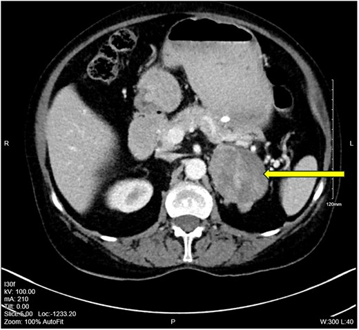
Computed tomography abdomen pelvis of clinical case with 8 cm heterogenous left-sided adrenal mass, with evidence of renal vein invasion indicating locoregional spread.

The initial biochemical results of clinical case demonstrating severe elevation of all serum androgens and excess circulating serum cortisol
| Results on presentation | |
|---|---|
| T | 288 ng/dL |
| (RR 0-40.4 ng/dL [0.1-1.4 nmol/L]) | (10.0 nmol/L) |
| DHEAS | >999.6 μg/dL |
| (RR 11-253 μg/dL [0.3-6.7 μmol/L]) | (>27.1 μmol/L) |
| A4 | 630 ng/dL |
| (RR 39.8-277.9 ng/dL [1.39-9.77 nmol/L]) | (22.0 nmol/L) |
| 17OHP | 184.8 ng/dL |
| (RR 19.8-72.6 ng/dL [0.6-2.2 nmol/L]) | (5.6 nmol/L) |
| Urinary free cortisol | 25.4 mcg/24 hours |
| (RR <5.3 mcg/24 hours [<146 nmol/24 hours]) | (701 nmol/24 hours) |
| Renin | 4.65 ng/mL |
| (RR 0.7-7.6 ng/mL [6.1-62.7 mIU/L]) | (38.2 mIU/L) |
| Aldosterone | 42.1 ng/dL |
| (RR 0-42.5 ng/dL [0-1179 pmol/L]) | (1168 pmol/L) |
Abbreviations: 17OHP, 17-hydroxy-progesterone; A4, androstenedione; DHEAS, dehydroepiandrosterone; RR, reference range; T, testosterone.
Given the presence of stage IV disease, she was deemed unsuitable for curative resection. Mitotane was commenced and titrated to achieve therapeutic drug target level of 14 to 20 mg/L. Metyrapone 500 mg four times daily was also initiated to suppress symptoms of hypercortisolism, with low-dose add-back hydrocortisone to prevent iatrogenic adrenal insufficiency. Restaging imaging demonstrated disease progression with enlarging liver metastases and new pulmonary nodules following 2 months of mitotane therapy. The patient completed 4 cycles of systemic gemcitabine and carboplatin, with resultant reduction in burden of metastases and size of primary tumor.
The patient experienced increasing symptoms of cortisol excess after 18 months following her original diagnosis. Extensive disease progression was identified, with significant enlargement of her original primary tumor and oligometastatic disease. The patient subsequently died due to complications secondary to metastatic ACC.
Female androgen physiology
Female androgen metabolism involves a complex interplay between the ovaries, adrenal glands, and peripheral tissues including, skin, adipose tissue, kidney, and liver (12, 13), as outlined in Fig. 2.
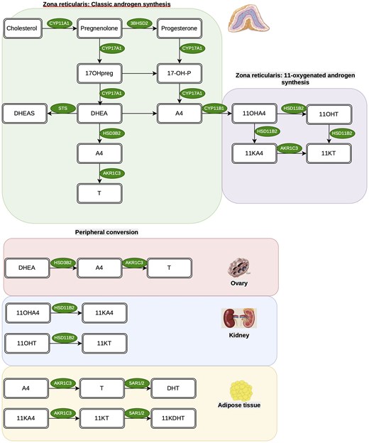
Pathways of androgen biosynthesis in the zona reticularis, including both classic and 11-oxygenated pathways. Peripheral activation to more androgens then occurs in tissues including the ovary, kidney, and adipose tissue. Abbreviations: 11KA4, 11-ketoandrostenedione; 11KT, 11-ketotestosterone; 11OHT, 11-β hydroxytesterone; 5AR1/2, 5-alpha reductase type 1/2 11OHA4, 11β-hydroxyandrostenedione; 17OHpreg, 17-hydroxy-pregnenelone; AKR1C3, aldo-keto-reductase type 1C3; CYP11B1, cytochrome P450 11B1; CYP17A1, cytochrome P450 17A1; DHEA, dehydroepiandrosterone; DHEAS, dehydroepiandrosterone sulfate; HSD11B2, 11β-hydroxysteroid dehydrogenase type 2; HSD3B2, 3-beta-hydroxysteroid dehydrogenase type 2; ; DHT, dihydrotestosterone; 11KDHT, 11-ketodihydrotestosterone; STS, steroid sulfatase.

The zona reticularis of the adrenal cortex is the major site of steroidogenesis of classic 19 carbon steroids. Dehydroepiandrosterone (DHEA) and its sulphated ester DHEAS are the primary androgens produced by the adrenals, with synthesis controlled by pituitary ACTH (14, 15). While DHEAS is the most abundant circulating androgen, it demonstrates significantly less biological potency at the androgen receptor compared with downstream metabolites such as androstenedione (A4) and T. The contribution of adrenal-derived T production from DHEA is negligible; however, A4 is synthesized from DHEA/DHEAS via the action of 3-beta hydroxysteroid dehydrogenase type 2 (HSD3B2). DHEA and DHEAS are therefore considered specific biomarkers of adrenal androgen output (16, 17).
The adrenal glands also produce a further subgroup of C-19 steroids, namely the 11-oxygenated androgen subclass (11oxyandrogens) (18). Previously, these were perceived as biologically inert byproducts of steroidogenesis. While recent work has highlighted their predominance in disorders of androgen excess, including PCOS and congenital adrenal hyperplasia (CAH) (19, 20), their true physiological role remains poorly understood. 11β-hydroxyandrostenedione is produced following catalysis of A4 by adrenal CYP11B1. The major contribution of more potent androgens to the circulating 11oxyandrogen pool occurs via peripheral activation of 11β-hydroxyandrostenedione to 11keto-androstenedione (11KA4) in the periphery, via the action of 11β-hydroxysteroid dehydrogenase type 2 within the kidneys and other mineralocorticoid target tissues (21). 11KA4 is then converted to the potent active androgen 11-ketotestosterone (11KT) by the activity of aldo-ketoreductase type 1C3 (AKR1C3) in peripheral tissues such as adipose (22). AKR1C3 catalyzes the conversion of 11KA4 to 11KT far more efficiently than its counterpart reaction in the classic pathway, namely A4 to T (18, 23).
Ovarian thecal cells are the principal site of ovarian androgen generation, with key steroidogenic enzymes upregulated by LH activation, primarily the 1720 lyase activity of CYP17A1 (17α-hydroxylase); responsible for the conversion of 17-hydroxyprogesterone (17OHP) to DHEA. Further catalysis of DHEA to A4 occurs via the action of HSD3B2. A4 is converted to T, both within the ovary and peripherally, via AKR1C3 and to a lesser extent 17β-hydroxysteroid dehydrogenase type 3. Thecal androgen production can also be enhanced by insulin-mediated activation of the LH receptor, a process upregulated in disorders of insulin resistance (IR), with the net effect of enhanced ovarian T production (24). The majority of circulating T is transported by SHBG, which is synthesized in the liver, alongside other binding proteins including albumin. Only the free component of T is biologically active. Conversion of T to the more biologically potent DHT occurs via the action of isoforms of 5α-reductase. 5α-reductase type 1 (SRD5A1) is highly expressed in liver, kidney, skin, and brain. Conversely, SRD5A2 encodes the type 2 isoform, which demonstrates enhanced ability to reduce T to DHT and is localized to genital skin (12, 13, 25).
Approximately 5% of the circulating androgen pool is maintained via peripheral activation of less potent androgens (26). Adipose tissue is a potent site of androgen activation via the action of AKR1C3, which is upregulated in women with simple obesity, and may account for reported rates of hyperandrogenemia in this cohort (27). This becomes particularly relevant beyond the menopausal transition (MT), when increasing adipose tissue activation can be a driver of mild androgen excess.
The menopausal transition
The MT is defined as the transitional phase preceding the final menstrual period. It is further subdivided into immediate and late MT based on development of menstrual cycle disturbance (28, 29). The most recognized hormonal shift during menopause is the cessation of ovarian production of 17β-estradiol. However, there is additional flux in the systemic hormonal milieu, with androgens profoundly impacted.
Longitudinal studies have demonstrated that circulating DHEA concentration declines beyond the third decade in both men and women, reaching a plateau in the eighth decade of life (30, 31). A 2002 study of 3029 women during the MT demonstrated a nonlinear decline, with a transient increase observed during the late MT (32). Longitudinal follow-up consolidated this relationship, suggesting that compensatory increases in DHEA during menopause may initially maintain the circulating pool of more potent downstream androgens including A4 and T (33).
In the immediate MT, ovarian androgen production persists, which may compound the more severe manifestation of menopausal symptoms experienced due to abrupt estrogen withdrawal following bilateral salpingoophorectomy (BSO) (34). In the early postmenopausal stage, T and A4 remain comparable to premenopausal concentrations. A combination of factors appears to contribute to this. The compensatory increase in FSH and midluteal LH during anovulatory cycles can drive thecal cell androgen generation during the MT. Additionally, with falling estradiol, there is a reduction in circulating SHBG with resultant increase in free androgen index. This persistence is not indefinite, with an observed reduction in T to 50% of premenopausal concentration subsequently observed 10 years following menopause (35-38). Beyond menopause, activation of DHEA to potent androgens including T and A4 is primarily mediated peripherally (39). In contrast to other androgens, 11oxyandrogens do not decline with physiological aging, representing the predominant circulating androgen in postmenopausal women (40), with 11KT demonstrating equivalent potency to T at the androgen receptor (18).
Clinical approach to postmenopausal androgen excess in women
A detailed clinical history is essential in the initial approach to a postmenopausal patient with androgen excess. A timeline of symptoms needs to be rigorously established as a signpost of potential malignant drivers of androgen excess, as rapidity of onset is a direct correlate of more sinister pathology. Prior reproductive history including menstrual history and symptoms of premenopausal androgen excess should be sought. Clinical stigmata of virilization including clitoromegaly, voice deepening, muscle hypertrophy, and breast atrophy should also prompt urgent work-up. Cardiovascular and metabolic risk profiling should be undertaken, considering preexisting features of cardiovascular disease, metabolic dysfunction associated steatotic liver disease (MASLD), impaired glucose tolerance, and prediabetes, features which are particularly prominent in patients with PCOS and ovarian hyperthecosis (OHT) (41, 42).
Examination should focus on detecting hirsutism, acne, and FPHL. Hirsutism is accepted as the most reliable indicator of female androgen excess and grading should be performed as per the modified Ferriman-Gallwey scale (43, 44). Clinical features suggestive of hypercortisolism or growth hormone excess should be identified, as well as evaluation for abdominal/pelvic masses. Examination should also include assessment for stigmata of severe IR (SIR).
Initial biochemical workup as a guide to further investigation
A comprehensive androgen profile to guide initial localization is an important first step in biochemical assessment. Knowledge of local assay method and performance is important for every clinician, with many institutions continuing to employ immunoassay-based measurements. Immunoassay performs poorly for quantification of steroids in low circulating concentrations, particularly serum androgen concentrations in women. Where possible, androgen quantification should be performed by gold-standard LC-MS/MS to improve sensitivity and reduce the risk of cross-reactivity observed with immunoassay-based methods (4, 45).
Androgen excess represents a spectrum, and absolute cutoffs correlating with significant pathology are not possible to implement in clinical practice. Interpreting results and biochemical patterns in tandem with the overall clinical picture remains the most important tool for detecting potentially malignant underlying conditions. T is the most reliable marker of ovarian androgen excess and should be measured in parallel with SHBG. Low SHBG is a surrogate marker of IR due to insulin-mediated suppression of hepatic SHBG output. In a postmenopausal patient, an isolated serum T above 2 nmol/L (57.7 ng/mL), with the previously outlined caveats surrounding absolute cutoffs, may be a reasonable threshold to institute investigations (2, 4). A4, DHEA, and 17-OHP are typically reflective of adrenal androgen production, although a significant proportion of A4 is also likely ovarian-derived (1). Significant elevations across all androgens, as outlined in our case due to malignant secretion and subsequent peripheral activation of adrenal androgen precursors, is indicative of ACC until proven otherwise and indicates an urgent need for adrenal imaging (2, 46).
Adjunctive measures in assessment include a full blood count. Erythrocytosis and an elevated hematocrit is supportive of severe androgen excess and suggestive of enhanced risk of venous thromboembolism (47, 48). Measurement of gonadotropins can demonstrate partial or indeed complete suppression in cases of severe androgen excess. This is likely due to peripheral aromatization of T to 17β-estradiol, which suppresses gonadotropin secretion through negative feedback.
Etiology of postmenopausal androgen excess
Polycystic ovary syndrome
PCOS is the most common cause of androgen excess in premenopausal women, with an estimated 10% to 15% of women affected during their reproductive years (7) (Table 2). PCOS is now understood as a lifelong disorder with implications beyond the reproductive lifespan, including a 2-fold increased risk of type 2 diabetes mellitus, hypertension, MASLD, and cardiovascular disease (49-52). Metabolic dysfunction and IR act synergistically with obesity to drive adrenal, ovarian, and peripheral androgen secretion, with severity of IR correlating directly with the magnitude of androgen excess (53, 54).
Etiology of androgen excess in postmenopausal women and estimated prevalence, in addition to associated clinical and imaging features
| Estimated prevalence in postmenopausal androgen excess (Elhassan et al, 2018 (2) N = 75) | Estimated prevalence in postmenopausal androgen excess (Luque-Ramirez et al, 2024 (9) N = 51) | Biochemical pattern: T | Biochemical pattern: DHEA | Biochemical pattern: A4 | Imaging findings | Clinical features | |
|---|---|---|---|---|---|---|---|
| PCOS | 29% | 10% | ↑ | ↑ | ↑ | May demonstrate minimal enlargement of postmenopausal ovary compared to non-PCOS women | Indolent androgen excess—symptoms over preceding decades; diagnosis of exclusion. |
| ACC | 14.7% | 2% | ↑↑↑ | ↑↑↑ | ↑↑↑ | Adrenal nodule—concerning features if >4 cm, heterogenous or contrast-enhancing | Rapid onset, virilization, symptoms of mass effect; symptoms of cortisol excess may coexist |
| CS | 4% | 2% | ↑ | ↑↑ | ↑↑ | Equivocal/pituitary vs adrenal lesion depending on ACTH dependent or ACTH independent | Cushingoid features coexist |
| OHT | 9.3% | 32% (combined OHT and functional gonadotrophin dependent androgen excess) | ↑↑ | — | — | Bilateral ovarian enlargement on US pelvis | Associations with IR, metabolic syndrome |
| VOTs | 2.4% | 20% | ↑↑↑ | — | ↑↑/– | Unilateral ovarian lesion on US pelvis/MRI pelvis. Lesions often subcentimeter so may not be visualized | Features of virilization; gonadotrophin suppression supportive |
| NCCAH | 0% | 14% | ↑↑ | ↑↑ | ↑↑ | Bilateral adrenal enlargement due to ACTH stimulation | Indolent androgen excess over preceding decades. History of subfertility |
| SIRS | 0% | 0% | ↑↑ | ↑↑/– | ↑↑/– | Variable androgen pattern dependent on subtype—TBIRs predominant T elevation. | Seek evidence of CTD or haematological malignancy if TBIRs |
| Lipodystrophy phenotypes associated with elevation of both T and A4/DHEA | |||||||
| Iatrogenic | — | — | Negative | Variable depending on causative agent |
Abbreviations: A4, androstenedione; ACC, adrenocortical carcinoma; CS, Cushing syndrome; CTD, connective tissue disease; DHEA, dehydroepiandrosterone; IR, insulin resistance; NCCAH, nonclassical congenital adrenal hyperplasia; OHT, ovarian hyperthecosis; PCOS, polycystic ovary syndrome; SIRS, severe insulin resistance syndromes; T, testosterone; TBIRs, type B insulin resistance syndrome; US, ultrasound; VOTs, virilizing ovarian tumors.
It has been suggested that age may ameliorate the PCOS phenotype, with a proposed delay in physiological menopause of 2 to 4 years (55-57). Overall, clinical androgen excess driven by PCOS tends to improve with age. Evidence regarding the effect of the postmenopausal PCOS phenotype suggests compounding of the adverse metabolic phenotype beyond that observed following normal menopause (50, 58-60).
No criteria currently exist for diagnosis of postmenopausal PCOS, and current guidelines advocate for consideration only following a preexisting premenopausal diagnosis or when symptoms of androgen excess and menstrual irregularity have been clearly established during reproductive years (7). New or evolving postmenopausal androgen excess, particularly with severe clinical and biochemical features, are not consistent with a diagnosis of PCOS. Given the high proportion of potentially sinister underlying pathology, a new diagnosis of PCOS should be considered solely as a diagnosis of exclusion. A prior diagnosis of PCOS also predisposes women to developing OHT, likely due to a persistent IR phenotype (61).
Ovarian hyperthecosis
OHT occurs due to persistent LH-mediated androgen production by ovarian thecal cells, with previously inert stromal cells developing steroidogenic capacity via enhanced LH and insulin stimulation following menopause (61). IGF-1 has also been identified as a potential costimulator of thecal androgen production (62). The estimated prevalence of OHT in postmenopausal women presenting with androgen excess is 9.8% in 1 series (2). PCOS shares some phenotypic associations with OHT, with IR and hyperinsulinemia frequently implicated in both conditions. OHT is typically observed in women with other features of the metabolic syndrome (63, 64).
Women will typically describe indolent symptoms of androgen excess. Biochemically, there is generally an isolated but significant elevation of serum T, with circulating concentrations often exceeding 288.8 ng/mL (10 nmol/L). DHEA is frequently normal, with variable A4 concentration. Features of severe androgen excess can develop, although overt virilization is rare, highlighting the challenges in differentiating from virilizing ovarian tumors (VOTs), given significant phenotypic overlap (64, 65). While OHT can only be confirmed histologically, imaging such as ultrasound and magnetic resonance imaging (MRI) can be a useful discriminator in identifying bilateral bulky adnexae from unilateral lesions. The treatment of choice once confirmed is a bilateral oophorectomy.
Virilizing ovarian tumors
Secretory ovarian tumors account for 10% of ovarian neoplasia. Of these, androgen-producing tumors account for less than 1%. VOTs can occur at any age, accounting for approximately 2.7% of postmenopausal androgen excess (2, 66). These tumors are predominantly benign and arise from nonepithelial ovarian structures, primarily the sex cord and stromal cells. Features of virilization are observed in approximately 50% of patients. Postmenopausal bleeding is reported in up to 50% of cases, due to peripheral aromatization of T to 17β-estradiol, with consequent endometrial thickening (67). T concentration is often markedly elevated, with 1 case series reporting a range of 112.3 to 3433 ng/mL (3.9-119 nmol/L) (68). Correspondingly, A4 and DHEA may demonstrate resultant suppression below age-related reference ranges (69). Features supportive of a diagnosis of VOT over OHT include unilateral imaging findings, severe T elevation above 288 ng/dL (10 nmol/L) and FSH <22 IU/L and LH <10 IU/L (70), the latter due to complete or partial loss of negative feedback.
Histologically, LCTs are the most common subtype of VOTs encountered in postmenopausal women, compared to SLCTs in premenopausal women. They are derived from stromal lines. Bilateral involvement is observed in 1.5%, with clinical and biochemical differentiation from OHT in this context posing a true diagnostic conundrum (71). Thecal cell tumors (Thecomas) are benign tumors encountered in postmenopausal women and generally secrete estrogens, with approximately 10% demonstrating androgen production. Additional stromal cell tumors include sclerosing cell and steroid cell subtypes, which typically present premenopausal. Granulosa cell tumors are a rare form of sex cord tumors, representing 5% of all malignant ovarian tumors. While predominantly estrogen-secreting, sporadic case reports have identified androgen-producing granulosa cell tumors (72).
Consensus regarding diagnostic workup for VOTs is lacking. Pelvic imaging with a transvaginal pelvic ultrasound is a prudent initial step, with color Doppler of particular value in identifying LCTs due to their inherent lipid content (73). These lesions are often small so may evade detection. MRI pelvis and 18-fluorodeoxyglucose-positron emission tomography imaging provides greater detection of smaller lesions; however, visualization of the postmenopausal ovary remains challenging, with robust supportive data lacking in the literature (69). Surgical intervention with BSO is the preferred treatment of choice. This contrasts with premenopausal VOTs, where unilateral oophorectomy is preferential for fertility preservation, with lateralization essential where possible (see the following discussion). Preoperative endometrial biopsy is recommended to ascertain risk of hyperplasia/malignancy, which may prompt extended surgery to include a hysterectomy. Androgens typically normalize 48 hours postoperatively (67, 74).
Discriminating benign from malignant VOTs preoperatively is challenging. The recent Society for Endocrinology Clinical Practice Guidelines on Evaluation of Androgen Excess in Women suggest that baseline gonadotrophin suppression may correlate with an increased risk of malignancy (1). Operative histology is ultimately required for definitive diagnosis.
Adrenocortical carcinoma
ACC is a rare cause of postmenopausal androgen excess but must be actively excluded in cases of rapidly progressive virilization. The incidence of ACC is approximately 0.2 to 0.7 million per year, typically arising in postmenopausal women in the fifth and sixth decade (46). Adrenal androgen excess is observed in 40% to 60% of patients (75).
Alarming features suggestive of underlying ACC include rapidly progressive symptoms of virilization, combined with significant elevations in A4, DHEA, and T. ACC exhibits a predilection for secreting immature precursors of multiple steroidogenic pathways, primarily glucocorticoid derivatives, as demonstrated in our case. Cortisol-producing tumors have been associated with increased risk of poor outcome (76). Concurrent elevations in products of more than 1 adrenal cell subtype raises suspicion for ACC. Presentation is often late and may be associated with symptoms of mass effect. Some cases of ACC are detected initially as incidental adrenal nodules, highlighting the importance of a cautious approach to workup for lesions with Hounsfield units > 20, heterogenous lesions or lesions >4 cm; particularly in younger patients (77).
When suspected, urgent adrenal imaging must be obtained, with expedited referral to a dedicated tertiary center for multidisciplinary management following identification of a suspicious adrenal mass. While trends in 5-year overall survival for stage IV disease have shown improvement in recent decades, this remains at around only 10% (78, 79). Marginal gains in overall survival are largely attributable to centralized treatment via high volume centers, use of adjuvant mitotane, and treatment with systemic chemotherapy as per the FIRM-ACT protocol (etoposide, doxorubicin, cisplatin, mitotane) for progressive or metastatic disease (46, 75, 80). Real-world data highlights mitotane is poorly tolerated due to systemic toxicity, with only 58% achieving therapeutic drug levels and 40% completing planned treatment course (81). ACC is the most critical diagnosis to exclude when approaching postmenopausal androgen excess.
Nonclassical congenital adrenal hyperplasia
CAH is a group of autosomal recessive defects in genes encoding adrenal steroidogenesis, with variable deficits in glucocorticoid and mineralocorticoid synthesis depending on the affected gene. CYP21A2 deficiency accounts for over 95% of cases (82, 83). Depending on the residual degree of enzymatic activity, patients may present with classic salt-wasting in infancy. Nonclassical CAH (NCCAH) typically presents during reproductive years with menstrual irregularity and androgen excess (84). While rare, cases can remain undetected until later life, particularly in women who never sought fertility. Luque-Ramirez et al described a prevalence of NCCAH of 14% in a cohort of 51 patients with postmenopausal androgen excess, contrasting with Elhassan's series of 324 consecutively recruited patients where no cases of NCCAH were identified (2, 9). NCCAH has been previously linked with rising serum androgens throughout the lifespan, particularly around the MT (85). Workup demonstrates elevations in 17OHP, T, A4, and DHEA, which may be biochemically and radiologically indistinguishable from ACC, highlighting the importance of a careful correlation with clinical history (86). Confirmatory testing includes a short synacthen test with serial measurement of 17OHP, and confirmatory sequencing of the CYP21A2 gene (82).
Cushing syndrome
Cushing syndrome (CS) can present with features of androgen excess in 50% of cases due to ACTH-dependent A4 and DHEA generation. Recently, elevated 11-oxyandrogens have been identified as the predominant biochemical signature of androgen excess in CS and correlates with the degree of androgen-mediated symptoms (87). Clinical features are nonspecific, including fatigue, proximal myopathy, central adiposity, and violaceous striae (88). Associated sequelae often overlap with those of the metabolic syndrome, which coexists with several causes of postmenopausal androgen excess, creating frequent diagnostic dilemmas. The 1 mg overnight dexamethasone suppression test is an excellent sensitive initial screen for patients with suspected CS, with a sensitivity and specificity of 95% and 80%, respectively, with a diagnostic threshold >1.8 mcg/dL (50 nmol/L) (89). Overt CS in the context of virilization raises significant concerns regarding an underlying ACC, particularly with ACTH suppression. Ectopic cortisol secretion by ovarian SLCTs is also a rare phenomenon previously described in the literature (90).
Iatrogenic
Exogenous T replacement is prescribed for hypoactive sexual desire disorder as an adjunct to hormone replacement therapy regimens (91). When titrated to achieve T in the physiological premenopausal range, women can develop mild androgen excess without virilization (92). Increasingly, a trend toward increased prescribing of transdermal T for postmenopausal women and iatrogenic androgen excess are likely to be encountered at higher rates in the future. Additional medications associated with androgen excess include sodium valproate and metyrapone (93). Metyrapone is utilized for pharmacological management of hypercortisolemia and reduces the activity of 11β-hydroxylase, which is responsible for conversion of 11-deoxycortisol to cortisol. This incurs androgen excess through upregulation of ACTH and resultant DHEA production (94).
Severe insulin resistance syndromes
SIRS encompasses a diverse group of disorders caused by defective insulin signaling pathways, manifesting as disproportionate indices of metabolic dysfunction and IR relative to adiposity. They are further subclassified into congenital or acquired subtypes and underlying pathophysiology relative to the pre/post insulin receptor abnormalities (1). Women may demonstrate features of profound IR including acanthosis nigricans and skin tags. Biochemical indicators include significant elevations in fasting serum insulin concentration (>25 μU/L/150 pmol/L). The etiology of SIRS can be subdivided into molecular defects in the insulin signaling pathway, including monogenic defects in the INSR and development of autoantibodies to the insulin receptor (type B insulin resistance syndrome). Conversely, lipodystrophy refers to defects in target tissues of insulin action, namely adipose tissue (95). Type B insulin resistance syndrome is a rare, acquired form of SIRS. This may be more relevant for consideration in the postmenopausal context than lipodystrophy or INSR variations, which typically present in premenopausal women. Variable phenotypes exist, with spectrum of presentation ranging from SIRS to life-threatening hypoglycemia, depending on the presence of agonistic or antagonistic anti-insulin receptor antibodies (96). Some patients report rapid onset androgen excess, coupled with clinical signs of IR. There is concomitant association between anti-INSR antibodies and underlying connective tissue disorders, primarily systemic lupus erythematosus (SLE) (1, 95, 97). Connective tissue disorders may not always present concurrently and can manifest following an interlude of several years. A subset of type B insulin resistance, namely isolated hypoglycemia with low insulin, represents a paraneoplastic phenomenon, associated with aggressive forms of lymphoma (98). Similarly, this may evolve sequentially, highlighting the need for a thorough diagnostic approach including screening for known associated pathology.
The role of dynamic testing
Biochemical patterns can be provided by initial androgen profiles, with additional adjunctive dynamic testing useful in refining the differential diagnosis, particularly in the context of equivocal imaging.
Gonadotropin-releasing hormone analog suppression testing
Gonadotropin-releasing hormone (GnRH) analog suppression testing involves administering a single dose of leuprorelin or triptorelin intramuscularly or subcutaneously. Following binding, there is initial activation with resultant desensitization and downregulation of GnRH receptors in pituitary gonadotroph cells. FSH and LH become suppressed 21 to 28 days postinjection, with a subsequent fall in T in scenarios of ovarian androgen excess where gonadotropin-mediated control of thecal cell function is preserved. A reduction in T of >50% strongly supports an ovarian source of androgen hypersecretion (99). Theoretically, androgen excess occurring secondary to VOTs demonstrates increased likelihood of developing autonomous function, so will not demonstrate GnRH analog suppression. Real-world data demonstrates significant overlap in observed response, so this remains an unreliable discriminator between VOTs and OHT in isolation (70, 100). Rarely, LH or GnRH-mediated adrenal hyperandrogenism has been described in isolated cases (101). In postmenopausal women who demonstrate androgen suppression following a GnRH analog, referral for BSO should be considered. In those unsuitable or unwilling to undergo surgery, long-term GnRH analog therapy may be considered as a therapeutic strategy (47). Certrorelix, a GnRH antagonist, can also be utilized to achieve similar effect, but without the initial surge in gonadotrophins and androgens. Androgen suppression can therefore be achieved within days (102).
Dexamethasone suppression testing
A 1 mg overnight dexamethasone suppression test has a primary role as an initial screening test for concomitant CS (88). Dexamethasone suppression testing may also have diagnostic utility in the workup of suspected adrenal androgen excess, especially in scenarios where an adrenal mass has not been identified and the likelihood of ACC is low. Due to the prolonged half-life of DHEAS, a 96-hour suppression test may be required (0.5 mg every 6 hours). Suppression of DHEAS >50% from baseline is suggestive of preserved ACTH-mediated hyperandrogenism, as seen in PCOS and idiopathic adrenal androgen hypersecretion (103, 104), inferring nonneoplastic pathology as the mediator of androgen excess (105).
Imaging for localization
Adrenal imaging
All postmenopausal women presenting with significant elevations in DHEA/DHEAS require urgent adrenal imaging. DHEAS concentration >736 μg/dL (20 μmol/L) have a high positive predictive value for ACC particularly in postmenopausal women; in premenopausal women, severe isolated DHEAS elevations may still be consistent with underlying PCOS (2). Unenhanced computed tomography of the adrenal gland is the preferred first-line modality and can detect nodules >5 mm. A density of >20 Hounsfield units has a sensitivity >99% for ACC, but poor specificity (46, 77). The incidence of benign nonsecretory adrenal lesions in this cohort is significant, with a reported prevalence in excess of 7% over the age of 70 (106). Adopting a multidisciplinary approach is crucial in striving for diagnostic consensus and avoiding unnecessary surgery for benign lesions. Adenomas are generally smaller (2-2.5 cm), whereas ACCs are typically larger (4-21 cm, median size 10 cm at diagnosis) (107, 108). In cases of equivocal imaging with an indeterminate adrenal nodule, 18-fluorodeoxyglucose-positron emission tomography imaging may be appropriate, as may close interval follow-up imaging or surgical resection following multidisciplinary team discussion (77).
Ovarian imaging
When ovarian hyperandrogenism is suspected, transvaginal ultrasound is the first-line investigation. The mean postmenopausal ovarian volume is estimated at 2.2 ± 0.01 cm3. Postmenopausal women with prior PCOS generally have larger ovaries than controls but remain smaller than those typically observed in OHT (109). Asymmetry of the ovaries or a hypervascularized area on Doppler imaging suggests a tumor. Homogeneous bilateral enlargement of the ovaries without hypervascularization is supportive of OHT. The absence of these findings does not exclude these diagnoses (70, 110). Ultrasound is a highly subjective, user-dependent imaging modality, with a low reported sensitivity for VOTs and OHT (111). Further investigation with MRI is often required, with higher positive and negative predictive values reported. Imaging is often negative, as a high proportion of VOTs are <1 cm (70).
Invasive testing: simultaneous ovarian and adrenal vein sampling (OVS and AVS)
There is limited evidence supporting the use of simultaneous AVS/OVS in postmenopausal patients. Clinical diagnostic utility is generally confined for maximizing fertility preservation in premenopausal women (112). Simultaneous catheterization of the adrenal, ovarian, and peripheral veins can demonstrate a gradient that localizes the androgen-producing tumor. However, success rates are poor, and this represents an invasive test with inherent associated risks (113, 114). It also requires a high level of operator expertise generally not accessible outside tertiary centers. The role of paired venous sampling is therefore limited in the workup of postmenopausal androgen excess and may be reserved for cases with coexistent adrenal and ovarian masses alongside an equivocal or mixed biochemical androgen picture following multidisciplinary consensus.
Sequelae of androgen excess
Metabolic risk
Androgen excess in women correlates positively with rates of IR, T2DM, MASLD, obesity, and incidence of cardiovascular disease (49-53). Evidence supporting increased prevalence of metabolic dysfunction relates to longitudinal follow-up of chronic disorders including PCOS and CAH, with limited evidence interrogating postmenopausal-onset androgen excess (115). OHT affects a similar patient cohort to metabolic syndrome, with IR hypothesized as a driving factor in pathogenesis (63-65). While collectively rare, VOTs appear to mirror increased rates of cardiovascular disease and metabolic dysfunction as seen with other disorders of androgen excess (116). Screening for associated metabolic comorbidities represents an important aspect of management.
Hormone-sensitive cancers
Persistent postmenopausal hyperandrogenism has been linked with endometrial hyperplasia and resultant carcinoma, due to both peripheral aromatization of androgens to 17β-estradiol and possibly a direct impact of androgen excess on endometrial proliferation (9, 110, 117). Preoperative endometrial biopsy has been proposed by some groups, particularly in the context of OHT given associations with obesity and metabolic syndrome (118). Prospective studies have also demonstrated a link between elevated T and both hormone-dependent and independent breast cancer due to increased lifelong estrogen exposure (119, 120). Knowledge of the unique steroid milieu in breast cancer has also gained traction, with an androgenic environment identified in subtypes of breast cancer, which has potential implications for patients with hyperandrogenemia (121).
Overall complexities and considerations in management
Postmenopausal androgen excess presents a challenging clinical conundrum to even experienced endocrinologists. Comparatively, there is a greater risk of neoplastic etiology in comparison to common disorders such as PCOS, but also a high prevalence of incidental imaging findings that can make securing an accurate diagnosis challenging. Additionally, with advancing age, patients present with increasing comorbidities making definitive management infeasible or undesirable to some individuals. Given this context, clinicians must develop an approach that acknowledges this complexity. Figure 3 summarizes 1 such pragmatic approach.
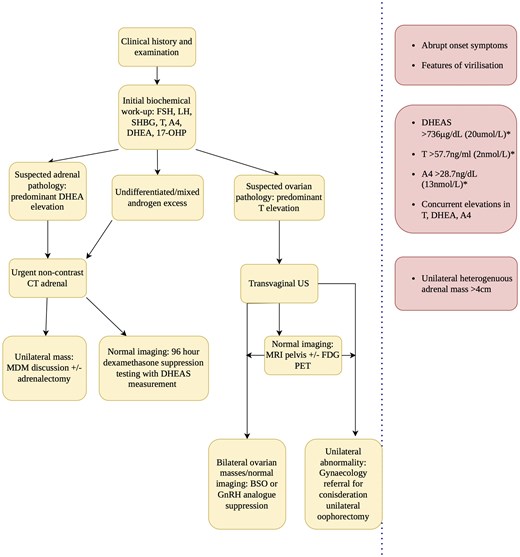
A suggested pragmatic approach to diagnosis and workup of postmenopausal androgen excess based on initial biochemical workup in yellow. Column in red on the left hand side indicates potential red flags at each stage of work-up prompting expedited investigation. Abbreviations: 17-OHP, 17-hydroxy-progesterone; A4, androstenedione; BSO, bilateral salpingoophorectomy; DHEA, dehydroepiandrosterone; GnRH, gonadotrophin releasing hormone; MDM, multidisciplinary meeting; ONDST, overnight dexamethasone suppression testing; PET, positron emission tomography; SHBG, sex hormone binding globulin; T, testosterone; US, ultrasound.
*Suggested cutoffs to prompt urgent investigation. A detailed knowledge of local assay and cutoffs is essential and should always be interpreted in the context of the overall clinical context.

Regarding management, both topical and systemic agents can be considered to address symptoms, including spironolactone and minoxidil. GnRH analogs are useful adjuncts in benign ovarian pathology, particularly those who represent poor surgical candidates due to preference or comorbidities (1, 9, 47).
Future directions
Unique metabolomic signatures have been identified using multisteroid profiling in primary aldosteronism, CS, and ACC, aiding diagnostic accuracy (122-124). Scant published exploratory mechanistic studies point to a potential use for identifying altered metabolic pathways in PCOS (125). A proof-of-concept study of nontargeted metabolomics and machine-learning in PCOS and NCCAH demonstrated ability to accurately identify NCCAH in 100% of cases amongst 283 patients (126). Theoretically, multi-steroid profiling may represent an adjunct for work-up of postmenopausal androgen excess based on detection of adrenal or ovarian-specific metabolites. Currently, predominant utility remains in workup of indeterminate adrenal nodules.
Conclusion
Postmenopausal androgen excess encompasses a heterogenous group of conditions, with potential for sinister underlying neoplastic pathology. Features of virilization coupled with significant biochemical hyperandrogenemia indicate a need for urgent diagnostic workup. Following a complete biochemical assessment to direct further diagnostics, noncontrast computed tomography adrenal and MRI pelvis remain the most sensitive modalities; however, there is an increasing burden of incidental pathology identified in this age cohort. Inclusion of dynamic testing may further aid localization.
Disclosures
The authors have no conflicts of interest to declare.
Data availability
No new data were generated or analyzed in support of this article.