 https://orcid.org/0000-0003-3421-7147
Abstract
Osteoporosis is associated with reduced active vitamin D, but the molecular basis remains incompletely understood. This study investigated how active vitamin D preserves skeletal homeostasis and prevents bone loss.
Experimental mechanistic study using genetically modified mouse models and bone marrow mesenchymal stem cells (BMSCs).
Mice with reduced active vitamin D production were examined for bone mass, skeletal aging, and osteogenic capacity. Mouse and human BMSCs were used to assess proliferation, osteogenic differentiation, and molecular signaling. Gene expression, protein turnover, and pathway analyses determined how active vitamin D regulates cell-cycle control in skeletal progenitors. Genetic deletion studies tested the functional contribution of P27 to bone loss.
Active vitamin D protected against osteoporosis by activating a vitamin D receptor (VDR)-dependent pathway that increased cyclin-dependent kinase 2 (CDK2) expression and promoted P27 degradation in BMSCs. Loss of this signaling caused P27 accumulation, reduced stem-cell proliferation, impaired osteogenic differentiation, and enhanced skeletal senescence, leading to bone loss. Active vitamin D promoted P27 phosphorylation at threonine 187 and its degradation via the ubiquitin-proteasome pathway. Importantly, P27 deletion partially rescued the osteoporotic phenotype in mice with reduced active vitamin D production.
Active vitamin D maintains bone integrity through a VDR-dependent mechanism that enhances CDK2 expression and promotes P27 degradation in BMSCs. These findings identify a previously unrecognized mechanism linking vitamin D signaling to skeletal aging and suggest that targeting P27 turnover may offer a therapeutic strategy, provided tissue-specific approaches mitigate the oncogenic risks of P27 manipulation.
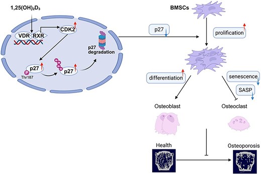

This study reveals a new mechanism by which active vitamin D protects the skeleton from osteoporosis. We show that vitamin D receptor signaling increases CDK2 transcription in bone marrow mesenchymal stem cells, promoting phosphorylation and degradation of the cell-cycle inhibitor P27. When active vitamin D is reduced, P27 accumulates, leading to impaired stem-cell proliferation, reduced bone formation, cellular senescence, and bone loss. Genetic deletion of P27 partially rescued osteoporosis caused by reduced active vitamin D, demonstrating its functional importance. These findings advance understanding of how endocrine vitamin D signaling preserves skeletal homeostasis and identify P27 turnover as a potential therapeutic target for vitamin D–related osteoporosis, provided bone-specific strategies are used to minimize cancer-related risks.
Introduction
Osteoporosis is a highly prevalent skeletal disorder and a leading cause of fragility fractures, imposing substantial individual and societal burdens.1 Vitamin D insufficiency/deficiency is widely recognized as a modifiable risk factor for impaired skeletal health, increased falls, and fracture risk,2,3. Beyond its classical role in calcium–phosphate homeostasis, active vitamin D signaling has increasingly been linked to cellular stress responses and aging-related bone phenotypes through vitamin D receptor (VDR)-dependent transcriptional programs,4,5. However, the molecular mechanisms by which reduced active vitamin D accelerates skeletal aging and bone loss remain incompletely defined.
Vitamin D is synthesized mainly in the skin from 7-dehydrocholesterol after ultraviolet exposure, converted in the liver to 25-hydroxyvitamin D [25(OH)D], and activated primarily in the kidney by 1α-hydroxylase [1α(OH)ase] to 1,25-dihydroxyvitamin D [1,25(OH)2D], the high-affinity ligand for VDR.4,6 Disruption of this activation step provides a model to examine the specific skeletal consequences of reduced active vitamin D. In particular, 1α(OH)ase+/− mice show reduced circulating 1,25(OH)2D with preserved 25(OH)D and mineral parameters under standard conditions and develop aging-like skeletal changes, making them useful for studying active vitamin D in bone maintenance.7-10 This heterozygous model avoids the severe hypocalcemia, hypophosphatemia, rickets/osteomalacia, and mineralization defects seen in homozygous 1α(OH)ase−/− mice, which can obscure cell-autonomous aging mechanisms. Previous work has also implicated 1,25(OH)2D–VDR signaling in limiting senescence in bone marrow mesenchymal stem cells (BMSCs) and osteocytes through epigenetic regulation, including EZH2–H3K27me3, and suppression of P16/P19 pathways.8,11,12
Cellular senescence and stem-cell dysfunction are increasingly recognized as drivers of age-related osteoporosis.13-16 Cellular senescence in bone primarily afflicts BMSCs and osteocytes. The impact of senescence on the aging skeleton is dual: it directly exhausts the stem cell pool by arresting BMSC proliferation and osteogenic differentiation and indirectly disrupts bone remodeling via the senescence-associated secretory phenotype (SASP), which paracrinally drives osteoclastogenesis and propagates senescence to neighboring cells. Although the P16–Rb and P53–P21 axes are canonical senescence regulators, P27Kip1 (CDKN1B) also regulates cell-cycle control and aging phenotypes.17-19 P27 is a cyclin-dependent kinase inhibitor mainly controlled post-transcriptionally through phosphorylation-dependent ubiquitination and proteasomal degradation.20-22 P27 deficiency enhances BMSC proliferation and osteogenic differentiation and increases osteoblast-mediated bone formation, suggesting that P27 restrains osteogenesis during stress and aging.22-25
Tissue-specific P27 regulation has been observed during 1,25(OH)2D insufficiency, but whether active vitamin D signaling regulates P27 in bone and BMSCs remains unclear.26 Moreover, links between 1,25(OH)2D–VDR signaling and ubiquitin–proteasome-mediated P27 control in skeletal cells have not been established. Because CDK2-dependent phosphorylation of P27 at Threonine 187 licenses its ubiquitination and proteasomal turnover, VDR-driven transcriptional programs may influence P27 stability through the CDK2/P27 axis.21,22,27
Here, we hypothesized that reduced active vitamin D accelerates skeletal aging by impairing VDR-mediated P27 degradation. We used global 1α(OH)ase (Cyp27b1) knockout mice, a model of active vitamin D insufficiency,28 crossed with P27 knockout mice to investigate genetic interactions between vitamin D signaling and cell-cycle control in vivo. We also examined how VDR regulates the CDK2/P27 axis and ubiquitin–proteasome system in human BMSCs under oxidative stress.
Materials and methods
Animal experiments
Global genetically modified VDR+/−, P27+/−, and 1α(OH)ase+/− mice were used.24,29 Heterozygous intercrosses generated double-heterozygous offspring, which were mated to obtain homozygous mutants. Experiments used male 2-month-old VDR−/− mice and 6-month-old P27−/−, 1α(OH)ase+/−, and P27−/−1α(OH)ase+/− mice. Genotyping followed established protocols.24,29 Only males were used to avoid estrous-cycle and estrogen-related effects on bone remodeling and BMSC senescence. As global knockout models, in vivo phenotypes reflected systemic rather than BMSC-specific effects. Sample sizes were based on power calculations from prior micro-CT data, requiring at least n = 5 mice per genotype/group. All procedures were approved by the Nanjing Medical University IACUC (IACUC-1802007).
Biochemical assays
Serum calcium and phosphorus were measured using an automated analyzer. Serum 1,25(OH)2D3 and 25(OH)D were quantified by radioimmunoassay, and intact PTH by ELISA.
Cell culture, osteogenic induction, and transfection
Mouse BMSCs were isolated from femurs and tibias as described,30 using three independent mice per genotype with triplicate technical wells. Human BMSCs were obtained with informed consent from four to five female osteoarthritis patients aged 58-72 years undergoing hip arthroplasty, under approved ethical guidelines. Experiments used passage-3 cells from at least three donors. BMSCs showed spindle morphology, CD73+/CD90+/CD105+/CD45−/CD34− marker profiles, and tri-lineage differentiation capacity.31 Cells were cultured in α-MEM with 10% FBS and antibiotics. Osteogenesis was induced with dexamethasone, β-glycerophosphate, and ascorbic acid for 7 days before ALP staining. Transfections used Lipofectamine 3000. VDR and P27 plasmids were previously described.11,30 siRNAs were si-VDR, 5′-UCAGACUCCAUUUGUAUUAUA-3′; si-P27, 5′-GCAACCGACGAUUCUUCUATT-3′.
Micro-CT and histology
Vertebrae were fixed in PLP, rinsed, and scanned using a SkyScan 1176 system at a 9 μm voxel size, 55 kVp, and 181 μA. Reconstruction used NRecon, with a fixed threshold of 55–255. Trabecular ROI included the L1 vertebral body excluding cortical shell. BMD, BV/TV, Tb.N, Tb.Th, and Tb.Sp were analyzed using DataViewer, CTAn, and ANT. Decalcified vertebrae were paraffin-embedded, sectioned at 5 μm, and stained with H&E, collagen, ALP, and TRAP. Immunohistochemistry examined SOD2, γ-H2A.X, p16, IL-6, IL-1β, and β-galactosidase.
Immunofluorescence and cell assays
Human BMSCs were fixed, permeabilized, blocked, incubated with anti-P27 antibody, stained with fluorophore-conjugated secondary antibody and DAPI, imaged by fluorescence microscopy, and quantified using ImageJ. MTT assays measured viability, and EdU incorporation assessed proliferation.
RNA and protein analyses
RNA from vertebrae or cells was extracted with TRIzol, reverse-transcribed, and analyzed by SYBR Green qPCR using the 2−ΔΔCt method with GAPDH normalization. GAPDH stability was confirmed after H2O2 or 1,25(OH)2D3 treatment. Western blotting was performed as described32 using antibodies against P27, P27(T187), CDK2, Osterix, Runx2, OPN, SOD2, γ-H2A.X, 8-OHdG, P16, and P53. β-actin served as loading control and was similarly validated.
ChIP, Co-IP, and statistics
ChIP used Magna ChIP™ with anti-VDR or IgG. Co-IP used cross-linked magnetic beads with anti-P27, followed by immunoblotting for ubiquitin and P27; inputs were blotted for P27 and β-actin. Experiments were repeated at least three times. Data were analyzed in GraphPad Prism 10.0 as mean ± SEM. Normality and variance were assessed by Shapiro–Wilk and Brown–Forsythe tests. Appropriate t-tests, Mann–Whitney U, ANOVA, Welch's ANOVA, Kruskal–Wallis, and post hoc tests were applied. P < .05 was significant.
Results
Reduced active vitamin D accelerates bone loss and is associated with elevated P27 protein expression
Serum levels of calcium, phosphorus, parathyroid hormone (PTH), and 25-hydroxyvitamin D [25(OH)D] did not differ significantly between 6-month-old 1α(OH)ase+/− mice and their wild-type (WT) littermates. However, circulating 1,25-dihydroxyvitamin D [1,25(OH)2D] levels were markedly reduced (Figure 1A–E). Full serum data including mean ± SEM, n, and exact P-values are provided in Table S1. Micro–computed tomography (micro-CT) analysis revealed an osteoporotic phenotype in 1α(OH)ase+/− mice, characterized by significantly decreased bone mineral density (BMD) and impaired trabecular microarchitecture, including reduced trabecular bone volume fraction (BV/TV), trabecular number (Tb.N), and trabecular thickness (Tb.Th), along with increased trabecular separation (Tb.Sp), compared with WT controls (Figure 1F–J). These findings indicate that 1α(OH)ase+/− mice represent a valid model of reduced active vitamin D–associated osteoporosis.
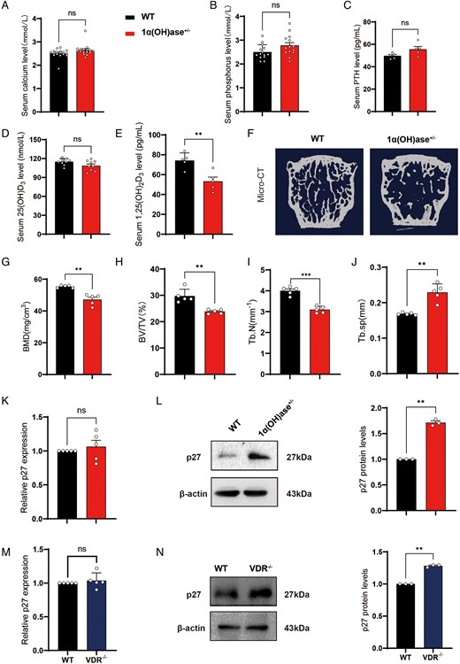
Reduced active vitamin D accelerates bone loss and is associated with increased P27 protein expression. Serum levels of (A) calcium (mmol/L), (B) phosphorus (mmol/L), (C) parathyroid hormone (PTH, pg/mL), (D) 25-hydroxyvitamin D (25(OH)D, mmol/L), and (E) 1,25-dihydroxyvitamin D (1,25(OH)₂D, pg/mL) in 6-month-old wild-type (WT) and 1α(OH)ase⁺/⁻ littermate mice maintained on a normal diet. (F) Representative three-dimensional micro-computed tomography (micro-CT) reconstructions of the first lumbar vertebra. Quantitative micro-CT analysis of (G) bone mineral density (BMD, mg/cm³), (H) trabecular bone volume fraction (BV/TV, %), (I) trabecular number (Tb.N, mm⁻¹), and (J) trabecular separation (Tb.Sp, mm). (K) P27 mRNA and (L) P27 protein expression in vertebral tissues of 6-month-old mice. (M) P27 mRNA and (N) P27 protein expression in bone tissues from 2-month-old WT and vitamin D receptor knockout (VDR⁻/⁻) littermate mice fed a normal diet. Serum sample sizes for panels A–E vary by assay and are detailed in Supplementary Table 1. For all remaining analyses, the sample size was n = 5 mice per group for micro-CT and qRT-PCR, and n = 3 mice per group for Western blotting. ns, not significant; *, P < .05; **, P < .01; ***, P < .001 versus WT littermate controls.

To determine whether reduced active vitamin D–induced bone loss is associated with altered P27 expression, we assessed P27 mRNA and protein levels in vertebral tissues from 6-month-old WT and 1α(OH)ase+/− littermates. Quantitative real-time PCR (qRT-PCR) revealed no significant difference in P27 mRNA levels between groups (Figure 1K), whereas immunoblotting demonstrated a marked increase in P27 protein in 1α(OH)ase+/− mice (Figure 1L). Similarly, 2-month-old vitamin D receptor knockout (VDR−/−) mice exhibited increased P27 protein levels without changes in P27 mRNA compared with WT controls (Figure 1M, N). These results suggest that 1,25(OH)2D suppresses P27 expression in bone primarily through a VDR-dependent post-transcriptional mechanism, thereby contributing to protection against osteoporosis.
1,25-dihydroxyvitamin D promotes P27 degradation via the ubiquitin–proteasome pathway under oxidative stress
Our previous study reported elevated superoxide anion levels in bone marrow–derived mesenchymal stem cells (BMSCs) from VDR−/− mice.8 Because our prior work identified elevated superoxide anion—a primary reactive oxygen species and key inducer of cellular senescence—in BMSCs lacking VDR, we sought to model this oxidative environment in vitro. To model oxidative stress associated with reduced active vitamin D, human BMSCs were exposed to 30 μM H2O2 for 24 h, followed by treatment with 10−8 M 1,25(OH)2D3 for an additional 12 h. A sublethal dose of 30 μM H2O2 for 24 h was used to induce stress-induced premature senescence (SIPS) rather than acute apoptosis, mimicking the chronic oxidative environment of aging bone 43. Consistent with a post-transcriptional regulatory mechanism, this dose significantly increased P27 protein but not mRNA levels. H2O2 did not significantly alter P27 mRNA levels but substantially increased P27 protein abundance; subsequent treatment with 1,25(OH)2D3 reduced P27 protein levels (Figure 2A, B). Cycloheximide (CHX) chase experiments showed that H2O2 treatment slowed P27 turnover, whereas 1,25(OH)2D3 accelerated P27 degradation (Figure 2C). Moreover, the proteasome inhibitor MG132 abolished the 1,25(OH)2D3-induced reduction in P27, supporting a proteasome-dependent mechanism (Figure 2D). Immunofluorescence staining confirmed that 1,25(OH)2D3 decreased nuclear P27 levels (Figure 2E). Collectively, these data indicate that 1,25(OH)2D3 facilitates ubiquitin–proteasome–mediated P27 degradation in hBMSCs under oxidative stress.
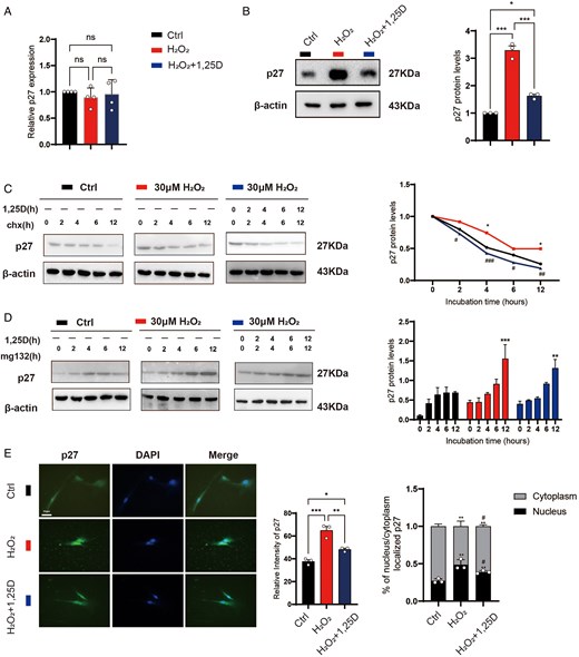
1,25-Dihydroxyvitamin D promotes proteasome-mediated degradation of P27 under oxidative stress. Human BMSCs were exposed to 30 μM H₂O₂ for 24 h, followed by incubation with 10⁻⁸ M 1,25(OH)₂D₃ for 12 h. (A) Levels of P27 mRNA. (B) P27 protein expression and corresponding quantitative analysis. (C) P27 protein levels after treatment with 10 μM cycloheximide CHX. (D) P27 protein levels after treatment with 10 μM MG132. (E) Immunofluorescence staining of P27, quantification of mean fluorescence intensity, and analysis of nuclear versus cytoplasmic P27 localization. Human BMSCs were derived from four independent donors. For qRT-PCR analysis, n = 4 independent donor-derived cell isolations per group; for Western blot and immunofluorescence analyses, n = 3 independent donor-derived cell isolations per group. ns, not significant; *, P < .05; **, P < .01; ***, P < .001 compared with the control group; #, P < .05 compared with the H₂O₂ group.

VDR knockdown increases P27 accumulation by inhibiting ubiquitin–proteasome–mediated degradation
To determine whether VDR is required for 1,25(OH)2D3-induced P27 degradation, we knocked down VDR in hBMSCs (Figure 3A) and examined P27 expression. VDR silencing did not affect P27 mRNA levels (Figure 3B) but significantly increased P27 protein abundance (Figure 3C), as confirmed by densitometric quantification derived from three independent technical replicates. CHX chase assays revealed that VDR knockdown markedly delayed P27 degradation (Figure 3D). Consistently, MG132 treatment resulted in greater P27 accumulation in VDR-knockdown cells than in control cells (Figure 3E). Immunofluorescence analysis showed increased overall P27 fluorescence intensity and enhanced nuclear localization following VDR knockdown (Figure 3F), which was confirmed by immunoblotting of nuclear extracts (Figure 3G). Furthermore, co-immunoprecipitation demonstrated that VDR knockdown reduced P27 ubiquitination while increasing total P27 protein levels; MG132 further amplified these effects (Figure 3H). These findings indicate that VDR deficiency promotes P27 accumulation by suppressing ubiquitin–proteasome–dependent P27 degradation.
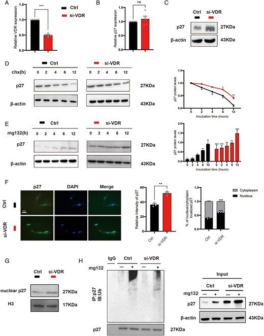
VDR knockdown increases P27 accumulation by inhibiting ubiquitin–proteasome–mediated degradation. Human BMSCs were transfected with si-VDR for 36 hours. (A) VDR knockdown efficiency. (B) P27 mRNA expression. (C) P27 protein expression. (D) Time-dependent changes in P27 protein levels were examined in control and VDR-knockdown cells treated with 10 μM cycloheximide (CHX). (E) P27 protein levels were analyzed in control and VDR-depleted cells following treatment with 10 μM MG132. (F) Immunofluorescence staining of P27 was conducted, followed by quantification of mean fluorescence intensity and assessment of nuclear versus cytoplasmic P27 distribution. (G) Nuclear P27 protein levels were determined. (H) Co-immunoprecipitation (Co-IP) assays were carried evaluating P27 ubiquitination. Left panel: Immunoprecipitation with anti-P27 antibody, followed by immunoblotting with anti-ubiquitin antibody to detect ubiquitinated P27 (high molecular weight smears). Right panel (Input): 5% of total cell lysate prior to IP, probed with anti-P27 to show total P27 protein levels. Human BMSCs were derived from five independent donors. For qRT-PCR analyses, n = 4 independent donor-derived cell isolations per group; for Western blot, immunofluorescence, nuclear protein extraction, and Co-IP analyses, n = 3 independent donor-derived cell isolations per group. Statistical significance is indicated as follows: ns, not significant; *P < .05; **P < .01; ***P < .001 compared with the control group.

1,25-dihydroxyvitamin D transcriptionally upregulates CDK2 via VDR to promote P27 phosphorylation
Given that CDK2-dependent phosphorylation regulates P27 ubiquitination and degradation,22,27,33 we hypothesized that 1,25(OH)2D3 promotes P27 phosphorylation and subsequent degradation by upregulating CDK2 expression through the vitamin D receptor (VDR). Treatment of hBMSCs with 10−8 M 1,25(OH)2D3 for 12 h significantly decreased P27 protein levels, increased P27 phosphorylation at Threonine 187, and upregulated CDK2 protein expression (Figure 4B), accompanied by increased CDK2 mRNA expression (Figure 4A). In contrast, treatment with the CDK2 inhibitor CVT-313 for 24 h reduced CDK2 mRNA and protein expression, decreased P27 Threonine 187 phosphorylation, and restored P27 protein levels (Figure 4A, B). EdU incorporation assays further showed that CVT-313 abrogated 1,25(OH)2D3-induced hBMSC proliferation (Figure 4C). Together, these findings demonstrate that 1,25(OH)2D3 promotes hBMSC proliferation through a CDK2-dependent mechanism involving P27 Threonine 187 phosphorylation and subsequent P27 degradation.
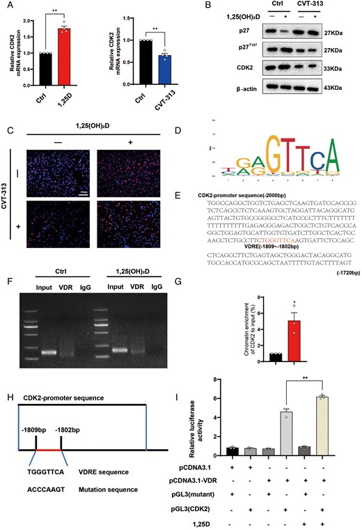
1,25-Dihydroxyvitamin D transcriptionally upregulates CDK2 via VDR to promote P27 phosphorylation. Human BMSCs were treated with 10⁻⁸ M 1,25(OH)₂D₃ for 12 hours, with or without pretreatment with the CDK2 inhibitor CVT-313 10 μM for 24 h. (A) CDK2 mRNA expression. (B) Western blot analysis of P27, phosphorylated P27 at Thr187, and CDK2. (C) EdU incorporation assays evaluating human BMSC proliferation. (D) Identification of putative VDRE-like sequences. (E) Predicted VDR-binding sites in the upstream promoter region of CDK2. (F) Agarose gel electrophoresis of PCR-amplified ChIP products. (G) Quantitative ChIP analysis of VDR enrichment at the CDK2 promoter. (H) Schematic representation of the pGL3-CDK2 promoter luciferase reporter construct and its corresponding VDRE-mutant construct. (I) Luciferase reporter assay assessing CDK2 promoter activity. Human BMSCs were derived from four independent donors. For qRT-PCR analyses, n = 4 independent donor-derived cell isolations per group; for all other quantitative analyses, n = 3 independent donor-derived cell isolations per group. *, P < .05; **, P < .01; ***, P < .001 versus control group.

Bioinformatic analysis revealed multiple putative vitamin D response elements (VDREs) within approximately 2 kb upstream of the CDK2 transcription start site (Figure 4D, E). Chromatin immunoprecipitation followed by PCR (ChIP-PCR) demonstrated direct binding of VDR to this promoter region, with 1,25(OH)2D3 treatment enhancing VDR enrichment (Figure 4F, G). In dual-luciferase reporter assays, 1,25(OH)2D3 significantly increased the transcriptional activity of the CDK2 promoter containing the wild-type VDRE, whereas mutation of the VDRE abolished this responsiveness (Figure 4H, I). Collectively, these findings indicate that 1,25(OH)2D3 activates CDK2 transcription via VDR binding to a functional VDRE in the CDK2 promoter, thereby promoting phosphorylation of P27 at Threonine 187 and facilitating its degradation.
P27 acts downstream of VDR to mediate the regulation of hBMSC proliferation and osteogenic differentiation
To determine whether P27 functions downstream of VDR in these processes, we modulated the expression of P27 and VDR using small interfering RNAs (siRNAs) and overexpression plasmids (Figure 5A–C). Knockdown of P27 significantly enhanced cell proliferation and osteogenic differentiation, whereas VDR knockdown produced the opposite effects, as evidenced by MTT assays, EdU incorporation, alkaline phosphatase (ALP) staining, and the expression of osteogenic markers (Figure 5D–H). Importantly, simultaneous knockdown of P27 partially reversed the inhibitory effects of VDR depletion on proliferation and osteogenic differentiation (Figure 5D–H). Conversely, overexpression of P27 suppressed both proliferation and osteogenesis, while VDR overexpression promoted these processes. Co-overexpression of P27 attenuated the pro-osteogenic effects induced by VDR overexpression (Figure 5I–M). These findings support a model in which P27 serves as a key downstream effector of VDR, mediating VDR-dependent control of hBMSC proliferation and osteogenic differentiation.
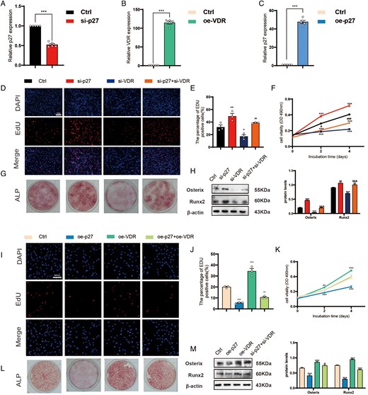
P27 acts downstream of VDR to mediate the regulation of hBMSC proliferation and osteogenic differentiation. (A) Transfection efficiency of si-P27 in human BMSCs at 36 h post-transfection. (B) Transfection efficiency of the VDR overexpression plasmid (oe-VDR) in human BMSCs at 36 h post-transfection. (C) Transfection efficiency of the P27 overexpression plasmid (oe-P27) in hBMSCs at 36 h post-transfection. hBMSCs were transfected with si-P27 and/or si-VDR for 36 h. (D) Representative images of EdU staining for the control, si-P27, si-VDR, and si-P27 + si-VDR groups. (E) Quantification of EdU-positive cells. (F) MTT assay results. (G) ALP staining. (H) Western blot analysis of the osteogenic marker proteins Osterix and Runx2; Western blot images are representative of at least three independent technical replicates, and densitometric quantification reflects the mean ± SEM of these replicates. hBMSCs were transfected with oe-P27 and/or oe-VDR for 36 h. (I) Representative images of EdU staining for the control, oe-P27, oe-VDR, and oe-P27 + oe-VDR groups. (J) Quantification of EdU-positive cells. (K) MTT assay results. (L) ALP staining. (M) Western blot analysis of the osteogenic marker proteins Osterix and Runx2; Western blot images are representative of at least three independent biological replicates, and densitometric quantification reflects the mean ± SEM of these replicates. For qRT-PCR-based transfection efficiency analyses, n = 5 independent donor-derived cell isolations per group. For MTT, EdU, Western blot, and ALP analyses, n = 3 independent donor-derived cell isolations per group. Statistical significance: ns, not significant; *, P < .05; **, P < .01; ***, P < .001 compared with the control group; #, P < .05; ##, P < .01; ###, P < .001 compared with the si-VDR group.

P27 knockout mitigates osteoporosis induced by reduced active vitamin D
To investigate whether P27 deficiency ameliorates osteoporosis resulting from reduced active vitamin D in vivo, we generated double-mutant mice lacking P27 and heterozygous for 1α(OH)ase (P27−/−1α(OH)ase+/−) and compared them with wild-type, P27−/−, and 1α(OH)ase+/− littermates using micro-computed tomography, histological and histochemical analyses, immunohistochemistry, and biomechanical testing. P27 knockout mice exhibited significantly higher bone mineral density, increased trabecular bone volume and number, enhanced type I collagen–positive area, and greater osteoblast numbers, whereas 1α(OH)ase+/− mice displayed opposing changes (Figure 6A–K). Notably, these parameters were substantially restored in P27−/−1α(OH)ase+/− mice relative to 1α(OH)ase+/− mice (Figure 6A–K). In contrast, trabecular separation and tartrate-resistant acid phosphatase (TRAP)-positive osteoclast surface were reduced in P27−/− mice, increased in 1α(OH)ase+/− mice, and significantly diminished in the double-mutant mice (Figure 6L, M). These results demonstrate that P27 deficiency partially reverses reduced active vitamin D–induced osteoporosis by promoting osteoblast-mediated bone formation and inhibiting osteoclast-mediated bone resorption.
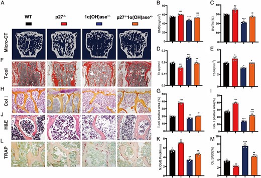
P27 knockout mitigates osteoporosis induced by reduced active vitamin D. Six-month-old littermate mice, including wild-type (WT), P27−/−, 1α(OH)ase+/−, and P27−/−1α(OH)ase+/− genotypes, were analyzed. (A) Three-dimensional reconstructions of micro–computed tomography (micro-CT) scans of the first lumbar vertebra. (B) Lumbar vertebral bone mineral density (BMD, mg/cm³). (C) Trabecular bone volume/total bone volume (BV/TV, %). (D) Trabecular number (Tb.N, mm⁻¹). (E) Trabecular separation (Tb.Sp, mm). (F) Microscopic images of paraffin sections stained for total collagen using histochemical methods. (G) Quantification of total collagen area. (H) Microscopic images of type I collagen (Col-I) immunohistochemical staining. (I) Percentage of Col-I–positive area (Col-I + area, %). (J) Hematoxylin and eosin (H&E)–stained images. (K) Osteoblast number per trabecular perimeter (N.Ob/B.Pm, #/mm). (L) Microscopic images of tartrate-resistant acid phosphatase (TRAP) histochemical staining. (M) TRAP-positive osteoclast surface per bone surface (Oc.S/BS, %). For all quantitative analyses, n = 4 mice per genotype. Statistical significance: *P < .05, **P < .01, ***P < .001 compared with WT; #P < .05, ##P < .01, ###P < .001 compared with the 1α(OH)ase+/− group.

P27 knockout prevents bone senescence induced by reduced active vitamin D
To determine whether the protective effects of P27 deletion are linked to reduced skeletal senescence, we assessed markers of oxidative stress, DNA damage, cellular senescence, and the senescence-associated secretory phenotype (SASP) in vertebral bone tissues across genotypes. The proportion of SOD2-positive osteoblasts and SOD2 protein levels were elevated in P27−/− mice but significantly reduced in 1α(OH)ase+/− mice; these alterations were largely reversed in P27−/−1α(OH)ase+/− double-mutant mice (Figure 7A, B, K). In contrast, the proportions of p16-, β-galactosidase-, IL-6-, and IL-1β–positive osteocytes, as well as protein levels of γ-H2A.X, p53 and p16, were decreased in P27−/− mice, increased in 1α(OH)ase+/− mice, and markedly reduced in the double mutants (Figure 7C–K). For markers of oxidative stress, senescence and SASP (SOD2, p16 and β-galactosidase, IL-6, IL-1β), positive cells were quantified specifically within osteocytes embedded in the cortical lacunae. These findings indicate that P27 deficiency mitigates skeletal senescence associated with vitamin D deficiency by attenuating oxidative stress, DNA damage, and SASP.
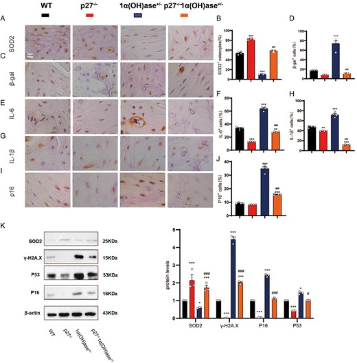
P27 knockout attenuates bone senescence induced by reduced active vitamin D. Cortical bone sections from the lumbar vertebrae of 6-month-old wild-type (WT), P27⁻/⁻, 1α(OH)ase⁺/⁻, and P27⁻/⁻1α(OH)ase⁺/⁻ littermate mice were analyzed. (A, B) Representative immunohistochemical images of the antioxidant protein SOD2 and quantification of SOD2-positive cells. (C, D) Representative images of senescence-associated β-galactosidase (β-gal) staining and quantification of β-gal–positive cells. (E, F) Representative immunohistochemical images of IL-6 and quantification of IL-6–positive cells. (G, H) Representative immunohistochemical images of IL-1β and quantification of IL-1β–positive cells. (I, J) Representative immunohistochemical images of p16 and quantification of p16-positive cells. (K) Western blot analysis and quantitative assessment of SOD2, γ-H2A.X, p53 and p16 protein expression levels in vertebral tissues across the four genotypes. For markers of oxidative stress, senescence and SASP (SOD2, IL-6, p16 and β-galactosidase, IL-1β), positive cells were quantified specifically within osteocytes embedded in the cortical lacunae. For immunohistochemical staining quantifications, n = 4 mice per genotype. For Western blot analyses, n = 3 mice per genotype. Statistical significance: *P < .05, **P < .01, ***P < .001 compared with WT mice; #P < .05, ##P < .01, ###P < .001 compared with the 1α(OH)ase⁺/⁻ group.

P27 knockout restores osteogenic function and alleviates senescence in BMSCs under conditions of reduced active vitamin D
To investigate whether P27 knockout prevents osteogenic impairment and senescence of BMSCs induced by reduced active vitamin D, we assessed proliferation, differentiation, oxidative stress, and senescence-related markers in ex vivo cultured BMSCs from genetically distinct mouse strains. Results demonstrated significantly increased proportions of EdU-positive cells, ALP-positive areas, and Osterix and Runx2 protein expression in P27−/− BMSCs, whereas these parameters were markedly reduced in 1α(OH)ase+/− BMSCs. In P27−/−1α(OH)ase+/− mice, these parameters showed marked recovery compared with the 1α(OH)ase+/− group (Figure 8A–D, I). Conversely, ROS levels and the proportion of SA-β-gal-positive cells decreased in P27−/− BMSCs, increased in 1α(OH)ase+/− cells, and were significantly reduced in the double-mutant group (Figure 8E–H). These data demonstrate that P27 knockout directly decreases senescence in BMSCs, expanding the functional stem cell pool (increased proliferation and osteogenic differentiation) to directly supply osteoblasts for bone formation, while simultaneously reducing SASP-mediated paracrine damage to the bone microenvironment.
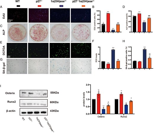
P27 knockout restores osteogenic function and alleviates senescence in BMSCs under conditions of reduced active vitamin D. BMSCs were isolated from wild-type (WT), P27⁻/⁻, 1α(OH)ase⁺/⁻, and P27⁻/⁻1α(OH)ase⁺/⁻ mice. (A) Representative images of EdU staining. (B) Quantification of EdU-positive cells. (C) Representative images of alkaline phosphatase (ALP) staining. (D) Quantification of ALP-positive area. (E) Representative images of DCFDA fluorescence staining. (F) Quantification of intracellular reactive oxygen species (ROS) levels. (G) Representative images of senescence-associated β-galactosidase (SA-β-gal) staining. (H) Quantification of SA-β-gal–positive cells. (I) Western blot analysis and quantification of osteogenic marker proteins—Osterix, Runx2—in BMSCs from the four genotypes. For all analyses, n = 3 independent mouse-derived BMSC isolations per genotype. Statistical significance: *P < .05, **P < .01, ***P < .001 versus WT; #P < .05, ##P < .01, ###P < .001 versus 1α(OH)ase⁺/⁻ group.

Discussion
In this study, we identified a novel mechanism by which active vitamin D preserves skeletal health and delays bone aging. We demonstrated that 1,25(OH)2D deficiency induces pathological accumulation of the cell-cycle inhibitor P27 in bone tissue, contributing to osteopenia, impaired BMSC function, and cellular senescence. Genetic deletion of P27 significantly ameliorated bone loss and senescence in 1α(OH)ase-deficient mice, establishing P27 as a critical downstream effector of VDR signaling in the skeleton. However, the partial rescue observed indicates that P27-independent mechanisms also contribute. Previous studies have identified additional VDR-dependent pathways in skeletal aging, including the EZH2–H3K27me3 axis, the Nrf2 antioxidant pathway, and the classical P16/Rb and P53/P21 senescence pathways.7,8 Thus, although targeting P27 turnover is promising, complete phenotypic rescue may require multi-targeted strategies addressing parallel senescence pathways.
Our findings agree with previous reports that VDR signaling maintains BMSC stemness and proliferative capacity.34 We extend this knowledge by defining a molecular hierarchy in which VDR transcriptionally activates CDK2, which phosphorylates P27 at Threonine 187 and promotes its ubiquitin-mediated proteasomal degradation. This is consistent with canonical P27 regulation, whereby SCFSkp2-mediated ubiquitination requires prior CDK-dependent phosphorylation.35,36 By sustaining VDR and CDK2 activity, active vitamin D maintains P27 turnover and prevents senescence-associated cell-cycle arrest. Conversely, 1,25(OH)2D deficiency permits P27 accumulation, locking cells in a senescent state characterized by elevated p16, p53, and SASP factors.37,38
A key advance is showing that oxidative stress and VDR signaling converge on post-translational P27 regulation. ROS does not increase P27 transcription but stabilizes P27 by inhibiting proteasome activity and impairing CDK2-mediated phosphorylation at Threonine 187. Active vitamin D counteracts this through VDR-dependent CDK2 upregulation, restoring P27 phosphorylation and ubiquitin–proteasome degradation. Because ROS can inhibit proteasomes independently of VDR, vitamin D alone may not clear P27 under high oxidative stress. Combining active vitamin D with antioxidants may therefore offer a synergistic strategy for age-related osteoporosis.
P27-associated senescence may contribute to osteoporosis through a dual hit: cell-autonomous arrest depletes the osteogenic BMSC pool, while the SASP creates a toxic microenvironment that promotes osteocyte senescence and stimulates osteoclastic bone resorption. The observation that P27 knockout attenuates oxidative stress and DNA damage during vitamin D deficiency is particularly intriguing. Although P27 is traditionally viewed as a cell-cycle brake, recent evidence suggests that it also regulates apoptosis and autophagy under stress.39 Our data suggest that persistent P27 accumulation may exacerbate oxidative stress-induced damage, possibly by impairing repair mechanisms or reinforcing the SASP feedback loop.40,41 Reduced SASP cytokines, including IL-6 and IL-1β, in double-mutant mice supports the concept that targeting senescent cells or their pathways through senolytic or senomorphic strategies may be useful for osteoporosis.42
Although our study highlights the VDR–CDK2–P27 axis in BMSCs, the global nature of the knockout models means that the osteoporotic phenotype likely reflects cumulative alterations across multiple skeletal cell types. Osteoclasts express VDR and may exhibit altered resorptive activity, while osteocytes, key regulators of bone remodeling, are susceptible to senescence under vitamin D insufficiency. In addition, systemic factors preserved in the 1α(OH)ase+/− model, such as normal PTH, may interact with skeletal outcomes. Therefore, our in vivo conclusions are framed at the level of skeletal integrity, with BMSC dysfunction representing a major, but not exclusive, driver of bone loss.
Clinically, these findings emphasize the importance of adequate vitamin D levels for preserving the regenerative capacity of bone stem cells. However, large randomized controlled trials, such as the VITAL trial, have not consistently shown that vitamin D supplementation reduces fracture risk in unselected, generally healthy populations.2 Our mechanistic data suggest that skeletal benefits mediated through the VDR–CDK2–P27 axis may be most relevant to specific high-risk groups, including individuals with documented 1,25(OH)2D insufficiency, VDR polymorphisms, or intrinsic BMSC senescence. Nevertheless, therapeutic targeting of this pathway requires caution. P27 is a canonical tumor suppressor, and systemic P27 depletion or uncontrolled CDK2 activation could predispose patients to oncogenic transformation.18,35 Thus, future translational efforts should prioritize bone-targeted delivery systems, such as bisphosphonate-conjugated compounds or bone-seeking nanoparticles, to ensure tissue specificity, or focus on senomorphic agents that modulate P27 activity rather than eliminate it.
This study has limitations. The genetically modified models provide a robust platform for dissecting molecular consequences of impaired vitamin D activation, but they represent an isolated perturbation and do not fully recapitulate the multifactorial etiology of common human osteoporosis. Age-related and postmenopausal bone loss involve intersecting pathways, including estrogen deficiency, metabolic stress, and systemic inflammation. Therefore, our findings may be most relevant to subpopulations with impaired vitamin D activation or VDR signaling rather than the general aging population. Another limitation is that in vitro mechanistic studies used BMSCs from a small cohort of older female osteoarthritis patients (n = 5). Although OA primarily involves cartilage degeneration rather than systemic osteoporosis, the skeletal microenvironment of OA patients may differ from that of individuals with osteoporosis. Future studies using BMSCs from osteoporotic donors are needed to validate generalizability. Finally, the E3 ligase responsible for VDR-mediated P27 degradation in bone remains to be confirmed, although Skp2 is the most likely candidate.36
Conclusion
In summary, our study identifies P27 as a key downstream effector of active vitamin D signaling in bone. We show that 1,25(OH)2D3 promotes VDR-dependent transcription of CDK2, enhances P27 phosphorylation at Threonine 187, and accelerates ubiquitin–proteasome-mediated P27 degradation. Through this VDR–CDK2–P27 axis, active vitamin D preserves BMSC proliferation and osteogenic differentiation while limiting oxidative stress, senescence, and bone loss. Genetic deletion of P27 partially rescues the osteoporotic phenotype caused by reduced active vitamin D production in vivo, although P27-independent mechanisms also contribute. These findings provide mechanistic insight into vitamin D-mediated skeletal protection and suggest that targeting P27 turnover may be a therapeutic strategy for osteoporosis associated with reduced vitamin D, provided that tissue-specific delivery approaches are developed to mitigate oncogenic risk.
Supplementary material
Supplementary material is available at European Journal of Endocrinology online.
Authors’ contributions
Fangrong Xu (Data curation, Formal analysis, Investigation, Methodology, Software, Validation, Visualization, Writing—original draft [equal]), Quan Liu (Data curation, Formal analysis, Investigation, Methodology, Software, Validation, Visualization [equal]), Mingxin Huang (Formal analysis, Investigation, Methodology, Validation [supporting]), Jing Wang (Data curation, Formal analysis, Funding acquisition, Investigation, Validation [supporting]), David Goltzman (Conceptualization, Writing—review & editing [equal], Resources [supporting]), Bingjie Gu (Conceptualization, Project administration, Supervision, Writing—review & editing [equal]), and Dengshun Miao (Conceptualization, Funding acquisition, Project administration, Resources, Supervision, Writing—review & editing [equal])
Funding
This work was supported by the National Natural Science Foundation of China (82172504 to DM; 82302935 to JW).
Data availability
All data and materials used in the study are available to any researcher for purposes of reproducing or extending the analysis.
Ethics approval and consent to participate
All animal procedures were approved by the Committee on the Ethics of Animal Experiments of Nanjing Medical University. Human bone marrow aspirates were obtained with written informed consent from patients undergoing total hip arthroplasty, and the protocol was approved by the Institutional Review Board of Nanjing Medical University. All human research was conducted in accordance with the Declaration of Helsinki.
References
Author notes
Fangrong Xu and Quan Liu contributed equally to this work.
Conflicts of interest: The authors declare no competing interests.